
Device and method for expanding the spinal canal with spinal column stabilization and spinal deformity correction

a technology of spinal column stabilization and spinal canal, applied in the field of spinal surgery, can solve the problems of spinal nerves, symptoms of leg or back pain, walking problems, and current techniques for correcting these deformities are either marginally effective or highly invasive, and achieve the effect of lengthening the spinal pedicl

Inactive Publication Date: 2015-06-02
INNOVATIVE SURGICAL DESIGNS
View PDF57 Cites 0 Cited by
  • Summary
  • Abstract
  • Description
  • Claims
  • Application Information

AI Technical Summary

Benefits of technology

[0011]The present invention comprises a novel medical device capable of lengthening the spinal pedicle and securing the pedicle to a longitudinal member such as a rod or plate to achieve fixation of two or more vertebrae. The current invention is capable of correcting a narrowed spinal canal, while allowing the vertebral segment to be incorporated into a spinal stabilization construct. In addition, the device is useful when treating deformed spines (particularly those spines with excessive kyphosis, scoliosis or a rotational deformity) and provides a biomechanically superior vertebral anchorage compared to the use of a traditional pedicle screw.
[0014]provide an improved ability to correct a spinal deformity (particularly scoliosis, kyphosis or a rotational deformity) while allowing longitudinal fixation of the spinal column;
[0015]allow a surgeon to connect a longitudinal member (such as a plate or rod) onto a pedicle lengthening device so that a stabilization procedure can be performed at the site of a prior pedicle lengthening without the need to remove or revise the pedicle lengthening device;
[0022](1) both the correction of spinal stenosis and stabilization of the spinal column can be performed through a single device allowing a much broader range of spinal pathology to be addressed;
[0026](5) a minimally invasive spinal device is provided that can produce both decompression of stenotic regions of the spinal canal in addition to spinal fixation for a spinal stabilization procedure; both procedure being achieve through small or percutaneous incisions.

Problems solved by technology

This disease leads to compression of the spinal nerves and produces symptoms of leg or back pain, walking problems, weakness and / or numbness in the affected portions of the body.
Many patients with spinal stenosis also have co-existent spinal problems such as a spinal deformity or spinal instability.
Current techniques for correcting these deformities are either marginally effective or highly invasive.
Current techniques for fixation of poor quality bone have major drawbacks and in many cases are marginally effective.

Method used


Image

  • Device and method for expanding the spinal canal with spinal column stabilization and spinal deformity correction
  • Device and method for expanding the spinal canal with spinal column stabilization and spinal deformity correction
  • Device and method for expanding the spinal canal with spinal column stabilization and spinal deformity correction

Examples


Embodiment Construction

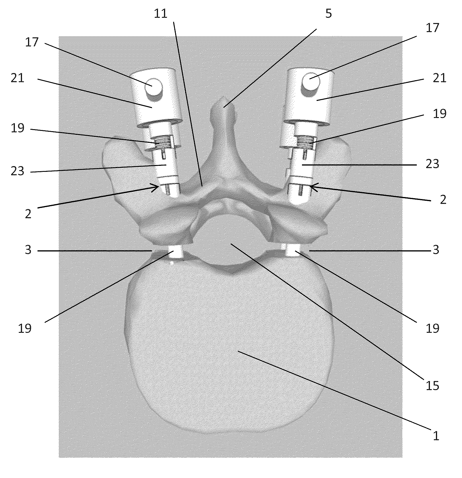
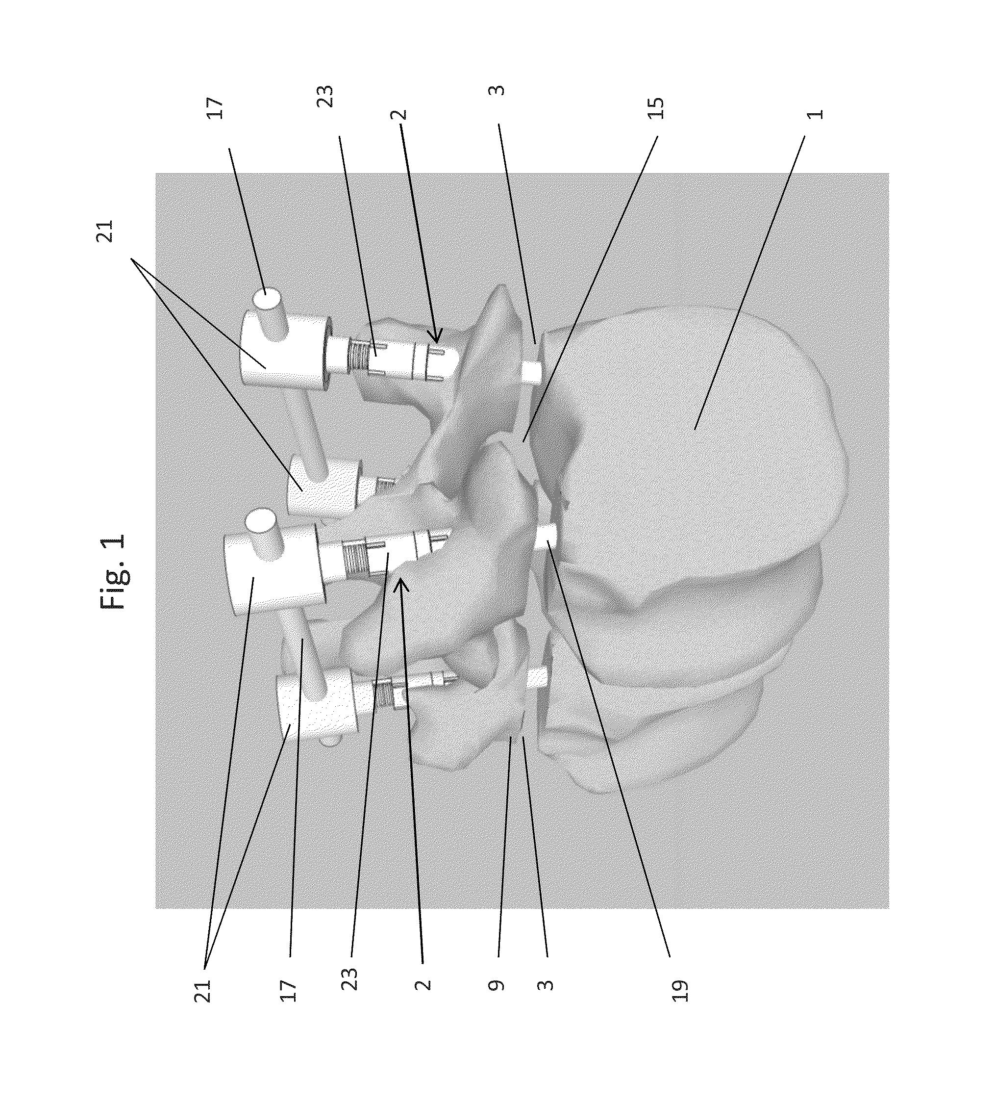
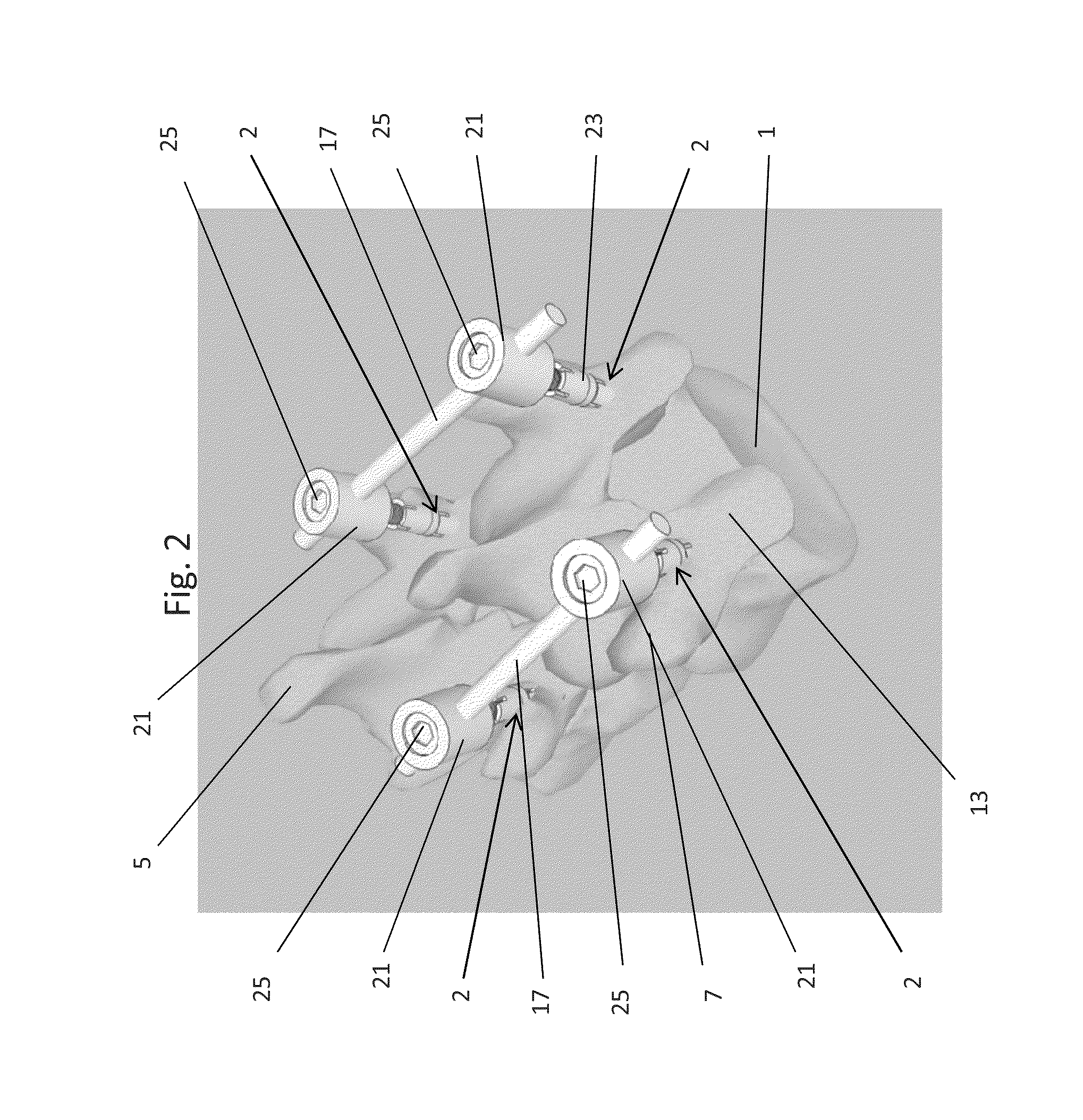
[0036]Referring now to the figures, where like numerals indicate like elements, there is shown in FIG. 1 a perspective view of a pedicle lengthening and stabilization construct, in accordance with one embodiment of the present invention, implanted within two adjacent vertebrae that have undergone pedicle lengthening. Pedicle lengthening implants 2 can be seen implanted into the pedicles 9 of the two adjacent vertebrae.

[0037]The pedicle lengthening implants 2 have been extended to create a gap 3 at the base of the pedicle 9 and thus expand the dimensions of the spinal canal 15. Techniques and implants (devices) for pedicle lengthening have been described in detail in U.S. Pat. No. 7,166,107, issued Jan. 23, 2007, entitled “Percutaneous Technique and Implant for Expanding the Spinal Canal;” and in U.S. application Ser. No. 12 / 624,946, filed Nov. 24, 2009, (US Publication No. 2010 / 0168751), entitled “Method, Implant & Instruments for Percutaneous Expansion of the Spinal Canal.” U.S. Pa...


PUM


Abstract

Spinal canal expansion through pedicle lengthening in combination with spinal column stabilization, to treat spinal stenosis, spondylolisthesis, kyphosis, scoliosis or a major spinal rotational deformity, with or without a spinal fusion. A device or implant that includes a pedicle lengthening implant to decompress (expand) the spinal canal, and a bridge to connect two or more pedicle lengthening implants and / or pedicle screws or bone anchors to achieve simultaneous spinal stabilization across one or more vertebral segments. The bridge across the vertebral segments can include a longitudinal member, such as a plate or rod. The pedicle lengthening implant is originally made, or can be modified to, connect to the longitudinal member. Spinal deformity can be manipulated, through operation of one or more pedicle lengthening implants in respective vertebral segments, to improve a relative relationship between the respective vertebra segments, the longitudinal member then fixating the vertebral segments into a multi-level spinal construct.

Description

RELATED APPLICATIONS[0001]This application is a continuation-in-part (CIP) of U.S. Ser. No. 12 / 624,946, filed Nov. 24, 2009; which application is a continuation-in-part (CIP) of U.S. Ser. No. 11 / 656,790, filed Jan. 22, 2007; which application is a continuation of U.S. Ser. No. 10 / 102,525, filed Mar. 19, 2002 (now U.S. Pat. No. 7,166,107).[0002]This application also claims benefit of U.S. Provisional Application Ser. No. 61 / 318,441, filed Mar. 29, 2010. U.S. Ser. No. 12 / 624,946, filed Nov. 24, 2009, claims benefit of U.S. Provisional Application Ser. No. 61 / 117,726, filed Nov. 25, 2008. All of the above-identified related applications are incorporated herein by reference.FIELD OF THE INVENTION[0003]The present invention relates generally to spinal surgery, and more particularly to a method and device for expanding the spinal canal when treating spinal stenosis and for simultaneously providing fixation between vertebrae for fusion and non-fusion applications; namely, for correction of...

Claims


Application Information

Patent Timeline
Patent Type & Authority Patents(United States)
IPC IPC(8): A61B17/56A61B17/70A61B17/86
CPCA61B17/7071A61B17/7001A61B17/8665A61B17/8685
Inventor ANDERSON, D. GREGTURNER, BARRYSPENCINER, DAVID
Owner INNOVATIVE SURGICAL DESIGNS
Learn More