
Multi-spectral tissue imaging

a tissue imaging and multi-spectral technology, applied in the field of tissue imaging, can solve the problems of inability to resolve depth, near impossible to separate spectral information from rgb image, and compromise of accuracy, so as to improve detection, classification and understanding of tissue pathologies.

Active Publication Date: 2014-09-30
CANFIELD SCI
View PDF26 Cites 38 Cited by
  • Summary
  • Abstract
  • Description
  • Claims
  • Application Information

AI Technical Summary

Benefits of technology

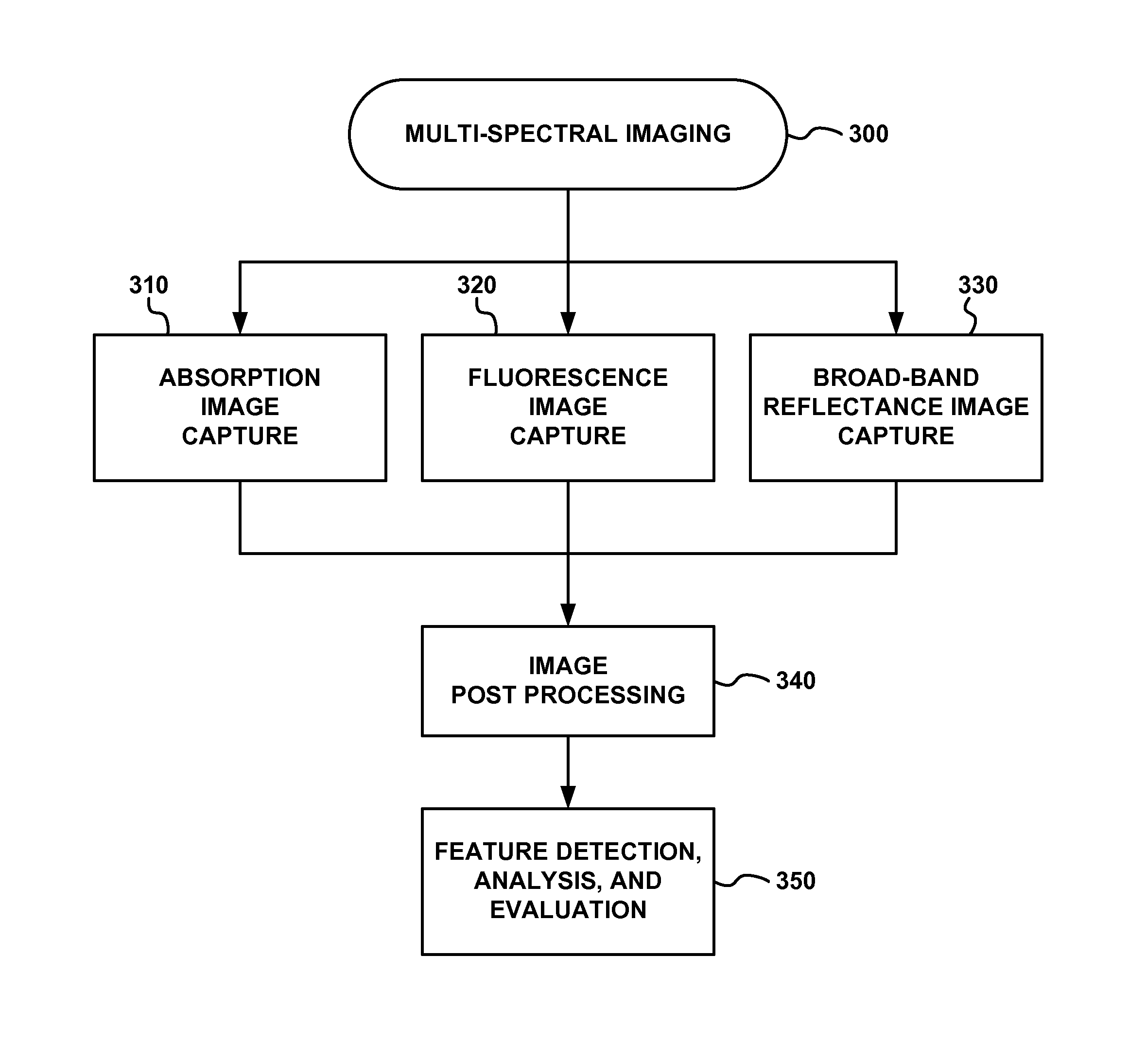
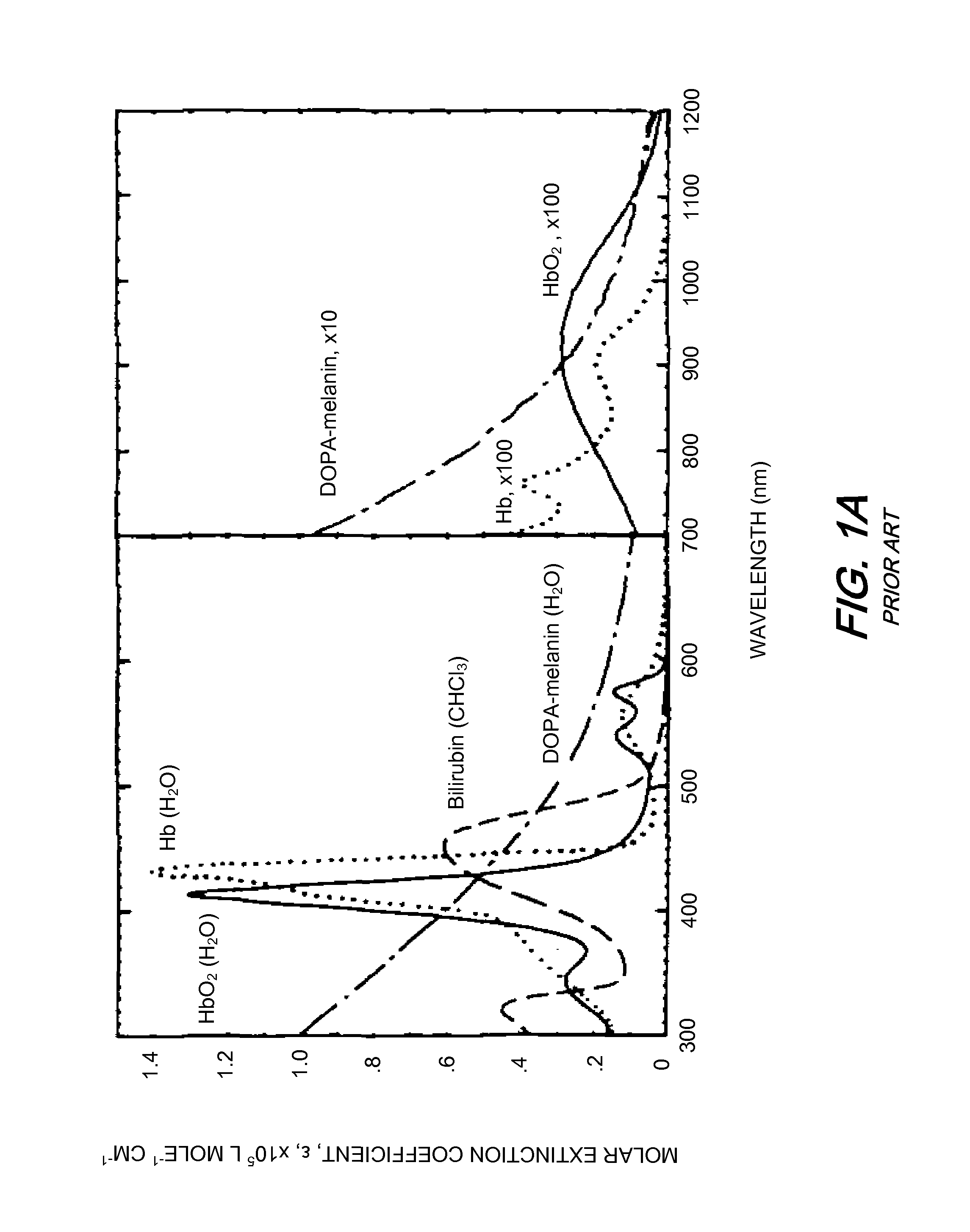
The patent describes an apparatus that can capture images by absorbing light at different wavelengths. This is done by selecting the illumination wavelength based on the absorption spectrum of a specific chromophore, such as melanin, oxy-hemoglobin, deoxy-hemoglobin, or water. This allows for the isolation, detection, and display of the chromophore. In simple terms, the patent describes how to capture and analyze different colors using a specialized device.

Problems solved by technology

A significant drawback of the planar imaging technique is that it cannot resolve depth and does not account for non-linear dependencies of the signal detected on propagation depth and the surrounding tissue.
Additionally, it is wrongly assumed that the camera provides the required spectral separation between the red, green and blue color bands without any overlap.
It is nearly impossible to separate the spectral information from the RGB image, even by using a very accurate camera model.
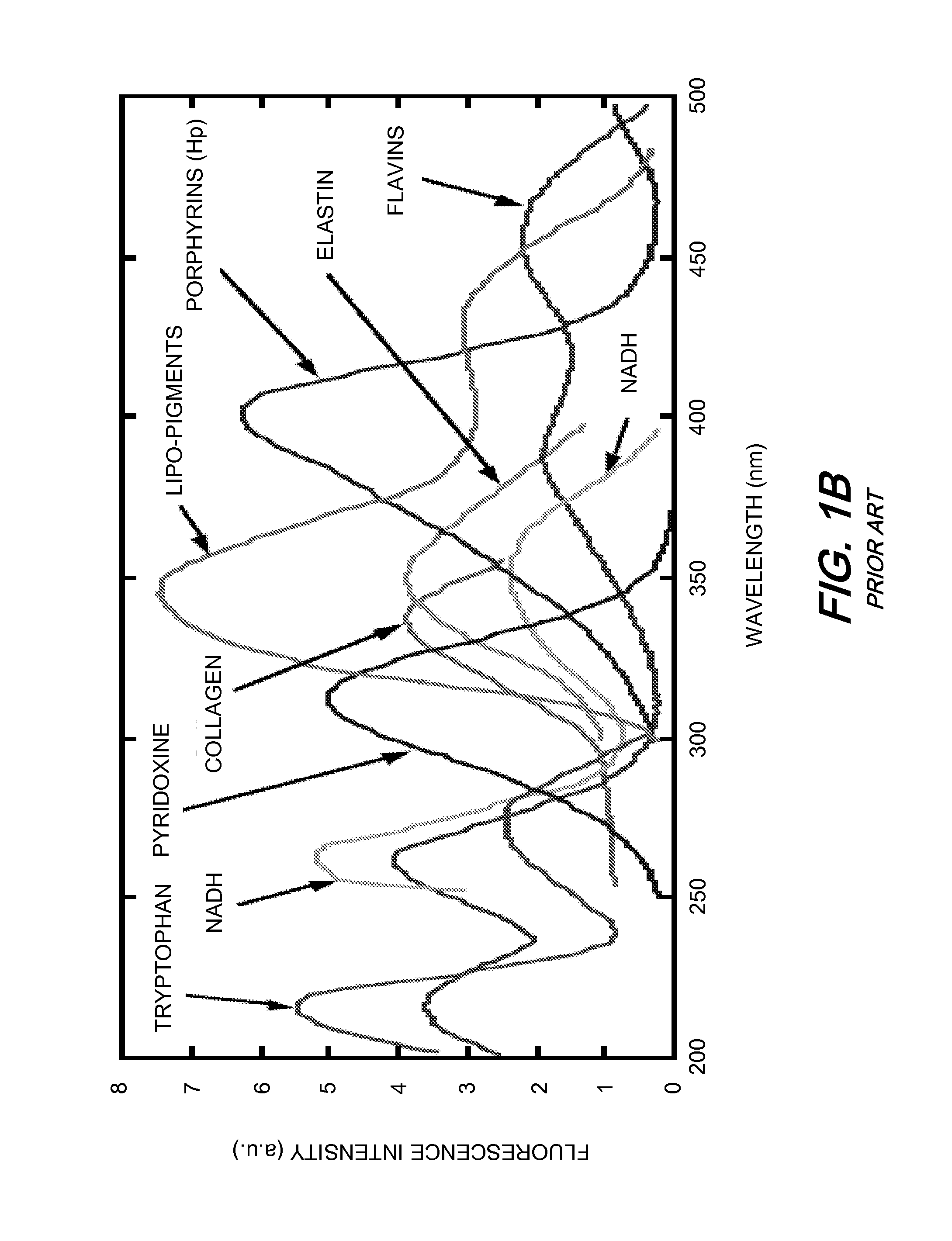
Without such separation, accuracy is compromised when attempting to analyze the blue, green, and red channels for collagen, horns and porphyrin fluorescence, respectively.
In doing so, scientifically significant spectral information within the data is corrupted.
Furthermore, superficial fluorescence activity may reduce the contrast of underlying activity from being detected owing to the simple projection viewing.
An additional limitation of known UV-excited fluorescence photography techniques is that they do not account for the non-uniformity of the illuminating light distribution in the fluorescence measurement and subsequent analysis.
Considering the fluorescence from a particular fluorophore of interest as the signal and the fluorescence from other fluorophores as the noise, the above-discussed problems of known UV-excited fluorescence photography techniques lead to poor signal-to-noise ratios in the individual R, G, and B channels of images captured using such techniques.
The simultaneous capture of various fluorescence spectral bands with overlapping detector response introduces noise.
Finally, the excitation (illumination) wavelength band selected is not optimized for imaging fluorophores of interest, such as collagen, horn, and porphyrin.

Method used


Image

  • Multi-spectral tissue imaging
  • Multi-spectral tissue imaging
  • Multi-spectral tissue imaging

Examples


Embodiment Construction

[0052]FIG. 2A schematically depicts an exemplary embodiment of a multi-spectral imaging system 200, in accordance with the present invention, for capturing images of tissue to be studied, such as skin. Illumination from one or more sources 210 is shone onto a subject's skin or a tissue sample 230 through a respective filtering element 215 and captured by a detector 220 through a filtering element 225. Each filtering element 215, 225 preferably comprises a plurality of filters, each of which can be selectively placed in the respective light path of the filtering element. (Note that the term “light” as used herein is not necessarily limited to humanly visible electromagnetic radiation, and may include portions of the electromagnetic spectrum above and below the visible range.) An exemplary embodiment of a filter wheel arrangement that may be used for filtering elements 215 and / or 225 is shown in greater detail in FIG. 2B, described below.

[0053]Preferably, the detector 220 comprises a ...


PUM


Abstract

Apparatus and methods are disclosed for multi-spectral imaging of tissue to obtain information about the distribution of fluorophores and chromophores in the tissue. Using specific spectral bands for illumination and specific spectral bands for detection, the signal-to-noise ratio and information related to the distribution of specific fluorophores is enhanced as compared to UV photography, which uses a single RGB image. Furthermore, the chromophore distribution information derived from the multi-spectral absorption images can be used to correct the fluorescence measurements. The combined fluorescence, absorption, and broadband reflectance data can be analyzed for disease diagnosis and skin feature detection.

Description

RELATED PATENT APPLICATION[0001]The present application claims priority from U.S. Provisional Patent Application No. 60 / 990,164, filed on Nov. 26, 2007, which is incorporated herein by reference in its entirety.FIELD OF THE INVENTION[0002]The present invention relates to the field of tissue imaging, and in particular, imaging the fluorescence or absorption of tissue such as skin. The present invention also relates to image processing, analysis, and segmentation, particularly as it relates to multi-spectral fluorescence and / or absorption images.BACKGROUND INFORMATION[0003]Light-induced fluorescence has been identified as a powerful noninvasive method for tissue pathology recognition and monitoring [1]. (Numbers in brackets refer to references listed under REFERENCES below.) A major advantage of fluorescence imaging is that fluorophores can be detected at low concentrations using harmless, non-ionizing radiation that can be applied repeatedly to the patient.[0004]In fluorescence imagi...

Claims


Application Information

Patent Timeline
Patent Type & Authority Patents(United States)
IPC IPC(8): A61B5/00
CPCA61B5/0059A61B5/0064A61B5/442A61B5/445A61B5/0075A61B5/443A61B5/444A61B5/0071A61B2576/00A61B5/742
Inventor PATWARDHAN, SACHIN, V.
Owner CANFIELD SCI
Learn More