
Method for ablation volume determination and geometric reconstruction

a volume determination and geometric reconstruction technology, applied in the field of electronic surgical equipment, systems and methods, can solve the problems of inaccurate reporting of achieved volumes, inexact measurements, inconsistent recording,

Active Publication Date: 2014-09-16
COVIDIEN LP
View PDF147 Cites 28 Cited by
  • Summary
  • Abstract
  • Description
  • Claims
  • Application Information

AI Technical Summary

Problems solved by technology

Conventional methods such as this often provide inexact measurements, inconsistent recordings, as well as inaccurate reporting of achieved volumes.
Further, conventional methods of volumetric calculation lack evaluative tools such as determining the effect of adjacent structures on the ablation volume, qualifying the completeness of the ablation volume, predicting specific volumes and / or shapes based on a given energy applicator configuration.

Method used


Image

  • Method for ablation volume determination and geometric reconstruction
  • Method for ablation volume determination and geometric reconstruction
  • Method for ablation volume determination and geometric reconstruction

Examples


example

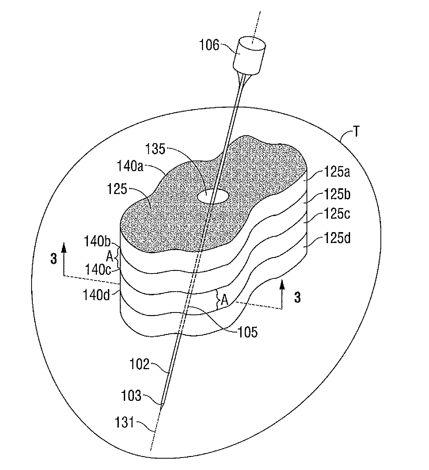
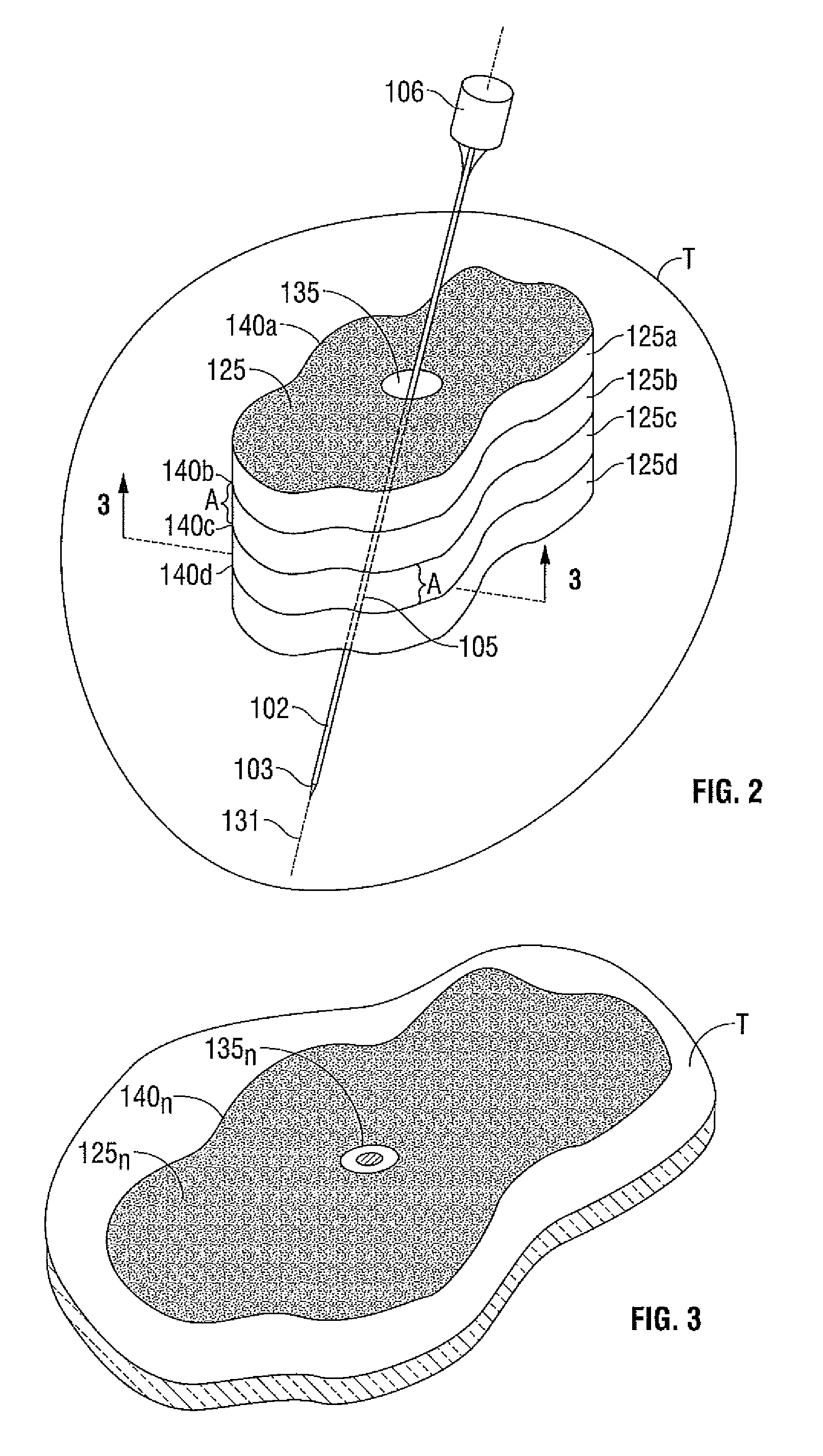
[0029]Trypan blue staining. An ablation lesion was created in porcine liver tissue. The ablation lesion was then excised and sliced into cross-sectional slices. A staining solution of trypan blue (0.4% trypan blue, 0.81% sodium chloride, 0.06% potassium phosphate dibasic) from Sigma-Aldrich of St. Louis, Mo. was placed in a beaker placed under a fume hood. The slice, with a pertinent surface containing a segment of the ablation volume was exposed to the solution in the beaker for approximately 5 minutes. The slice was then removed and was washed vigorously in a rinsing solution of 1× phosphate buffered saline solution. The rinsed and stained slice was then dried by absorbing the staining and rinsing solutions. The dried slice was then imaged by scanning the slice. The staining procedure was repeated for each of the slices of the ablated lesion.


PUM

PropertyMeasurementUnit
temperatureaaaaaaaaaa
thickaaaaaaaaaa
volumeaaaaaaaaaa

Abstract

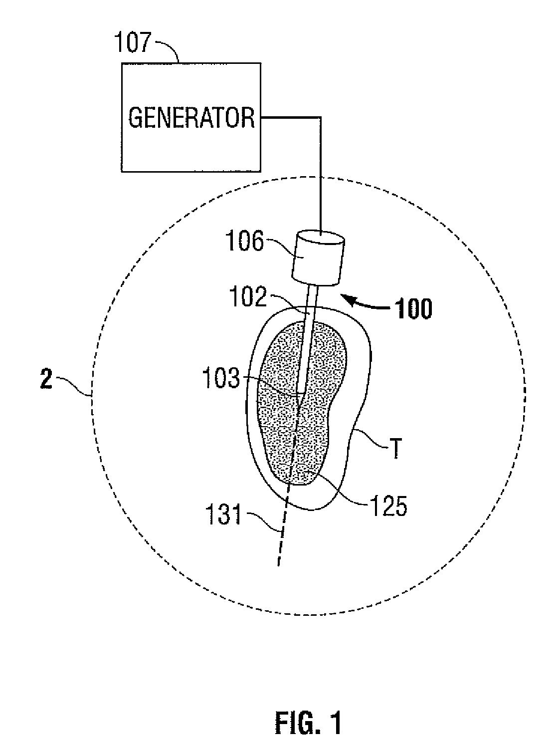
A method for determining a volume of ablated tissue is provided. The method includes the steps of: supplying energy to tissue to create an ablation volume therein; excising one or more slices of the tissue having a portion of the ablation volume therein; and subjecting at least a portion of the slice to a first contrast agent for a predetermined period of time, wherein the contrast agent is configured to color dead cells within the ablation volume.

Description

CROSS-REFERENCE TO RELATED APPLICATIONS[0001]This application claims the benefit of priority to U.S. Provisional Application Ser. No. 61 / 084,489 entitled “METHOD FOR ABLATION VOLUME DETERMINATION AND GEOMETRIC RECONSTRUCTION” filed Jul. 29, 2008 by Prakash Manley, which is incorporated by reference herein.BACKGROUND[0002]1. Technical Field[0003]The present disclosure relates to electrosurgical apparatuses, systems and methods. More particularly, the present disclosure is directed to a system and method for determining the volume of an ablation lesion after a tissue ablation procedure utilizing electrosurgical electrodes.[0004]2. Background of Related Art[0005]Energy-based tissue treatment is well known in the art. Various types of energy (e.g., electrical, ultrasonic, microwave, cryogenic, heat, laser, etc.) are applied to tissue to achieve a desired result. Electrosurgery involves application of high radio frequency electrical current to a surgical site to cut, ablate, coagulate or...

Claims


Application Information

Patent Timeline
Patent Type & Authority Patents(United States)
IPC IPC(8): A61B18/04A61B18/18G01N33/50
CPCG01N33/5091A61B18/18A61B18/1815A61B2018/00577A61B18/14G01N33/5005
Inventor MANLEY, PRAKASH
Owner COVIDIEN LP
Learn More