Imaging method and use thereof

a technology of atomic force microscopy and imaging method, which is applied in the field of imaging method and use thereof, can solve the problems of difficult reproducible quantification, difficult identification of distinct structures, and hampered fluorescence identification

Active Publication Date: 2014-08-05
RIETHMULLER CHRISTOPH
View PDF2 Cites 2 Cited by
  • Summary
  • Abstract
  • Description
  • Claims
  • Application Information

AI Technical Summary

Benefits of technology

[0015]It is intended that the analysed area is less than the surface of one cell, wherein the cell is preferably an analysed eukaryotic, more preferably a mammalian cell. Using overlapping areas for the analysis makes it possible to obtain the overview over a larger area by assembling them.

Problems solved by technology

But distinct structures are difficult to identify, especially on whole cells, where only cytoskeletal structures are obviously recognisable.
However, since morphological features are difficult to classify, a reproducible quantification is hard to perform.
However, the fluorescence identification is hampered by mutually disturbing fixation protocols for AFM and FM.

Method used


Image

  • Imaging method and use thereof
  • Imaging method and use thereof

Examples


Embodiment Construction

[0037]The invention will be further described by figures and examples without being limited to the described embodiments:

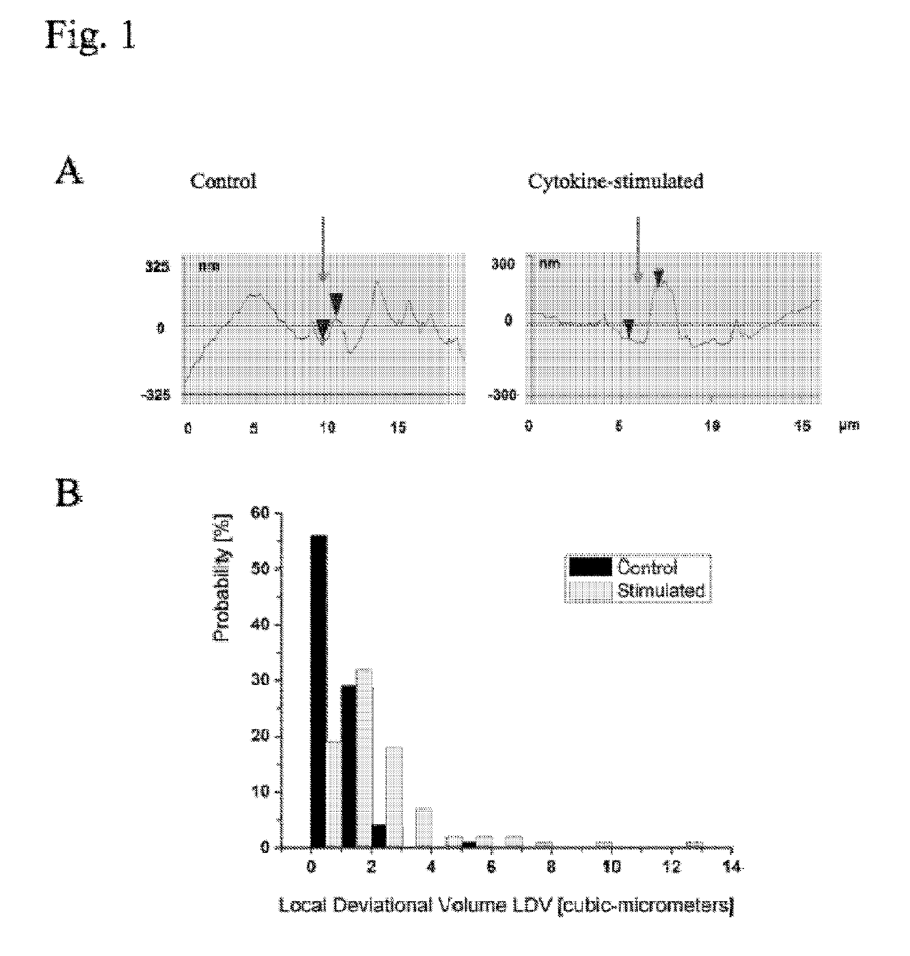
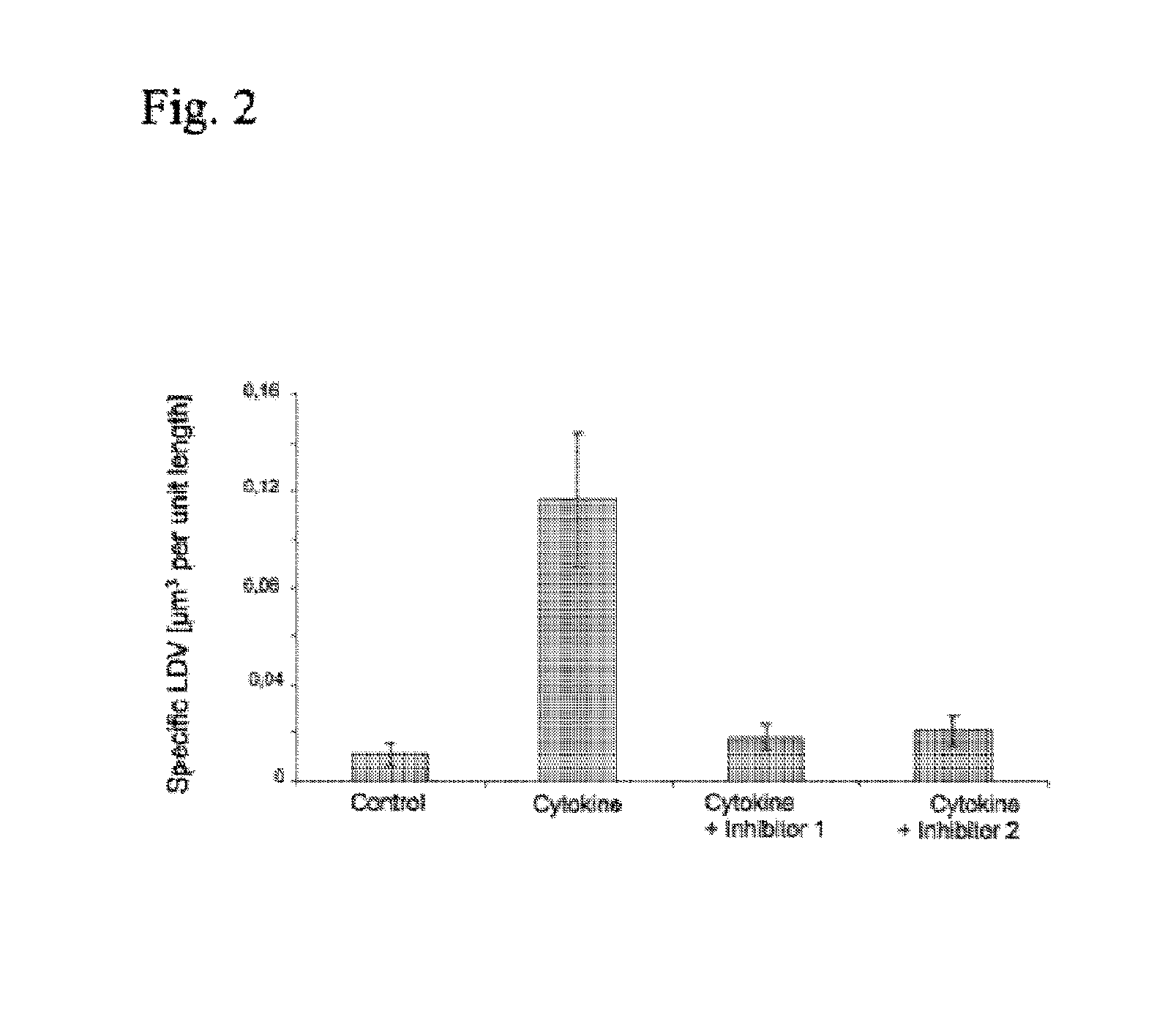
[0038]The invention is based on the experimental results, that the surface texture changes on the nanoscale, when cells are developing, are being stressed or undergo a transformation. Using the method according to the invention it was for the first time possible to show that the type of stimulus leads to distinct alterations in target cell models.

[0039]Samples and AFM was performed as follows:

Sample Preparation

[0040]Living cells were subjected to AFM-imaging either directly in growth medium or in physiological HEPES buffer (in contact mode using gold coated standard AFM tips) without further preparation.

Fixed Samples

[0041]After time intervals determined by the experimental model and specific question, the cell samples were fixed with glutardialdehyde (0.05% to 5% final concentration for 1-100 min in growth medium under physiological conditions (37° C., 19% O2, 5% ...


PUM

PropertyMeasurementUnit
lengthaaaaaaaaaa
lengthaaaaaaaaaa
temperatureaaaaaaaaaa

Abstract

The present invention relates to a method based on atomic force microscopy and the use thereof on biological surfaces. A method is provided to detect the Local Deviational Volume (LDV) of defined subcellular structures irrespective of a biochemical characterization.

Description

FIELD OF THE INVENTION[0001]The present invention relates to a method based on atomic force microscopy and the use thereof on biological surfaces.BACKGROUND OF THE INVENTION[0002]Several imaging methods are known for diagnostic purposes, including the use of radioisotopes or x-ray imaging. Besides these methods, diagnostic imaging is performed on biopsies of tissues or liquids from the living body to determine the existence or cause of a disease. In the last decade imaging systems were developed using cell cultures of cells taken from a patient, in order to detect markers that are correlated to diseases. These systems are mostly based on the coupling of biological samples to defined epitopes wherein the biological samples are labelled with a dye, preferably a fluorescent dye.[0003]Pharmacological testing and low throughput screening assays are also increasingly based on mammalian cell culture (High content screening, HCS). As compared to biochemical affinity tests used in molecular ...

Claims


Application Information

Patent Timeline
Patent Type & Authority Patents(United States)
IPC IPC(8): G01N33/48B82Y15/00B82Y35/00G01N33/50G01Q30/04G01Q60/42
CPCG01N33/5026G01Q60/42B82Y35/00G01Q30/04B82Y15/00
Inventor RIETHMULLER, CHRISTOPH
Owner RIETHMULLER CHRISTOPH
Why Eureka
Try Eureka