
Methods and devices for delivery of radiation to the posterior portion of the eye

a radiation delivery and posterior portion technology, applied in the field of methods and devices for introducing radiation to the posterior portion of the eye, can solve the problems of progressive damage to the retina, blindness or death through direct disruption of tissues, and difficulty in recognizing faces and other detailed tasks

Active Publication Date: 2013-12-10
PARK ROBERT +2
View PDF259 Cites 32 Cited by
  • Summary
  • Abstract
  • Description
  • Claims
  • Application Information

AI Technical Summary

Benefits of technology

The patent is about methods and devices for introducing radiation to the posterior portion of the eye for treating and managing eye conditions such as macular degeneration and intraocular tumors. The invention aims to improve vision and reduce vision loss by selectively damaging abnormal vessels and tissues without causing damage to the normal retina and choroid. The patent discusses the limitations of current treatments such as thermal laser photocoagulation and anti-VEGF therapy, and proposes a new approach using suprachoroidal methods of introducing radiation to the posterior portion of the eye. The technical effects of the patent include improved vision and reduced vision loss through selective damage to abnormal vessels and tissues, as well as reduced risk of complications and improved treatment outcomes.

Problems solved by technology

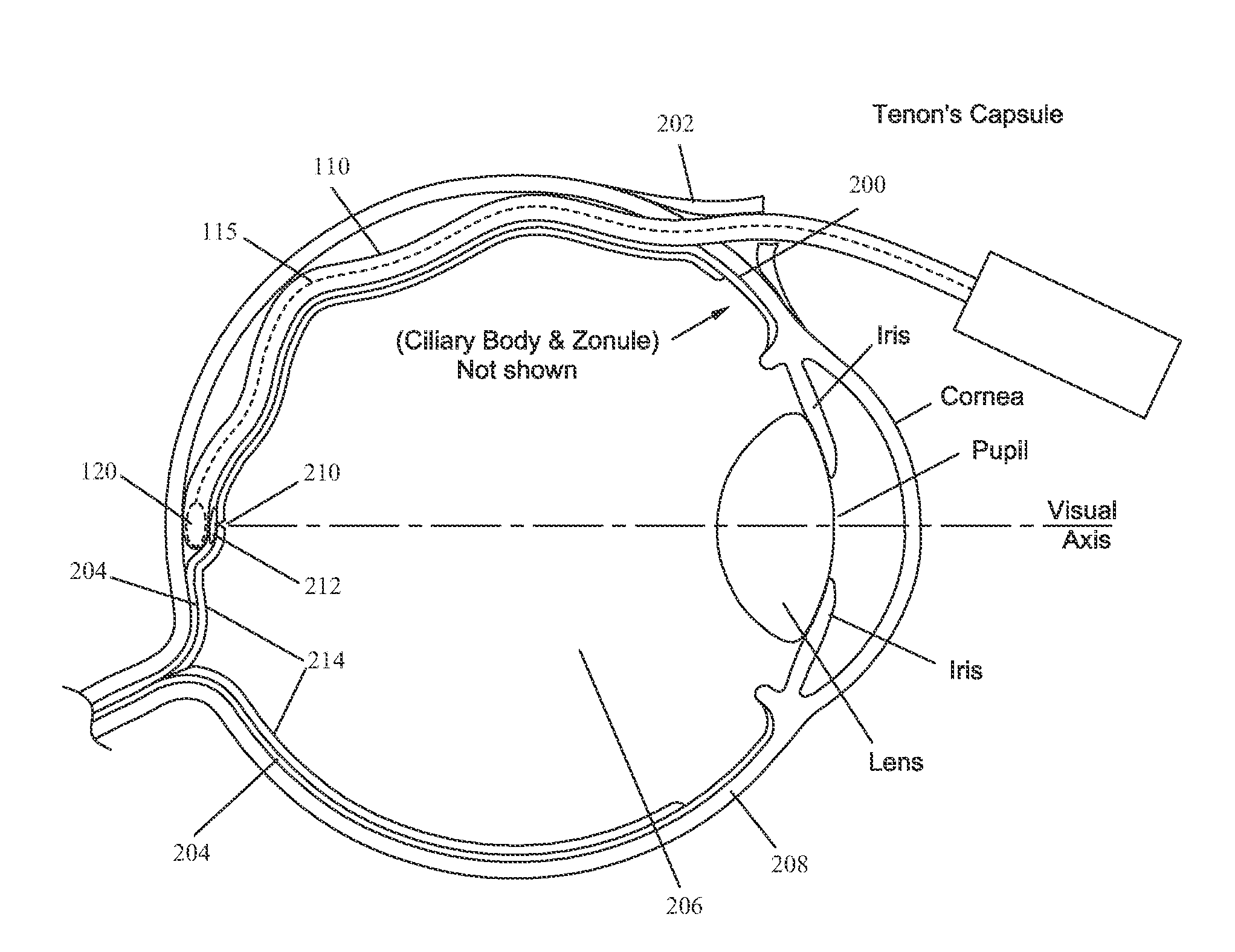
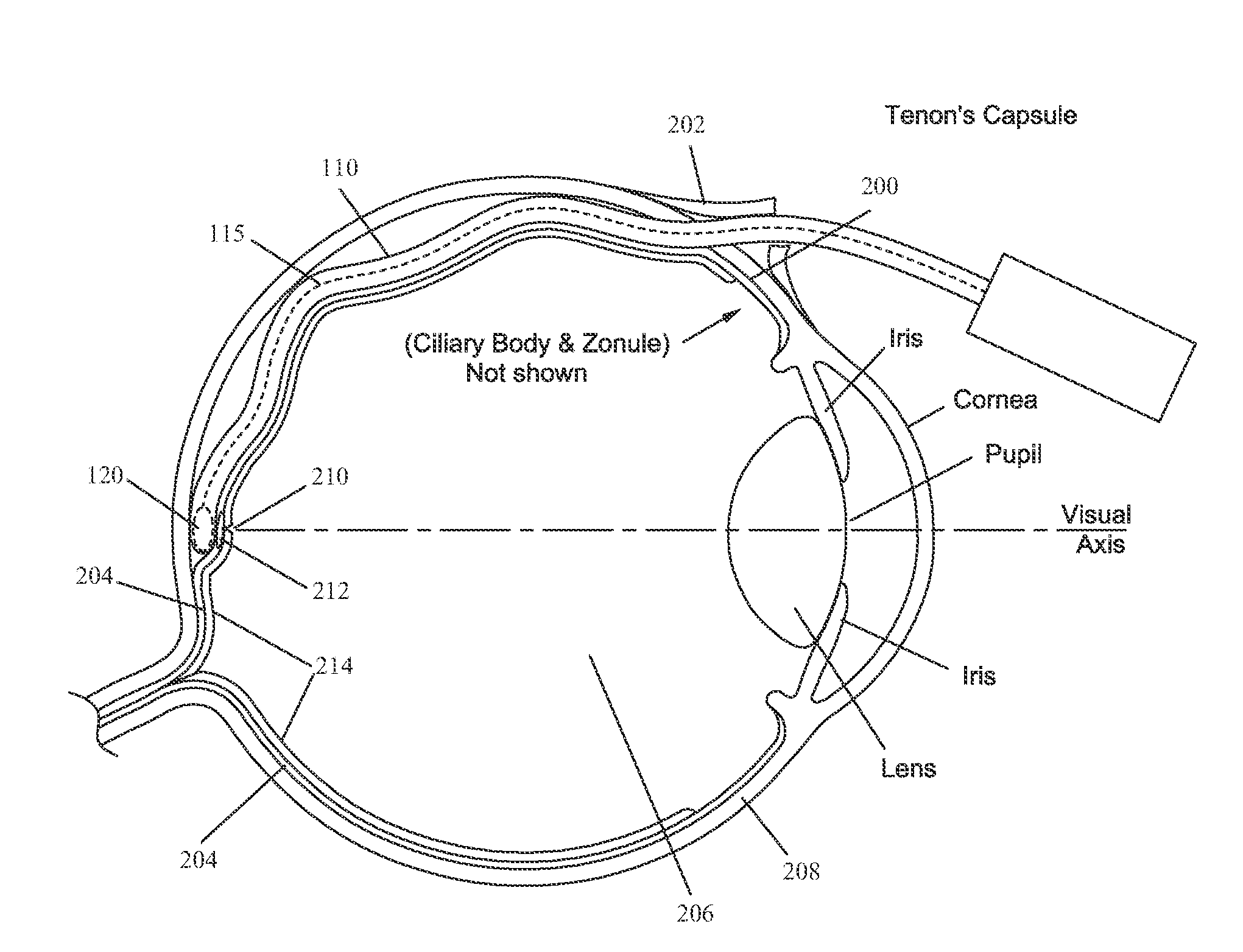
ARMD attacks the center region of the retina 214 (i.e., macula), responsible for detailed vision and damages it, making reading, driving, recognizing faces and other detailed tasks difficult or impossible.
In wet ARMD (e.g., WAMD), newly formed choroidal blood vessels (choroidal neovascularization (CNV)) leak fluid and cause progressive damage to the retina.
Intraocular tumors can cause blindness or death through direct disruption of tissues or metastatic spread.
A drawback of laser photocoagulation is that the laser ablates all structures in its path, not just the abnormal vessels but also the normal retina and choroid, resulting in vision loss at the location of laser application.
When this treatment is applied to subfoveal membranes (membranes located below the fovea 210, the very center of the macula), the result is an immediate, permanent, severe central vision loss.
The dye is activated at the site of the laser application resulting in localized damage to the neovascular membranes.
The result is localized damage to the young, abnormal, photosensitized vessels but not the normal vessels and retina tissue.
While PDT has yielded better visual results than thermal photocoagulation or observation alone (e.g., no treatment) it does not usually lead to an improvement of vision.
A drawback of anti-VEGF therapy is a relatively short duration of action, requiring monthly injections.
A limitation of anti-VEGF treatment is a limited effect of anti-VEGF agents against developed choroidal neovascular membranes (CNVMs).
In some cases, cessation of monthly injections of anti-VEGF agents can cause a gradual decline in vision as the CNVMs are reactivated.
External beam radiation therapy disrupts the reproducing cells (e.g., tumor cells) and can cause regression of the tumor.
However, the high-energy radiation used with external beam radiation therapy has the potential risk to induce radiation retinopathy, which may cause long-term retinal vascular damage and / or decreased vision.
Compared to external beam radiation therapy, brachytherapy uses a small, less powerful radiation source placed in close proximity to the tumor, which limits the radiation exposure to surrounding tissues.
A pars plana vitrectomy presents risks associated with manipulation of the vitreous and placement of a probe on the retinal surface.
Risks associated with the procedure include endophthalmitis, retinal detachment, cataract formation, bleeding, glaucoma and mechanical retinal damage (from direct contact) and those of general anesthesia, often required for this type of surgery.

Method used


Image

  • Methods and devices for delivery of radiation to the posterior portion of the eye
  • Methods and devices for delivery of radiation to the posterior portion of the eye

Examples


example 1

Example of a Surgical Procedure

[0033]The following example describes a surgical procedure utilizing methods of the present invention. The present invention is not limited to this example.

Preparation

[0034]The eye being operated is dilated, e.g., by instillation of tropicamide and epinephrine. The patient is brought to the operating room where cardiac monitoring and an intravenous line are established. A retrobulbar injection of a mixture of xilocaine and marcaine is given. Standard ophthalmic prepping and draping is performed, and a lid speculum is placed.

Incisions

[0035]A conjunctival incision is performed at the superotemporal quadrant about 3-4 mm posterior to the limbus, followed by an incision in the underlying Tenon's capsule to expose bare sclera. A bipolar cautery is used to cauterize episcleral vessels and render a vascular a patch of scleral bed. An initial, partial thickness scleral incision is performed followed by a series of shallower incisions to deepen the initial one....


PUM


Abstract

Methods and devices for delivering radiation to a target at a posterior portion of an eye using a brachytherapy-administering device adapted to be inserted into a suprachoroidal space of the eye including a brachytherapy source and a means of advancing the brachytherapy source from a storage position to a radiation position corresponding to a position in a distal portion of the device, positioning the device appropriately such that the distal portion of the device is in close proximity to the target, exposing the target to the brachytherapy source for a predetermined length of time then removing the device and closing incisions made during a surgical procedures to reach the suprachoroidal space of the eye.

Description

CROSS REFERENCE[0001]This application is a non-provisional application which claims priority to U.S. provisional Ser. No. 61 / 347,233 filed May 21, 2010, the specification of which is incorporated herein by reference in its entirety.FIELD OF THE INVENTION[0002]The present invention is directed to methods and devices for introducing radiation to the posterior portion of the eye for treating and / or managing eye conditions including but not limited to macula degeneration, more particularly to suprachoroidal methods of introducing radiation to the posterior portion of the eye.BACKGROUND OF THE INVENTION[0003]Several intraocular diseases and conditions of the eye threaten vision. Age related macular degeneration (ARMD), choroidal neovascularization (CNV), retinopathies (e.g., diabetic retinopathy, vitreoretinopathy), retinitis (e.g., cytomegalovirus (CMV) retinitis), uveitis, macular edema, ocular tumors, and glaucoma are several examples.[0004]Age related macular degeneration (ARMD) is t...

Claims


Application Information

Patent Timeline
IPC IPC(8): A61N5/00
CPCA61N5/1017A61N5/00A61N2005/1094
Inventor PARK, ROBERTBRIGATTI, LUCAHAMILTON, RUSSELL J.
Owner PARK ROBERT
Learn More