
Methods and compositions for culturing cardiomyocyte-like cells

a cardiomyocyte and composition technology, applied in the field of mammalian cell culturing methods, can solve the problem of engrafting cells not being immunologically rejected, and achieve the effect of enhancing the ability of clcs, optimal phenotypic qualities, and low efficacy

Inactive Publication Date: 2013-08-20
NAT HEART CENT OF SINGAPORE
View PDF10 Cites 0 Cited by
  • Summary
  • Abstract
  • Description
  • Claims
  • Application Information

AI Technical Summary

Benefits of technology

[0040]An advantage of the present invention is that CLCs can be produced with relatively simple defined cell culture medium, and without the use of toxic differentiating agents such as 5-azacytidine. Furthermore, there is no need to co-culture the cells with other cells of a cardiac lineage (such as rat myocytes) to stimulate differentiation. Co-culturing technique and 5-aza treatment are not likely to be applicable clinically due to their low efficacy and potentially harmful effects arising from nonspecific demethylation effect of 5-azacytidine. Liu, Y., Song, J., Liu, W., Wan, Y., Chen, X. & Hu, C. Growth and differentiation of rat bone marrow stromal cells: does 5-azacytidine trigger their cardiomyogenic differentiation? Cardiovasc Res. 2003; 58:460-8. Our study shows that human adult bone marrow cells can be successfully differentiated towards CLCs with defined myocardial characteristics using a simple culture medium independent of 5-aza treatment or co-culturing technique. Furthermore, the intrinsic cardiomyocyte-like characteristics may enhance the ability of CLCs to directly engraft as cardiomyocytes in transplanted myocardium.
[0041]One particularly useful base culture medium is Dulbecco's Modified Eagle's Medium-Low Glucose (DMEM-LG), augmented with foetal bovine serum. Another useful base medium is MCDB201. In a highly preferred form of the invention the base culture medium comprises about 60% DMEM-LG and 28% MCDB-201. For cells to attain optimal phenotypic qualities it is desirable to add serum to the base culture medium. The addition of serum provides hormones, transport proteins, trace elements and the like, and it is possible to ascertain the optimum concentration of serum in the medium purely by routine experimentation. In one form of the method the serum is present at a concentration of about 10%.
[0042]Applicants have found that certain supplements may be added to the base medium to improve the quality of the cells. Suitable supplements include insulin, an iron source, a trace element, a fatty acid, dexamethasone, and an antioxidant. In a highly preferred form of the method the iron source is transferrin, the fatty acid is linoleic acid, the trace element is selenium and the antioxidant is ascorbate. Of course, the skilled artisan could determine that the addition of other supplements or the substitution with similar supplements may give advantageous results. Such routine modifications to the culture medium are included within the scope of this application. For example, ethanolamine or oleic acid may be used as the fatty acid in place of linoleic acid. Similarly, sodium pyruvate could be added as an additional energy source, or further buffers may be added to maintain pH at an optimum level.
[0043]The culture can continue for any period of time so long as the medium is changed whenever necessary to ensure the viability of the cells. In one form of the method, the cells are cultured for about 7 days to about 10 days at 37° C. in 5% CO2 before further use. More preferably, the cultures are maintained until confluence is reached in about 14 to 21 days after the initial seeding.
[0044]In another aspect, the present invention provides a cardiomyocyte-like cell produced according to the methods described herein. As used herein, the term “cardiomyocyte-like cell” is intended to include any cell that is able to fulfill a function in myocardial tissue. The cell may be able to fulfill the function directly after culturing, or after some further treatment (e.g. exposure to a differentiation agent, passaging, engrafting to the heart). The cardiomyocyte-like cell may express one or more of the following cardiomyocyte-specific proteins at levels sufficient to be detected immunologically: sarcomeric α-actin, sarcomeric α-actinin, desmin, skeletal / cardiac specific titin, sarcomeric α-tropomyosin, cardiac troponin I, sarcomeric MHC, SERCA2 ATPase and connexin-43. The CLCs of the present invention show extensive expression of cardiac proteins and therefore clearly differ from previously reported undifferentiated human MSCs that showed no myocardial gene expression. Rangappa, S., Entwistle, J. W., Wechsler, A. S. & Kresh, J. Y. Cardiomyocyte-mediated contact programs human mesenchymal stem cells to express cardiogenic phenotype. J Thorac Cardiovasc Surg. 2003; 126:124-32. Furthermore, expression of α-MHC, a specific marker for adult cardiomyocytes, together with lack of skeletal MHC and MyoD expression indicate that CLCs are different from skeletal muscle stem cells that were previously reported to reside in bone marrow. Bossolasco, P., Corti, S., Strazzer, S., Borsotti, C., Del Bo, R., Fortunato, F. et al. Skeletal muscle differentiation potential of human adult bone marrow cells. Exp Cell Res. 2004; 295:66-78. In addition, the formation of cross-striated Z-bands also indicates a nascent cardiomyocyte phenotype. However, the absence of spontaneous beating in culture suggests the CLCs have not fully differentiated into mature cardiomyocytes. Similar observations have been reported in cultured Lin− / c-Kit+ cardiac-derived stem cells, Beltrami, A. P., Barlucchi, L., Torella, D., Baker, M., Limana, F., Chimenti, S. et al. Adult cardiac stem cells are multipotent and support myocardial regeneration. Cell. 2003; 114:763-76, suggesting that mature cardiomyocyte phenotype may require further differentiation in a proper cardiac milieu.
[0045]In one form of the invention the CLCs express one or more of the following markers: CD44, CD90, CD105 and CD106. Furthermore, the CLCs may exhibit adherent-dependent growth or a spindle morphology. Importantly, the cardiomyocyte-like cells produced by the present methods are capable of forming a viable engraftment of cardiac myofibers when administered to the heart.

Problems solved by technology

This entire method can be performed percutaneously resulting in reduced trauma to the subject, and obviates the problem of immunological rejection of the engrafted cells.

Method used


Image

  • Methods and compositions for culturing cardiomyocyte-like cells
  • Methods and compositions for culturing cardiomyocyte-like cells
  • Methods and compositions for culturing cardiomyocyte-like cells

Examples


example 1

Proliferation of Cardiomyocyte-Like Cells in Culture

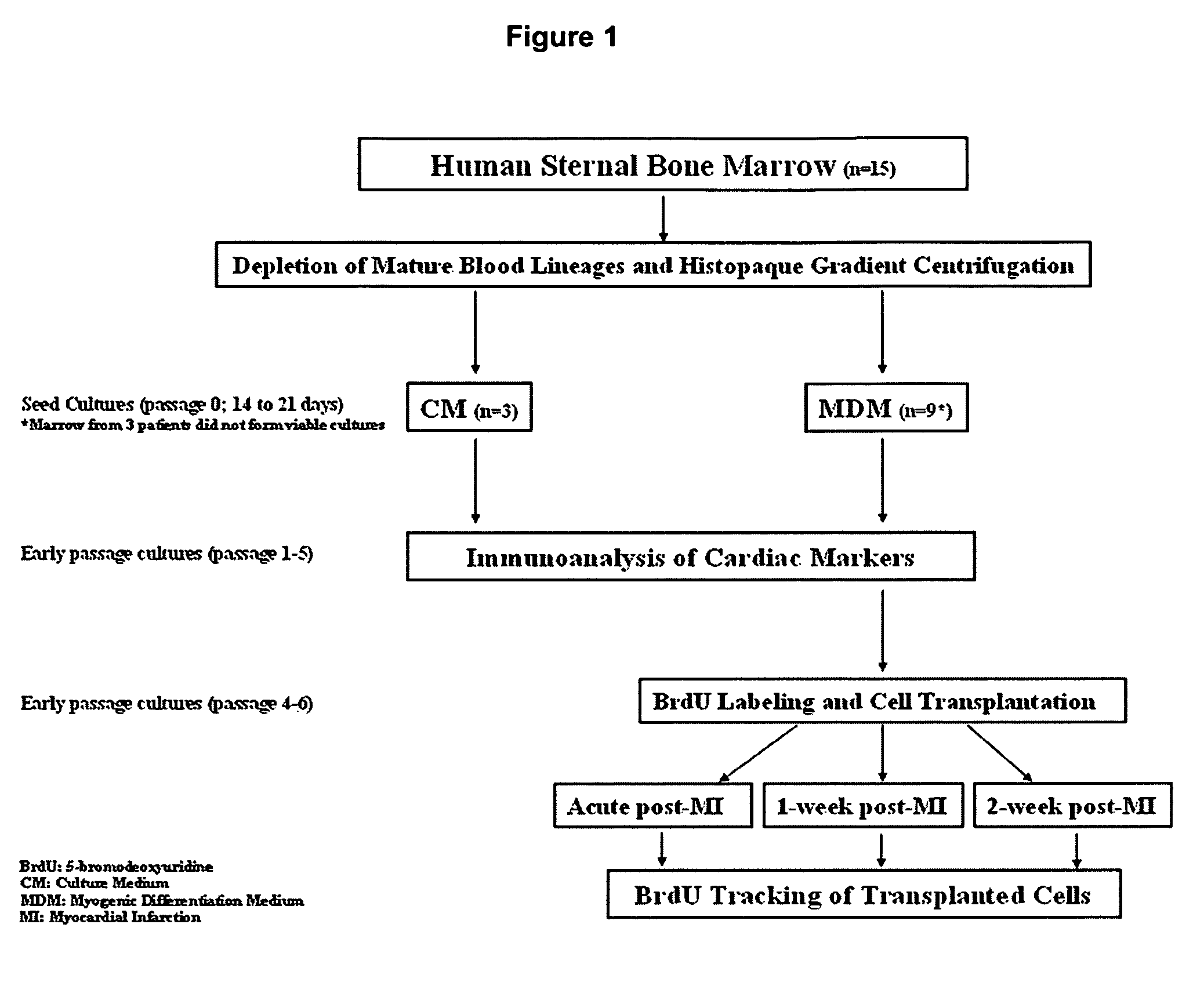
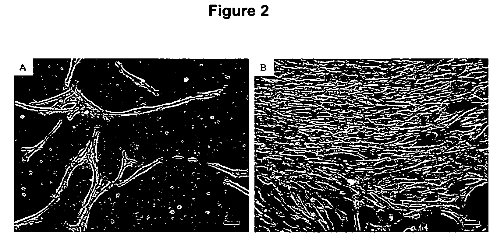
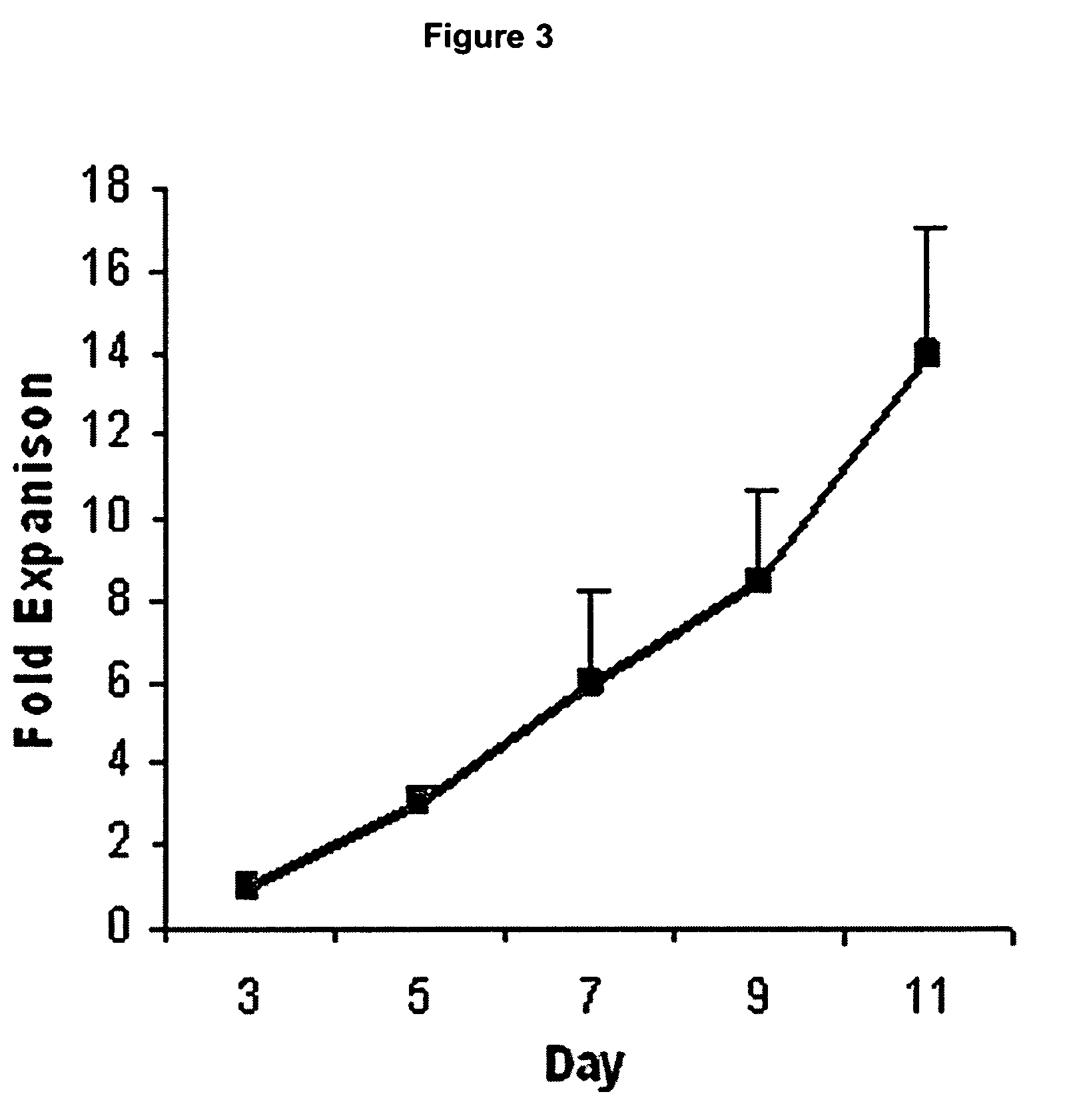
[0073]Cells from a total of 12 patients were successfully expanded and used for subsequent analyses (FIG. 1). Marrow from 3 other patients failed to form viable cultures due to sparse starting cell numbers. Single cells of spindle morphology were observed 3 to 4 days after the initial seeding in culture. They rapidly expanded into colonies of confluent spindle cells with approximately 1 to 2 thousand cells in the next 7 days (FIG. 2A and B). Most of the loosely attached round hematopoietic cells were removed following medium change. Cultured cells were mainly of spindle morphology with an average doubling time of 48 to 72 hours (FIG. 3) but 5 to 10% of the colonies consisted of cells of polygonal morphology. After prolonged passages (>10 passages) in culture, the cells progressively lost their spindle morphology and the cultures were increasingly dominated by cells with polygonal morphology with an average doubling time of more tha...

example 2

Cardiomyocyte-Like Cells Are Negative for Endothelial Phenotypes

[0074]The MDM cultured cells showed positive staining for markers such as CD44, CD90, CD105 and CD106 (FIG. 4A to D) but were negative for CD45 and CD117 (Table 1). The cells showed about 30% positive staining for CD34 and FLK1 markers (FIGS. 4E and F) but were negative for Tie2, von Willebrand factor (vWF), CD31 and FLT1 endothelial markers (Table 1). These may represent residual hematopoietic cells from the initial cultures or they may be a subpopulation of bone marrow cells that expresses both mesenchymal and hematopoietic markers, since over 99% of the cells were stained positive for CD44, CD90 and CD105 mesenchymal markers.

example 3

Cardiomyocyte-Like Cells Exhibit Cardiac but not Skeletal Muscle Characteristics

[0075]Approximately 1% of the cells in the colonies were stained positive for cardiac troponin I 2 weeks after first seeding in MDM. After 4 to 5 passages, more than 90% of the cells were consistently stained positive for sarcomeric α-actin, sarcomeric α-actinin, desmin, skeletal / cardiac-specific titin, sarcomeric α-tropomyosin, cardiac troponin I, sarcomeric MHC, SERCA2 ATPase and connexin-43 (FIG. 5A to J). Expression of troponin I and SERCA2 ATPase was detected early (passage 1-2) followed by titin and desmin (passage 2-3) and then tropomyosin and actinin (passage 3-5) indicating differentiation towards CLCs. Approximately 60% of the CLCs also stained positive for cardiac-specific α / β MHC and cardiac transcription factor GATA-4 (FIGS. 5, K and L) suggesting a developing cardiomyocyte phenotype. There was weak immunoreactivity with slightly above background staining against ANP and cardiac troponin T a...


PUM

PropertyMeasurementUnit
densityaaaaaaaaaa
thicknessaaaaaaaaaa
doubling timeaaaaaaaaaa

Abstract

The present invention provides cardiomyocyte-like cells that have been cultured from populations of bone marrow cells. The culture method includes the use of defined media that are free of agents such a 5-azacytidine. The cardiomyocyte-like cells can be engrafted to heart tissue to repair damage resulting from an ischaemic episode.

Description

[0001]This application claims benefit of U.S. Provisional Patent Application Ser. No. 60 / 604,651, filed Aug. 26, 2004, which is hereby incorporated by reference in its entirety.FIELD OF THE INVENTION[0002]The present invention relates to methods for culturing mammalian cells for use in therapeutic applications. More specifically the present invention is concerned with methods and compositions for producing cell populations useful in the treatment of damaged myocardial tissue.BACKGROUND TO THE INVENTION[0003]The heart is a vital organ, being responsible for pumping oxygenated and nutrient rich blood to the body. Efficient functioning of the heart can be compromised by trauma, infection and ischaemia. Ischaemic heart disease is the leading cause of death in industrialised countries, often manifesting clinically as a myocardial infarction (MI). The management of ischaemic heart disease essentially relies upon one of three strategies, including medical therapy, percutaneous coronary int...

Claims


Application Information

Patent Timeline
Patent Type & Authority Patents(United States)
IPC IPC(8): C12N5/00A61K35/12C12N5/077
CPCA61K2035/124C12N5/0657C12N2500/25C12N2500/36C12N2500/38C12N2501/39
Inventor SHIM, WINSTON SE NGIEWONG, PHILIP EN HOU
Owner NAT HEART CENT OF SINGAPORE
Learn More