
Medical imaging device and method to evaluate a test bolus image series

Inactive Publication Date: 2013-07-09
SIEMENS HEALTHCARE GMBH
View PDF3 Cites 0 Cited by
  • Summary
  • Abstract
  • Description
  • Claims
  • Application Information

AI Technical Summary

Benefits of technology

[0016]In this way the method according to the invention not only simplifies the detection of the test bolus in the time series, but also it precludes error sources that are present in the manual procedure. In particular the fully automatic procedure additionally allows a more exact and more precise determination of the time window for the subsequent image acquisition, such that high-contrast exposures of the blood vessels can be generated in high quality.
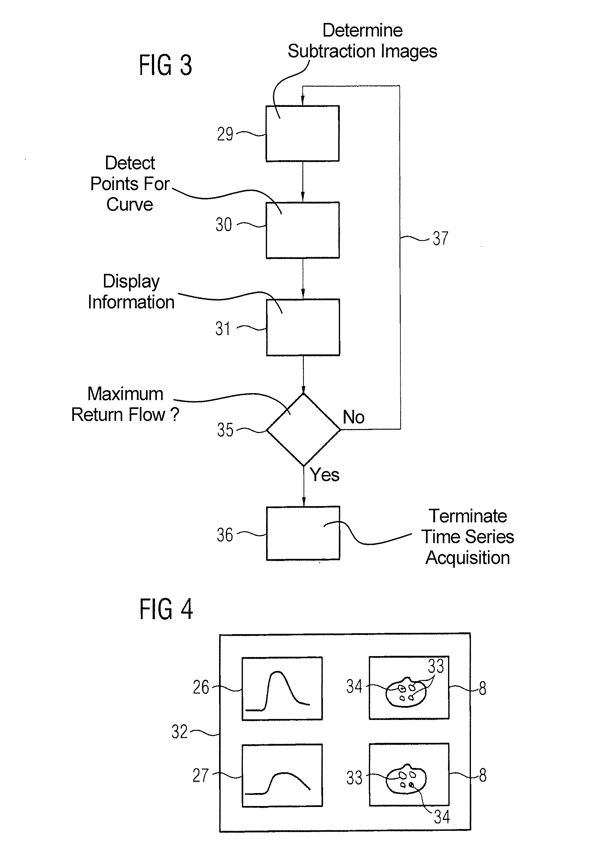
[0022]The automatic procedure and the time savings that this entails can also advantageously be, after acquisition of a new image, continuously to automatically seek new points exhibiting a contrast agent signal and not already associated with a blood vessel after determining the corresponding evaluation image, and / or to update the at least one contrast agent course curve. In such updating, for at least one blood vessel, the contrast agent course curve up to the current image and / or an image that is expanded with information regarding the determination of the contrast agent course curve, is displayed on a display device. It is thus possible to already produce contrast agent course curves while the time series is still being acquired. This can be advantageously used in various ways. For example, by display of the contrast agent course curve and / or display of an image in which the point or region forming the basis of the contrast agent course curve, a user can assess whether the method determines suitable contrast agent course curves. For example, a manual intervention can then ensue if necessary. The acquisition of the time series can be manually or automatically terminated upon detection of a maximum of a return flow in a vein. The maximum of the return flow is typically the chronologically last item of information that should be obtained from the test bolus measurement. Since any images that were acquired after the determination of this maximum are no longer necessary: time, patient stress and costs can thus be saved by an early termination. Instead of the large number of images of the time series that is typical acquired as a precaution in order to be sure an adequate analysis can ensue, this number can consequently be reduced. This will typically occur by a manual procedure if the user detects that the return flow maximum was correctly acquired. However, an automatic evaluation (and thus an automatic termination) is also conceivable.

Problems solved by technology

This procedure is disadvantageous because the segmentation of particularly small blood vessels is time-consuming and error-prone.

Method used


Image

  • Medical imaging device and method to evaluate a test bolus image series
  • Medical imaging device and method to evaluate a test bolus image series
  • Medical imaging device and method to evaluate a test bolus image series

Examples


Embodiment Construction

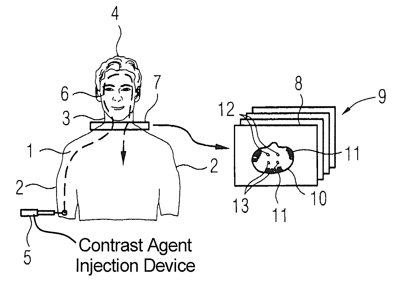
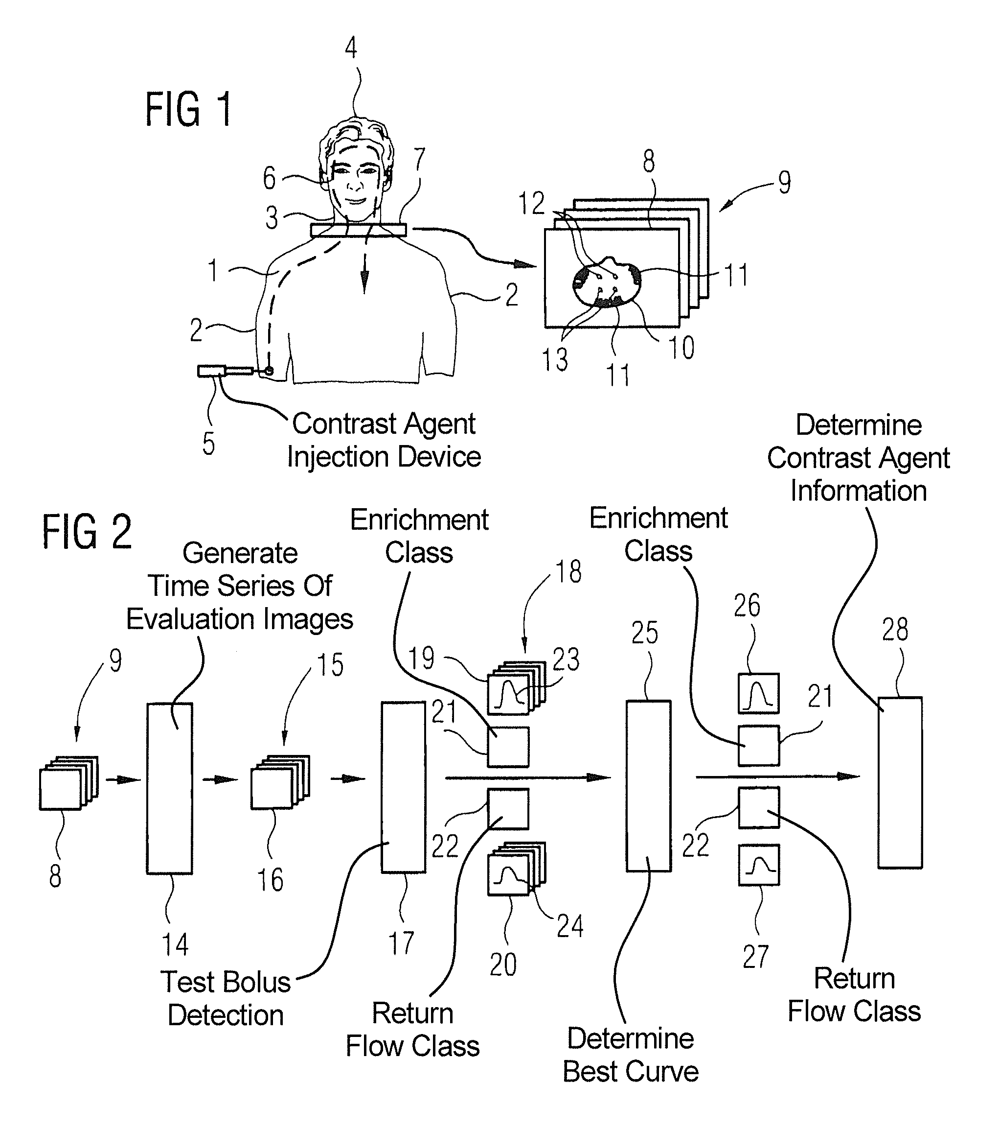
[0033]FIG. 1 shows a possible geometry implemented for a test bolus measurement before a magnetic resonance image acquisition of the blood vessels of the head with contrast agent administration. FIG. 1 shows is a patient 1 having the arms 2, a neck 3 and a head 4. The test bolus—and later also the actual bolus for subsequent measurement—are injected into the arm 2 of the patient 1 via a corresponding injection device 5. As is schematically indicated by the dashed line 6, the contrast agent is then transported through the arteries into the head (enrichment) and transported through the veins away from the head 4 again (return flow). The contrast agent crosses respective large blood vessels in the neck 3.

[0034]In order to determine the time window in which only the arterial blood vessels are emphasized by the contrast agent in a magnetic resonance exposure of the head 4, a test bolus measurement is first implemented in which only a small fraction of the contrast agent that is later to ...


PUM


Abstract

In a method and device to evaluate a time series of in particular two-dimensional images of a contrast agent flow in at least one blood vessel of the human body within the scope of a test bolus measurement, wherein the acquisition of the time series begins with administration of the contrast agent, the time series of two-dimensional images or a time series of images derived from this is used as a time series of evaluation images, and in at least one evaluation image exhibiting a contrast agent signal, at least one point associated with a blood vessel is determined semi-automatically or automatically and transferred to further evaluation images. For every blood vessel discovered in this way, a temporal contrast agent course curve is determined (in particular via a fit) automatically from the image signal of the evaluation images at the at least one point and / or in a region around the marked point in the blood vessel and at least one item of contrast agent information that affects the temporal workflow of a subsequent measurement is determined from at least one contrast agent course curve.

Description

BACKGROUND OF THE INVENTION[0001]1. Field of the Invention[0002]The invention concerns a method and a medical image acquisition device to evaluate a time series, in particular of two-dimensional images of a contrast agent flow in at least one blood vessel of the human body within the scope of a test bolus measurement, wherein the acquisition of the time series begins with administration of the contrast agent.[0003]2. Description of the Prior Art[0004]Contrast agent-enhanced magnetic resonance tomography is a known examination method to show the arterial or venous vessel system. A contrast agent that intensifies the signal of the blood vessels relative to the surrounding tissue is administered. By data acquisition before and after administration of the contrast agent, the surrounding tissue can be almost completely eliminated by subtraction. In this imaging method the most significant requirement is to synchronize the contrast agent administration exactly with the data acquisition so...

Claims


Application Information

Patent Timeline
Patent Type & Authority Patents(United States)
IPC IPC(8): G06K9/00
CPCA61B5/055G01R33/5635A61B5/7207
Inventor BLUM, THOMASSCHMIDT, MICHAELASCHMITT, PETERZENGE, MICHAEL
Owner SIEMENS HEALTHCARE GMBH
Learn More