
Extension of truncated CT images for use with emission tomography in multimodality medical images

a multi-modality, ct image technology, applied in the field of medical diagnostic imaging, can solve the problems of not using iterative techniques in ct, the co-alignment of images from different modalities is not straightforward or very accurate, and the method of extending fov is improved, so as to reduce the computation time for the extension of ct image, reduce the area, and improve the effect of extending the method

Active Publication Date: 2012-04-10
SIEMENS MEDICAL SOLUTIONS USA INC
View PDF6 Cites 21 Cited by
  • Summary
  • Abstract
  • Description
  • Claims
  • Application Information

AI Technical Summary

Benefits of technology

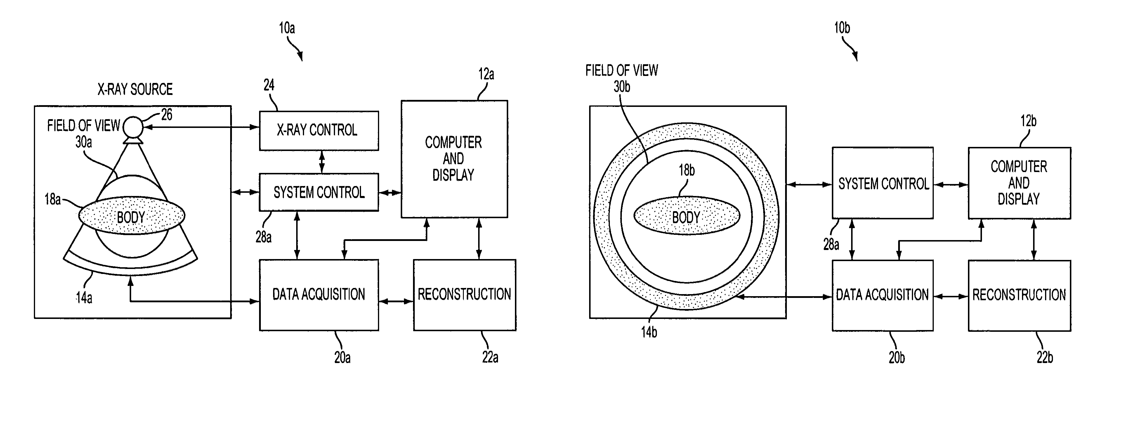
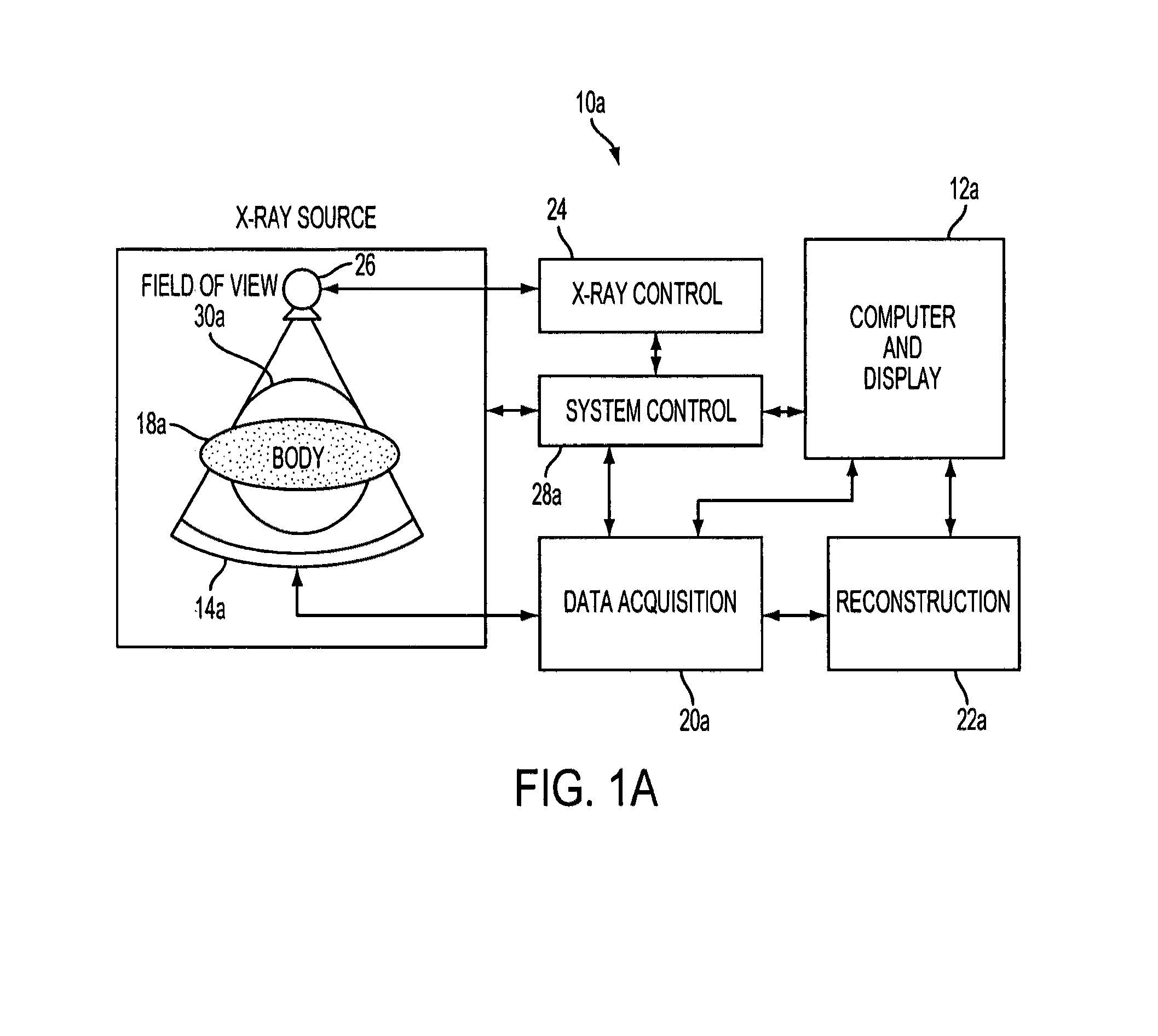
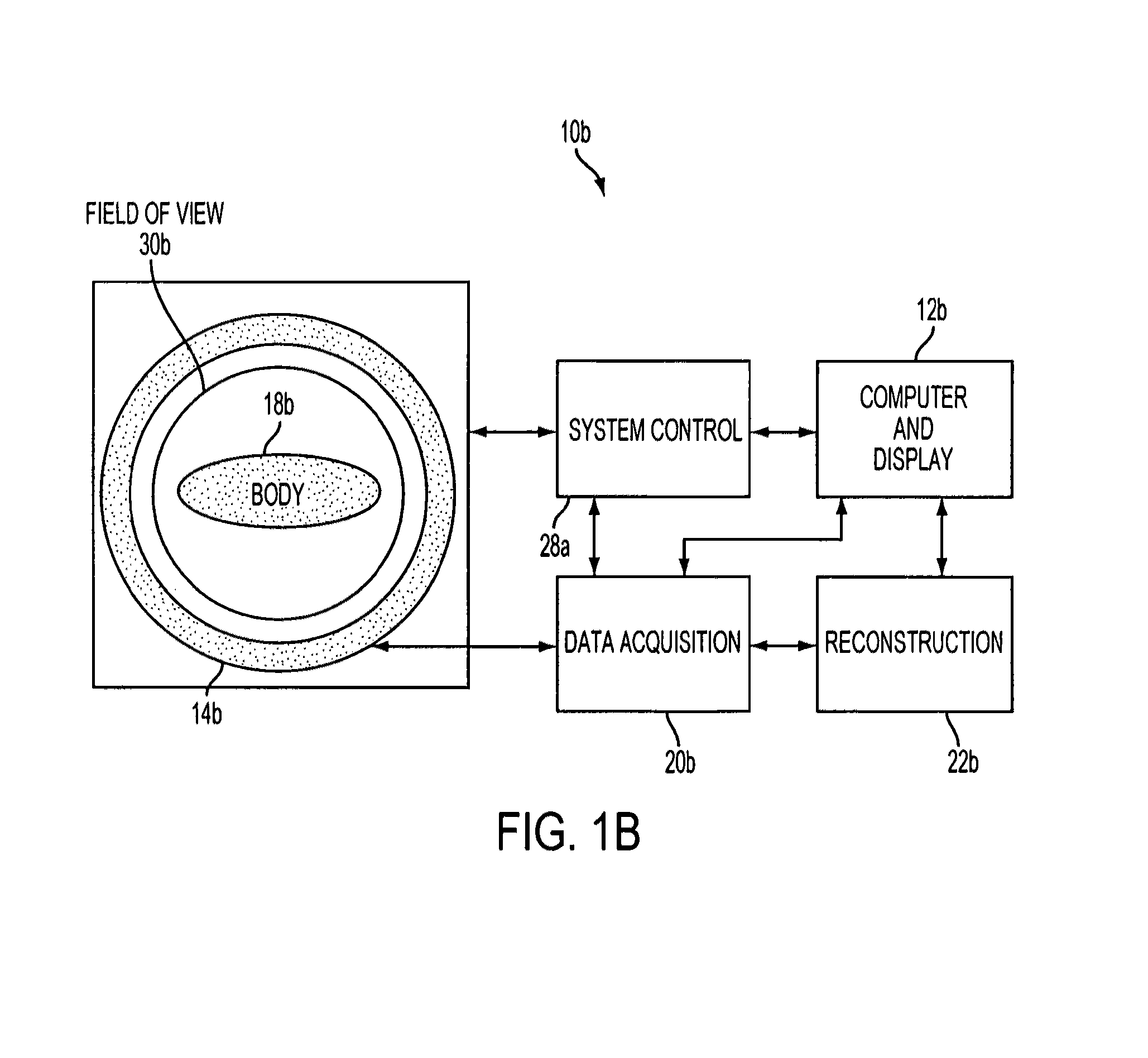
The present invention provides an apparatus and method for extending the field of view (FOV) of a computed tomography (CT) scan using a mask to determine the data to be used in the calculation. The mask can extend from an inner boundary around the image center to an outer boundary encompassing truncated portions of an imaged object. The technique uses the reconstructed CT image data within the inner boundary of the mask without change and extends the reconstructed CT image to areas outside the mask and, further, to areas outside the CT scanner FOV. The invention also includes a method for improving the accuracy of the CT image FOV when the CT system is part of a combined PET / CT scanner or SPECT / CT scanner by reducing the computation time for the extension of the CT image FOV and improving the accuracy of the PET and SPECT attenuation correction with respect to the prior art.

Problems solved by technology

However, with the exception of the brain, the co-alignment of images from different modalities is not straightforward or very accurate, even when surface markers or reference points are used.
However, iterative methods are generally more computationally intensive and more time-consuming than non-iterative methods.
Consequently, in the past iterative techniques were not used in CT as sufficient computational power was cost prohibitive for the resolution and contrast requirements of that modality.
According to U.S. Pat. No. 5,376,795, photon attenuation constitutes a major deficiency in diagnosis of heart disease with SPECT and is a major source of error in the measurement of tumor metabolism using radionuclide techniques.
One limitation of attenuation maps created by CT transmission data is the relatively small usable FOV (typically on the order of 50 cm diameter) compared to the FOV of a PET scanner (typically on the order of 70 cm diameter or more).
However, for various reasons, these attempts either do not extend the FOV far enough or introduce artifacts and / or distortion in the images.
However, these prior art methods have proven unacceptable for reconstructing truncated views.

Method used


Image

  • Extension of truncated CT images for use with emission tomography in multimodality medical images
  • Extension of truncated CT images for use with emission tomography in multimodality medical images
  • Extension of truncated CT images for use with emission tomography in multimodality medical images

Examples


Embodiment Construction

[0041]As required, disclosures herein provide detailed embodiments of the present invention; however, the disclosed embodiments are merely exemplary of the invention that may be embodied in various and alternative forms. Therefore, there is no intent that specific structural and functional details should be limiting, but rather the intention is that they provide a basis for the claims and as a representative basis for teaching one skilled in the art to variously employ the present invention.

[0042]A process according to the present invention uses an imaging system as a means to reveal the presence of defects in the structures, organs or tissues of a patient who exhibits symptoms of an undesirable condition. The imaging system first requires that the patient adopt a position for collection of data from the organ or area of tissue under study, also referred to herein as the imaged object. The process in accordance with the invention can be used to extend the FOV of a computed tomograph...


PUM


Abstract

An apparatus and method for expanding the FOV of a truncated computed tomography (CT) scan. An iterative calculation is performed on the original CT image to produce an estimate of the image. The calculated estimate of the reconstructed image includes the original image center and a estimate of the truncated portion outside the image center. The calculation uses an image mask with the image center as one boundary.

Description

CROSS-REFERENCE TO RELATED APPLICATION AND CLAIM FOR PRIORITY[0001]This application is a continuation-in-part of copending U.S. application Ser. No. 11 / 428,456, filed Jul. 3, 2006, which claimed priority under 35 U.S.C. §119(e) of Provisional Application Ser. No. 60 / 696,307 filed Jul. 1, 2005.BACKGROUND OF THE INVENTION[0002]1. Technical Field[0003]The present invention relates generally to medical diagnostic imaging and to use of medical images in one imaging modality with medical images from another imaging modality. More specifically the invention relates to extension or extrapolation of image data from a Computed Tomography (CT) transmission scan having a limited Field of View (FOV) so as to extend the CT image to a larger FOV. The FOV extension increases the anatomic information provided by the CT scan, and it makes the attenuation data provided by the CT image more accurate for correcting radionuclide emission data obtained from an emission computed tomography scan such as Pos...

Claims


Application Information

Patent Timeline
Patent Type & Authority Patents(United States)
IPC IPC(8): G06K9/00
CPCA61B6/032A61B6/037A61B6/5235G06T11/008A61B6/5241
Inventor FAUL, DAVID D.MICHEL, CHRISTIAN J.
Owner SIEMENS MEDICAL SOLUTIONS USA INC
Learn More