
Percutaneous transluminal endarterectomy

a transluminal endarterectomy and percutaneous technology, applied in the field of medical devices, can solve the problem of plaque separation along a plane of relative weakness

Inactive Publication Date: 2012-03-27
BOSTON SCI SCIMED INC
View PDF44 Cites 10 Cited by
  • Summary
  • Abstract
  • Description
  • Claims
  • Application Information

AI Technical Summary

Benefits of technology

[0014]In another aspect of the invention, fine mesh stents are provided for use after endarterectomy. In one method, which may be suitable for use with percutaneous endarterectomy, the stent is self-expanding and can be delivered intravascularly through a delivery device. In another method, suitable for use after conventional endarterectomy, a stent is provided together with outwardly protruding, open staples. The stent may be inserted through the incision used to perform the endarterectomy, and the vessel walls on either side of the incision closed over the sharp staple ends, forcing the ends through the vessel walls, and the staples closed to both secure the stent and to close the incision.

Problems solved by technology

The carbon dioxide pressure can cause the plaque to separate from the vessel wall along a plane of relative weakness, with the separation occurring ahead of the catheter distal end in some methods.

Method used


Image

  • Percutaneous transluminal endarterectomy
  • Percutaneous transluminal endarterectomy
  • Percutaneous transluminal endarterectomy

Examples


Embodiment Construction

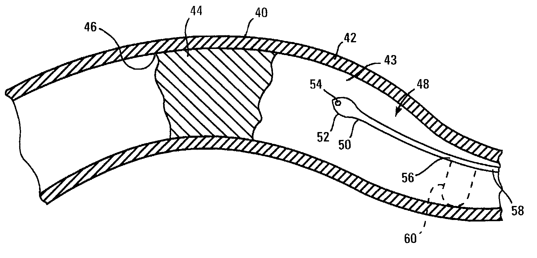
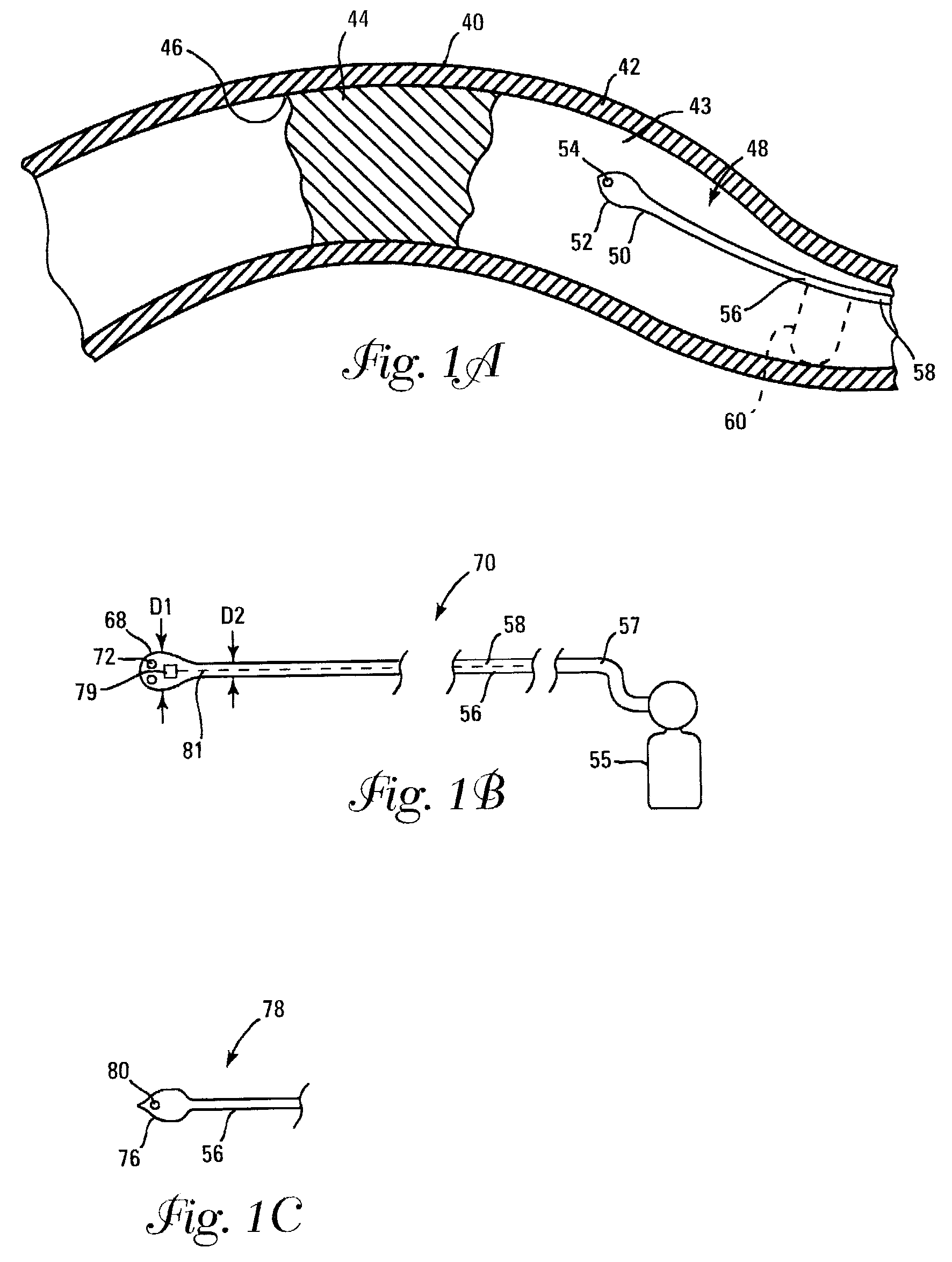
[0039]FIG. 1A illustrates a vessel 40 occluded by plaque 44 being approached by a carbon dioxide infusing catheter 48 having a bulbous distal head 52. Vessel 40 includes a vessel wall 42 and has plaque 44 attached to the vessel wall along an interface or separation plane 46. Catheter 48 has a distal region 50, a shaft 56, and a lumen 58 extending therethrough in fluid communication with a distal orifice 54. Catheter 48 can be formed of any suitable material, including metals and polymers. Some catheters are formed of Nitinol, while others are formed of stainless steel.

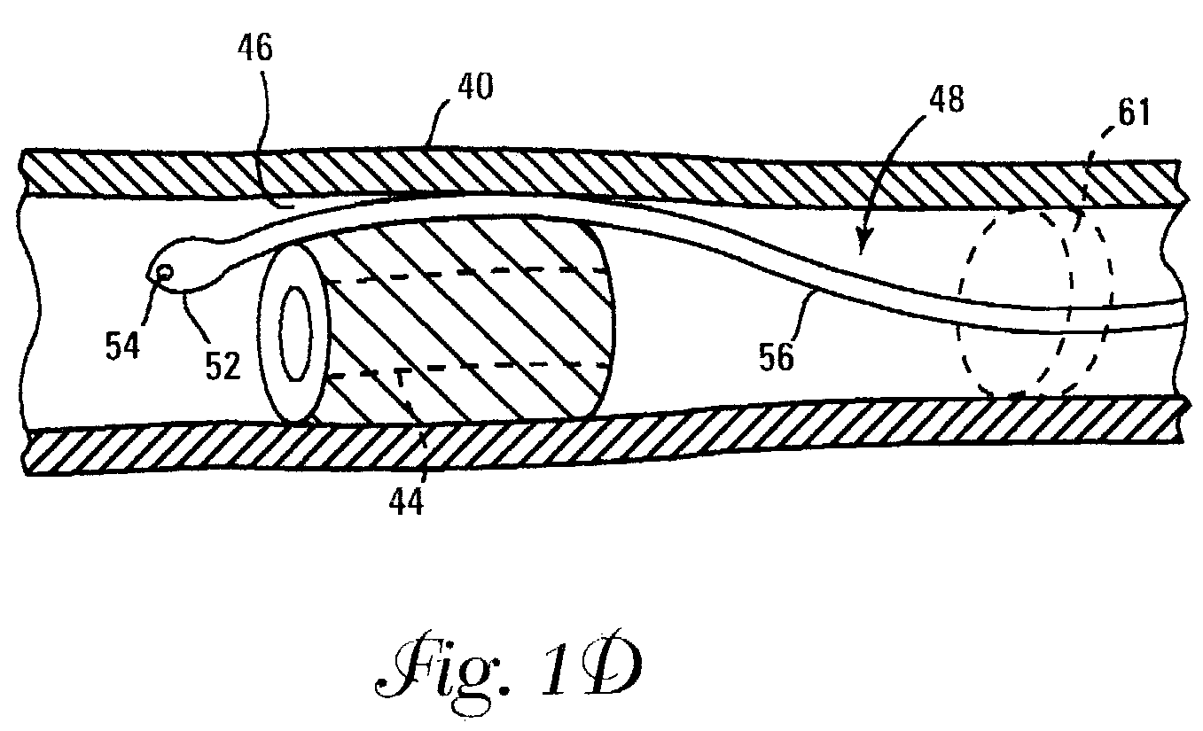
[0040]Catheter 48 may optionally be used with an occlusion device 60, shown in phantom in FIG. 1A. Occlusion device 60 may include an expandable or inflatable balloon or cuff. Occlusion device 60 may be configured to have a central orifice therethrough, disposed to allow passage of catheter 48 through the central orifice, followed by inflation, as illustrated by balloon 61 of FIG. 1D. In the embodiment illustrated in F...


PUM


Abstract

Methods and devices for performing intravascular endarterectomy. Methods includes intravascularly advancing a catheter having a carbon dioxide delivering distal end past a plaque-occluded vessel site, passing the distal end between the plaque and vessel wall. Other methods include intravascularly advancing paddles between plaque and vessel walls, including ultrasonic imaging paddles, ultrasonic vibrating paddles, and mechanically vibrating paddles. Some methods include providing intravascular devices having radially expandable jaws or paddles, and advancing those jaws or paddles along vessel walls to separate plaque from vessel walls. Still other methods include providing an anchoring guide catheter adapted to establish a suction grip around the left coronary artery ostium, and to use the anchored guide catheter to support intravascularly introduced endarterectomy devices operating on plaque in the left main coronary artery. One method utilizes retroperfusion of the coronary arteries to allow the anchoring guide catheter to remain in position for longer periods. Scissors-type expanding devices and stents for use after endarterectomy procedures are also provided.

Description

FIELD OF THE INVENTION[0001]The present invention is related generally to medical devices. More specifically, the present invention includes intravascular devices and catheters for performing endarterectomy.BACKGROUND OF THE INVENTION[0002]Arteriosclerosis is a common vascular disease in which blood vessels become hardened and blocked or partially blocked by plaque that can substantially reduce blood flow. The accumulation of plaque is often a major factor in the occurrence of myocardial infarction, stroke, and high blood pressure.[0003]To treat arteriosclerosis, minimally invasive techniques have been developed. One minimally invasive technique utilizes percutaneous transluminal coronary angioplasty (PTCA), which can include expanding a balloon under pressure within a coronary artery, and dilating a narrowed area of that artery. While PTCA procedures can significantly dilate narrowed coronary arteries, the treated vessel region may become narrow again in a process known as restenos...

Claims


Application Information

Patent Timeline
Patent Type & Authority Patents(United States)
IPC IPC(8): A61B17/22A61B17/00A61D1/02A61B17/30A61B17/32A61B19/00A61F2/90
CPCA61B17/22A61B17/32037A61B17/3207A61B17/22012A61B17/320708A61B17/320725A61B2017/00243A61B2017/22042A61B2017/22094A61B2017/306A61B2017/320044A61B2017/320741A61B2019/4889A61B2019/5278A61F2/90A61B2090/3782A61B2090/0817A61B2017/22095
Inventor LAFONTAINE, DANIEL M.
Owner BOSTON SCI SCIMED INC
Learn More