
Toolkit for implanting an intracorporeal lead such as for cardiac pacing or sensing

Active Publication Date: 2012-03-27
SORIN CRM
View PDF14 Cites 58 Cited by
  • Summary
  • Abstract
  • Description
  • Claims
  • Application Information

AI Technical Summary

Benefits of technology

The present invention proposes a toolkit for setting in place an intracorporal lead, including a guide-catheter and a hemostatic valve. The valve is frangible, meaning it can be separated from the guide-catheter, allowing for easy removal after use. The guide-catheter has a sheath with an internal lumen opened at its respective ends, and the valve can be opened and closed without needing an insertion tool. The valve is designed to allow for direct insertion of the lead into the guide-catheter without contacting any other element. The valve is also designed to freely rotate around the guide-catheter, allowing for easy orientation during the insertion process. The toolkit is simple and effective, with no need for additional accessories.

Problems solved by technology

The insertion of the latter type of leads, carried out through endocavitary approaches, is a particularly tricky intervention, taking into account the difficult access to the coronary sinus entrance via the right atrium, and also the required accuracy for the pacing sites once the lead is guided to its desired location and immobilized within the coronary network.
The extraction procedure is tricky because the lead must not be displaced, or its position or orientation altered as a result of the extraction.
One other difficulty of this extraction step is due to the presence of the electrical connector, at the proximal end of the lead: the diameter of this connector being greater than that of the internal lumen of the guide-catheter, it prevents the guide-catheter from being withdrawn by being simply slid backwardly along the lead.
It has already been proposed in the prior art, in order to avoid resorting to a sharp cutting tool, to use a non-reinforced sheath that is simply strippable; however, cutting of such a sheath along its whole length leads to a weakness and a lower rigidity of the guide-catheter, with a risk of folding and lower transmission of efforts (forces) during its setting up preparatory to an intervention event.
This method however leads to an increasing number of steps required for setting up and assembling the different elements, and also renders the intervention procedure more complicated.
One additional difficulty of this type of intervention comes from the presence, at the proximal end of the guide-catheter, of a hemostatic valve allowing to obdurate at will the internal lumen emerging from the proximal end, opened, of the guide-catheter.
This hemostatic valve must meet several requirements, adding to the difficulties explained above.
However, the procedure is rendered more complicated due to the need for resorting to an additional accessory known as TVI or dilator, in order to open the filling element of the valve and protect the lead during the passing through thereof.
All the various devices that have been proposed so far, however, suffer from several difficulties, particularly among them are the following:there is always an existing significant risk, during the intervention, of a lead displacement at the moment when the valve is removed and the guide-catheter extracted, especially with those leads that are provided with an electrical connector having a large diameter, as all the accessories that are part of the introducing toolkit have to be removed over this connector;the valves that are driven by a screw system require that the surgeon has a very accurate dexterity in order to prevent from damaging the lead by squeezing it too much and, reversely, from creating a lack of tightness during the intervention, due to an insufficient squeezing;in the case of a divisible coupling bell, cutting thereof by means of a standard slitter is difficult, due to the brutal variation of the cutting force during the transition between the material of the bell and the reinforced sheath, with a risk of displacing the lead if the forces exerted by the surgeon are not well accurately balanced;due to their simplicity, the frangible systems allow an easy removal of the valve, but have the drawback of not comprising any practical mechanism for driving the valve, thus oblige resorting to a dilator for introducing the lead in the valve; this accessory is “floating around” along the lead conductor during the intervention, and anyhow will have to be removed by means of peeling at the end of the intervention;the frangible valves that are made as a single block with the guide-catheter, do not allow the surgeon to turn the lateral way around the guide-catheter;all the solutions proposed up to now require a certain number of accessories or separate elements, which renders the surgeon's operation more complicated, during the critical step of the guide-catheter extraction, when the surgeon has to be focused on maintaining the position of the lead;as the lead has to pass through all these accessories, notably the dilator, the lead electrodes may become polluted, especially by the lubricant usually utilized with the valve.

Method used


Image

  • Toolkit for implanting an intracorporeal lead such as for cardiac pacing or sensing
  • Toolkit for implanting an intracorporeal lead such as for cardiac pacing or sensing
  • Toolkit for implanting an intracorporeal lead such as for cardiac pacing or sensing

Examples


Embodiment Construction

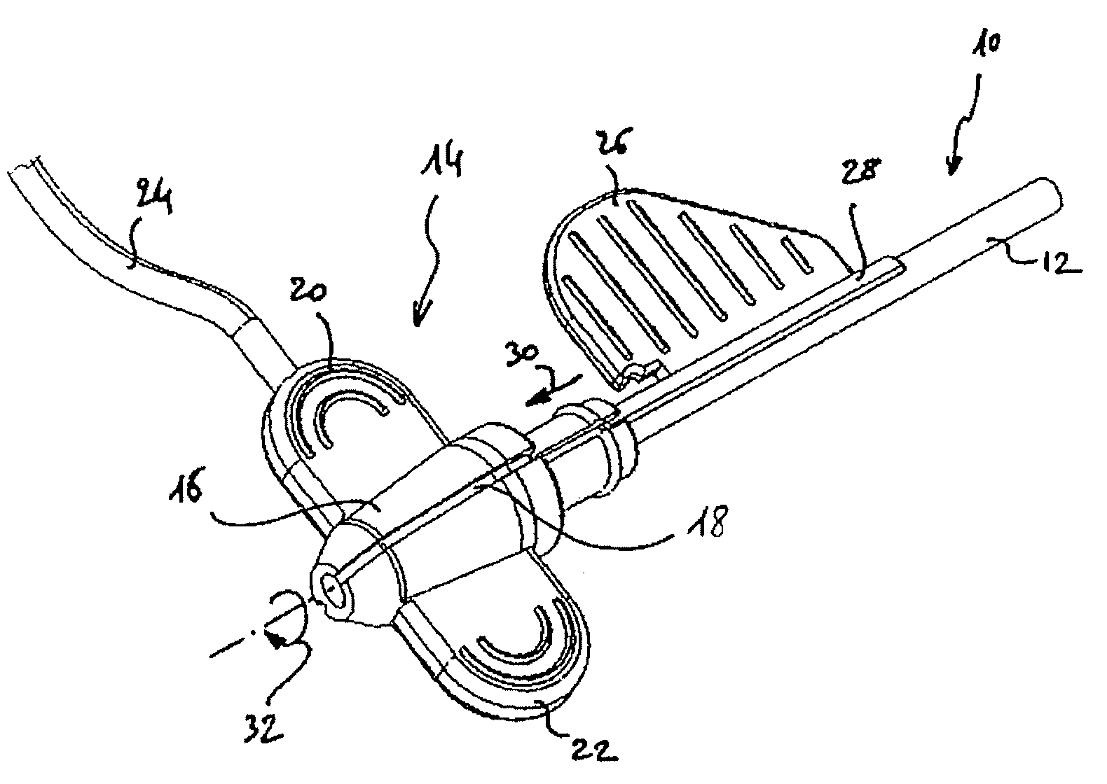
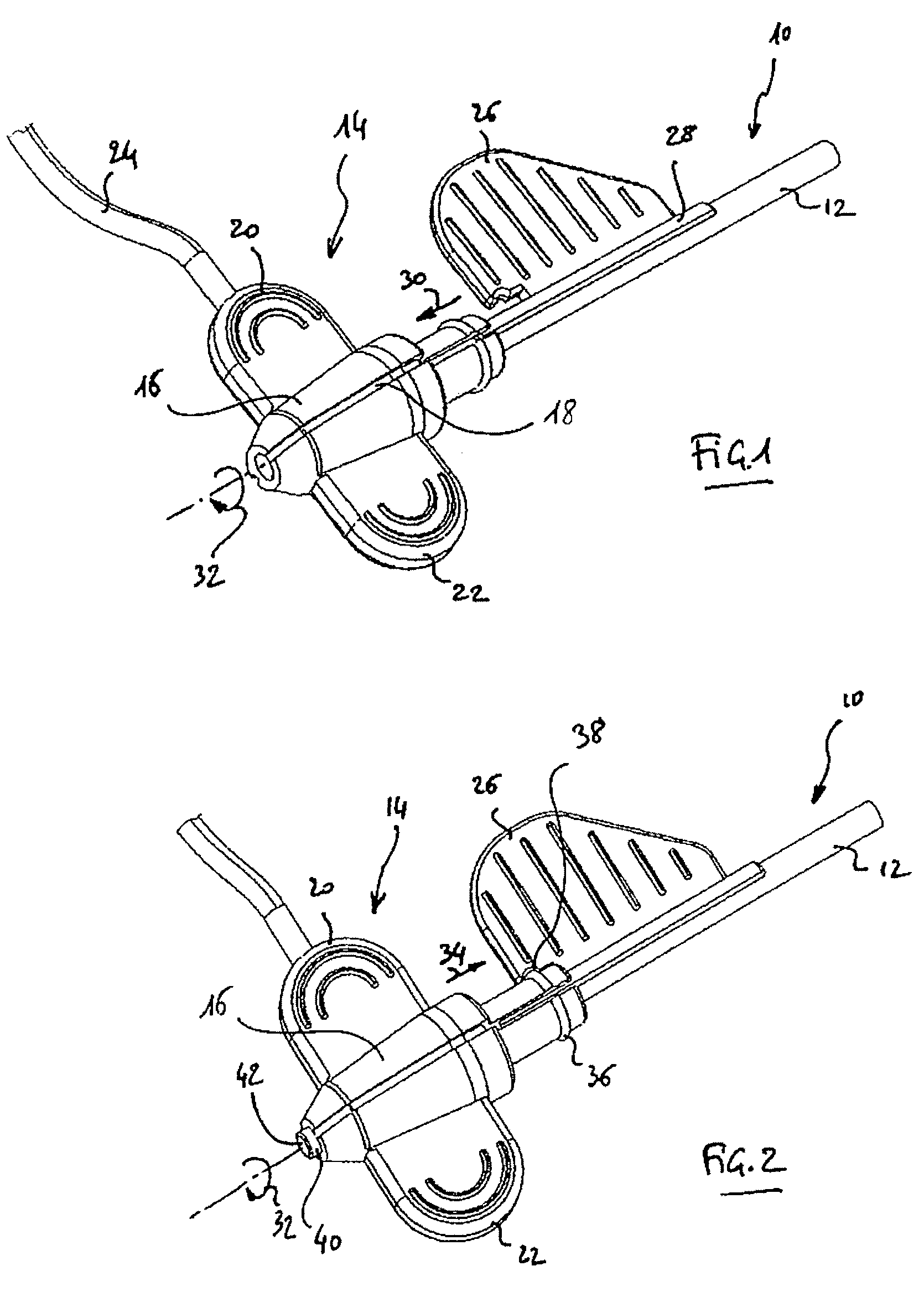
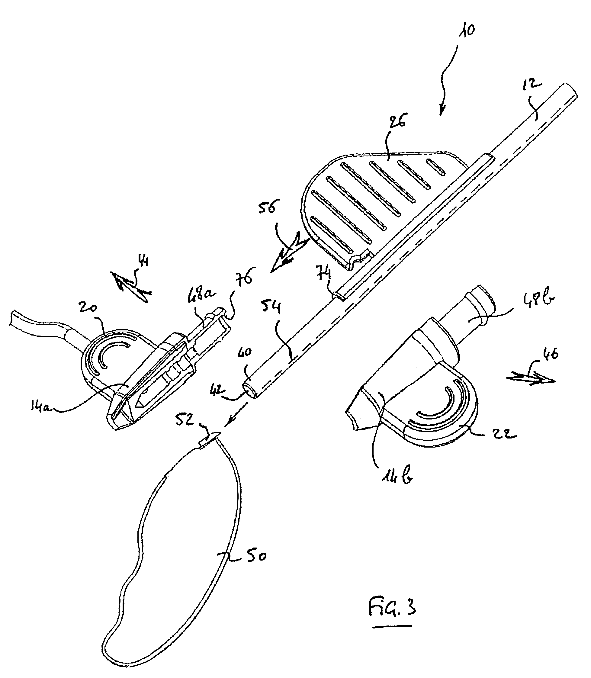
[0055]One will now describe an example embodiment of a toolkit in accordance with the present invention. With reference to the drawings, FIGS. 1 to 7 show the different aspects of an exemplary set comprising a guide-catheter and associated hemostatic valve, the valve being represented either in “closed” position (FIGS. 1 and 7) or “open” position (FIGS. 2, 4, 5 and 6), or after separating the valve from the guide-catheter, right after cutting thereof (FIG. 3).

[0056]The guide-catheter 10 comprises a hollow sheath 12 reinforced with a wire mesh in order to improve its rigidity; once the guide-catheter is in place, its proximal end (the one that is visible on the figures) is emerging from the introduction site, the surgeon using this emerging part for introducing, driving and positioning the lead at its final target site.

[0057]The proximal end of the guide-catheter 10 is provided with a hemostatic valve 14 allowing to selectively fill the internal lumen of sheath 12 at its input end.

[0...


PUM


Abstract

A toolkit for implanting an intracorporal lead, preferably a cardiac sensing / pacing lead. This toolkit includes a guide-catheter (10), having a sheath (12) with an internal lumen (42) opened at its distal and proximal ends, and cuttable along a generatrix in order to allow extracting of the guide-catheter after use. A hemostatic valve (14) is mounted at the proximal end (40) of the guide-catheter, for selectively filling, or not, the internal lumen of the guide-catheter at its input end. The valve is frangible in at least two parts, each dissociable from the guide-catheter at both sides around a median axial plane. The valve comprising a mobile element sliding on the guide-catheter between two extreme positions, with an opened position where the proximal end (40) of the guide-catheter (10) freely emerges out of the valve so as to allow access to said internal lumen, and a closed position where this proximal end of the guide-catheter is filled in a tight manner.

Description

FIELD OF THE INVENTION[0001]The present invention is directed to the implantation of intracorporal leads, such as leads for cardiac sensing / pacing that are usually associated with “active implantable medical devices” as such devices are defined by the Jun. 20, 1990 directive of the Counsel of European Communities, and more specifically to intracorporal leads for implantable devices for heart pacing, resynchronization, cardioversion and / or defibrillation.BACKGROUND OF THE INVENTION[0002]Cardiac leads may be endocardial leads, such as leads for sensing / pacing in right heart cavities, or leads introduced in the coronary network, notably leads comprising an electrode positioned in front of a left cavity of the myocardium.[0003]The insertion of the latter type of leads, carried out through endocavitary approaches, is a particularly tricky intervention, taking into account the difficult access to the coronary sinus entrance via the right atrium, and also the required accuracy for the paci...

Claims


Application Information

Patent Timeline
Patent Type & Authority Patents(United States)
IPC IPC(8): A61B19/00
CPCA61M25/0662A61M39/0693A61M25/0097A61N1/372
Inventor SHAN, NICOLAS
Owner SORIN CRM
Learn More