
Models and methods of using same for testing medical devices

a technology of medical devices and models, applied in educational models, additive manufacturing apparatus, instruments, etc., can solve the problems of reducing product quality, increasing development costs, and lengthening product timelines, so as to reduce product quality, increase development costs, and lengthen product timelines

Active Publication Date: 2011-08-09
SAKEZLES CHRISTOPHER
View PDF11 Cites 281 Cited by
  • Summary
  • Abstract
  • Description
  • Claims
  • Application Information

AI Technical Summary

Benefits of technology

"The patent text describes a new model for testing medical devices that can replace traditional methods such as live animal models and human clinical trials. These models are designed to mimic the target anatomy and the physical properties of living tissues. They can be used early in the development process to predict the effect of the device on the target tissue and reduce the risk of design errors. The models are also cost-effective and can be used at body temperature and in the presence of fluids. They can be used for an unlimited number of tests and can help to improve the quality of data collected. Overall, these models provide a better understanding of device performance and can help to protect the patient from harm during the development process."

Problems solved by technology

The use of a poorly conceived model in development testing will lead to reduced product quality, increased development costs, and greatly lengthened product timelines.
These models are unique in possessing a level of complexity that allows them to be substituted for either a live animal, an animal cadaver, or a human cadaver in the testing of these devices.

Method used


Image

  • Models and methods of using same for testing medical devices
  • Models and methods of using same for testing medical devices
  • Models and methods of using same for testing medical devices

Examples


example 1

Testing of a Guidewire Exchange Catheter

[0169]This following experiment describes a simple Animal Replacement Model used in the testing of a guidewire exchange catheter. The testing described includes the simulation of worst-case conditions to provide an estimate of device performance and reliability when misused in a clinical setting. This data was used to determine the suitability of the device for clinical trials. The materials required appear in Table III and the fabrication process for the tissue analog materials appears in Table IV.

[0170]

TABLE IIIMaterials and devices used in simulation example.Device or Material UsedQuantityDevice CodeBalloon Guidewire131Guidewire Exchange Catheter132AVE GT1 Floppy Guidewire13AVE Microstent II Stent Catheter14ACS RX Multilink Stent Catheter15Scimed Niron Ranger Stent Catheter16Scimed Magic Wallstent Stent Catheter17Hotplate Stirrer1—Animal Replacement Model1—Dimethyl SulfoxideA / R—Polyvinyl Alcohol (Mw = 130k-150k)A / RCompletelyhydrolysed

[0171]...

example 2

Rapid production of Neurovascular Model Employing a Monomer Containing Tissue Analog Liquid

[0193]According to one specific embodiment, the subject invention relates to the production of an anatomical neurovascular brain model comprising hydrogel materials and constructed using modified stereolithic techniques. Upon obtaining the anatomical images, converting them to CAD format and then converting to STL format, the model is built upon a platform situated just below the surface in a vat of tissue analog liquid. The liquid comprises water (50%-90% by mass), a monomer (10%-50% by mass), and an initiator (0.1%-1.0% by mass). In this example, a monomer such as N-vinyl pyrrolidone is implemented and a UV initiator such as ethyl 2,4,6-trimethylbenzoylphenylphosphinate is utilized. In this embodiment, the equilibrium water content of the tissue analog liquid closely approximates that of the intended hydrogel material of the artificial anatomical structure. Preferably, the water content of t...

example 3

Rapid Production of Neurovascular Model Employing a Polymer Containing Tissue Analog Liquid

[0196]According to this example, an artificial anatomical model is constructed utilizing modified stereolithography techniques that employ a tissue analog liquid comprising water, a polymer, and a cross-linking agent. The polymer agent in this example is poly-vinyl alcohol and the cross-linking agent is glutaraldehyde. The basic methodology is similar to that described in Example 1, except that instead of the tissue analog liquid comprising a monomer that forms a polymer network, the tissue analog liquid comprises a polymer that is cross-linked to achieve the desired gelling and other properties of the tissue analog material.


PUM


Abstract

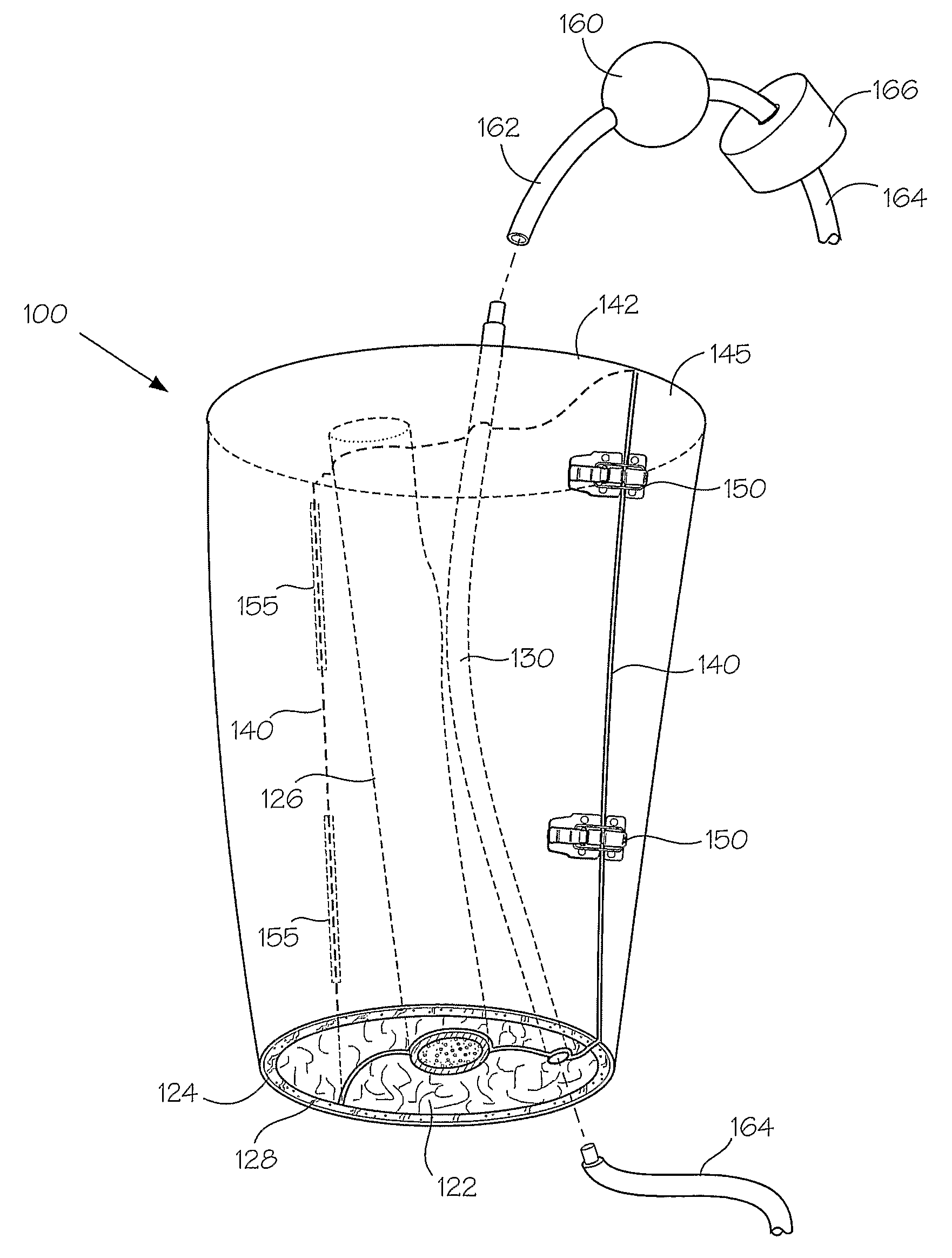
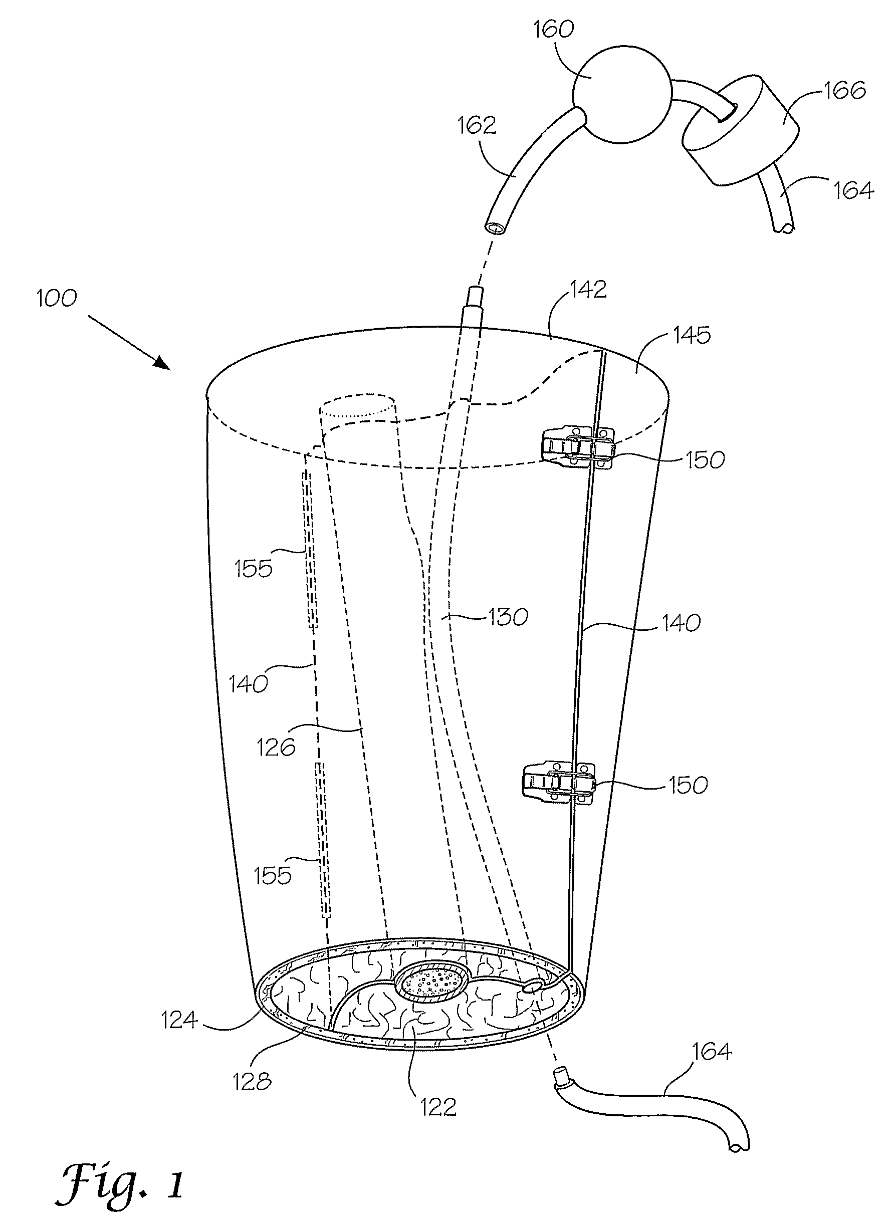
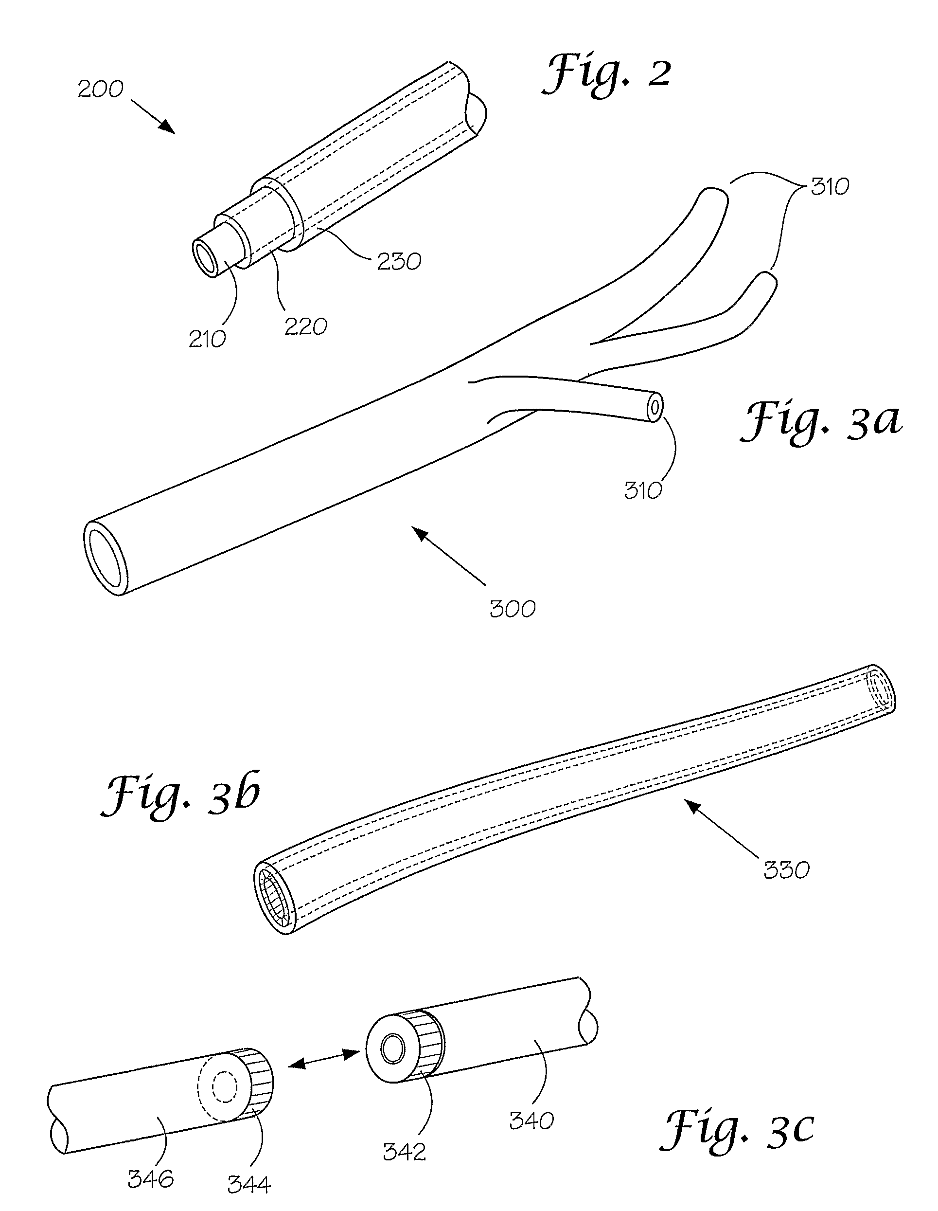
Disclosed herein are synthetic anatomical models that are designed to enable simulated use testing by medical device compam'es, medical device designers, individual inventors, or any other entity interested in the performance of medical devices. These models are unique in possessing a level of complexity that allows them to be substituted for either a live animal, an animal cadaver, or a human cadaver in the testing of these devices. These models are further characterized by a similarity of geometry, individual component physical properties, and component-to-component interfacial properties with the appropriate target tissue and anatomy.

Description

RELATED APPLICATIONS[0001]This application claims the benefit of U.S. application Ser. Nos. 11 / 050,161; filed Feb. 3, 2005, 11 / 098,248 filed Apr. 4, 2005; and 11 / 172,575 filed Jun. 30, 2005, and 60 / 749,972 filed Dec. 13, 2005, respectively, which are incorporated herein in their entirety.BACKGROUND OF THE INVENTION[0002]During the development of any new medical device, various tests may be required, including the characterization of physical properties (geometric, mechanical, electrical, electromagnetic, thermal, chemical, etc), the evaluation of overall device performance (numerical simulation or simulated use testing), or testing to determine the effect of the device on living tissues. These development tests may be broadly classified as either biological tests, theoretical tests, or physical tests, although there are areas where these testing classes overlap one another.[0003]Biological testing generally involves an analysis of the interaction between the device and human or anim...

Claims


Application Information

Patent Timeline
Patent Type & Authority Patents(United States)
IPC IPC(8): G09B23/28
CPCG09B23/306B33Y80/00
Inventor SAKEZLES, CHRISTOPHER
Owner SAKEZLES CHRISTOPHER
Learn More