
Method of recording two-dimensional images of a perfused blood vessel

a two-dimensional image and perfusion technology, applied in the field of two-dimensional images of perfused blood vessels, can solve the problems of affecting the quality of blood flow, the risk of tearing of the inner vascular wall, and the interruption of the incoming or outgoing blood supply for the duration of the investigation, so as to achieve a large mixing area and sufficient quantity

Inactive Publication Date: 2010-09-14
SIEMENS HEALTHCARE GMBH
View PDF26 Cites 6 Cited by
  • Summary
  • Abstract
  • Description
  • Claims
  • Application Information

AI Technical Summary

Benefits of technology

[0007]An object of the invention is therefore to specify a method that enables imaging to be improved whilst at the same time ensuring adequate blood supply to the organ area supplied by the vessel being investigated.
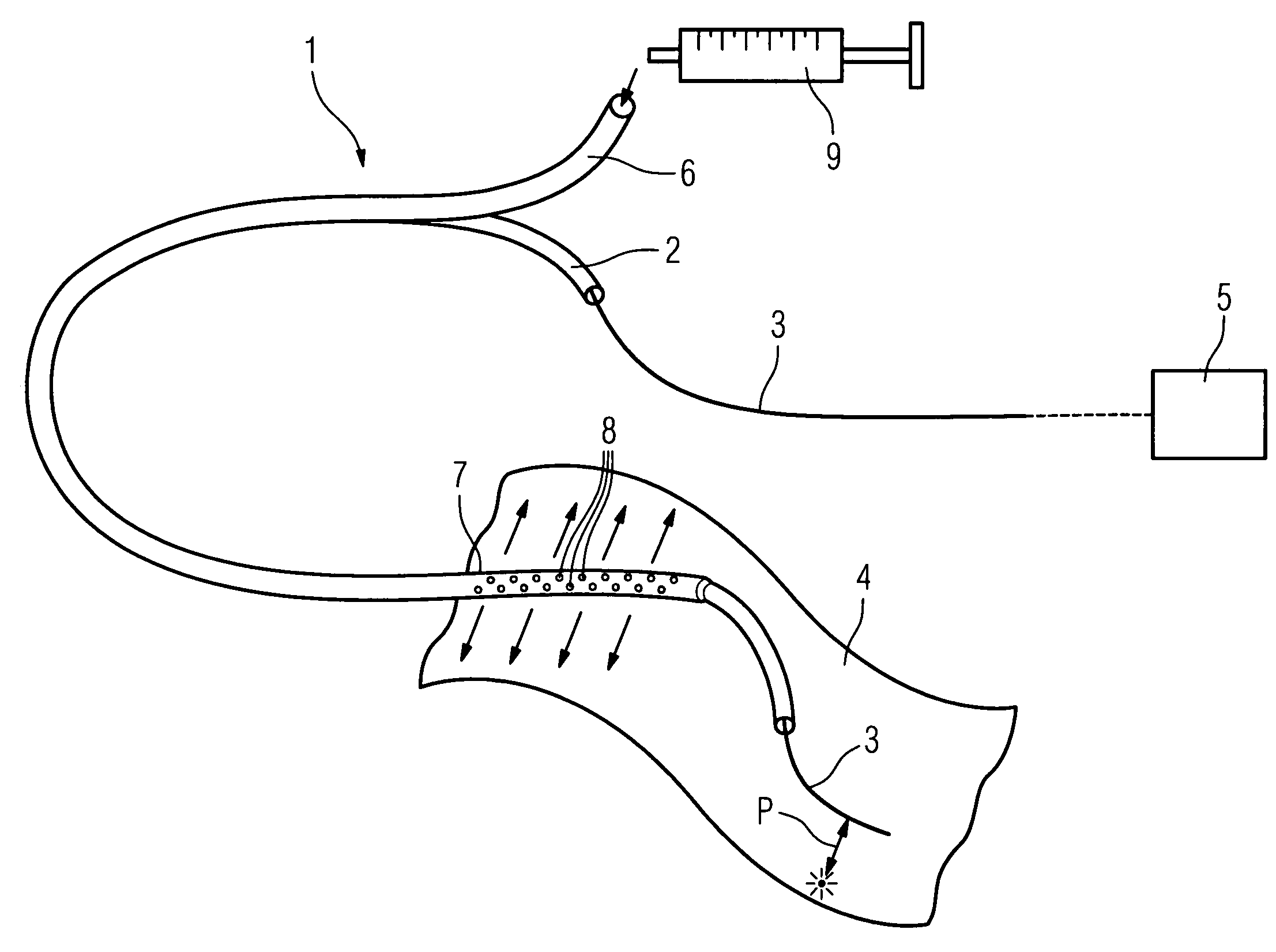
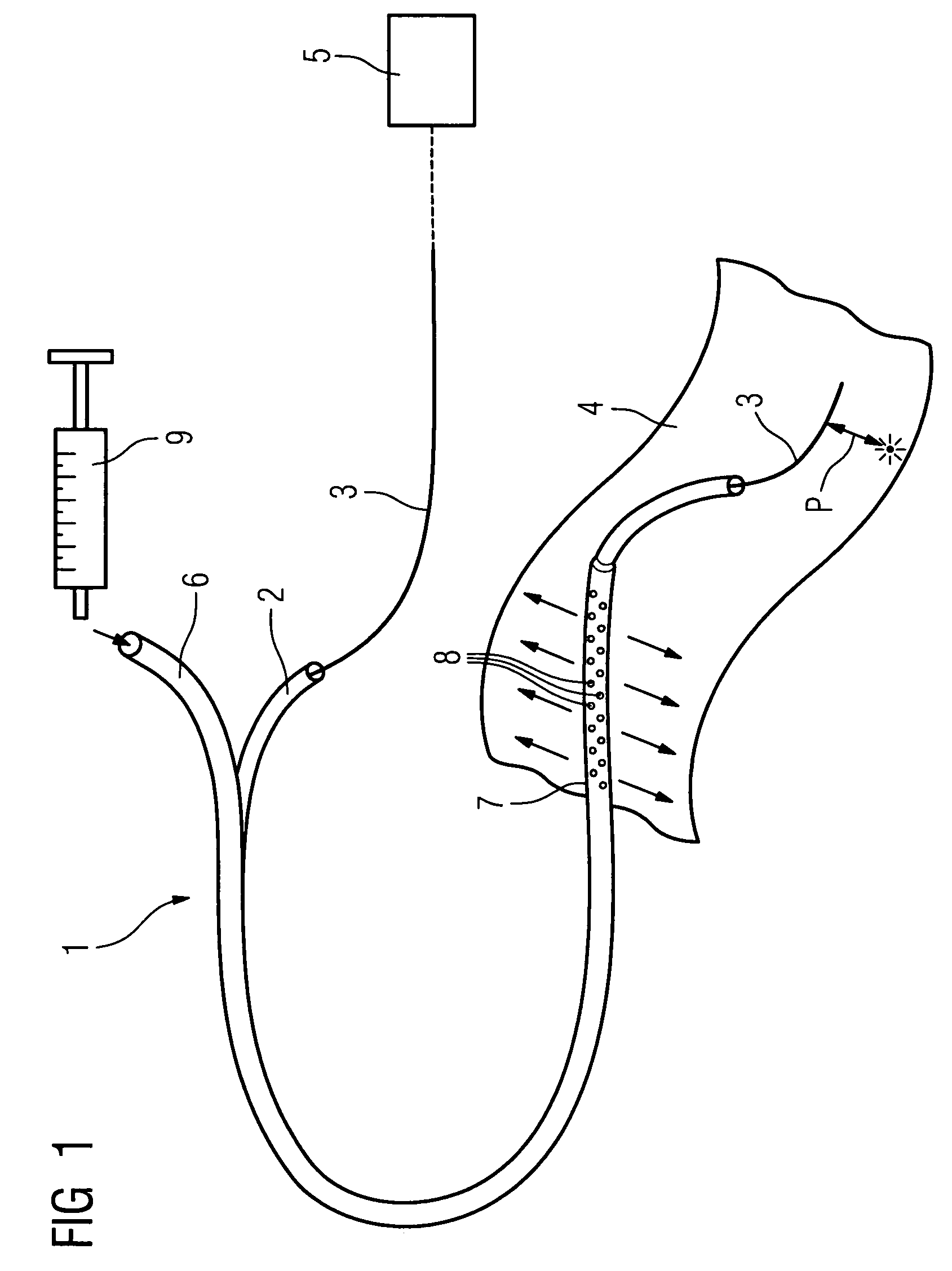
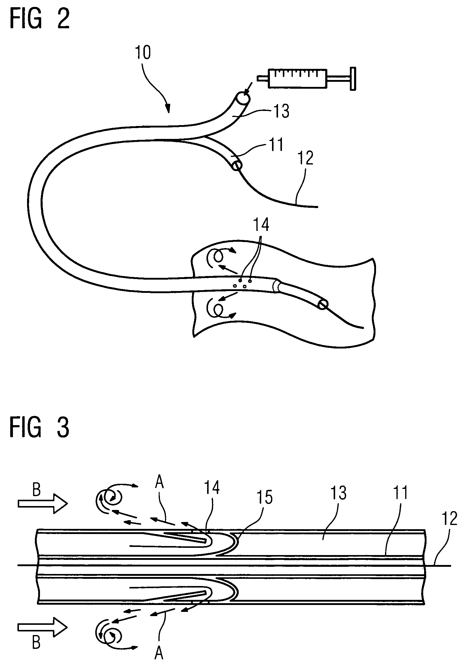
[0018]The outlet openings can be configured such that the fluid can be released outward in a direction essentially perpendicular to the longitudinal direction of the catheter. Alternatively, they may also be arranged or configured such that the delivered fluid is deflected and can be released outward in a direction that is essentially contrary to the supply direction. The second lumen actually surrounds the first lumen completely and in an essentially symmetrical manner, to advantageous effect.

Problems solved by technology

The disadvantage, however, is that the incoming blood supply (in the case of an artery) or outgoing blood flow (in the case of a vein) is interrupted for the duration of the investigation.
It is usually arteries that are being investigated, whereby the interruption of the blood supply is particularly problematic since organs arranged downstream of the point of interruption are no longer supplied.
Moreover, the inflation of the balloon in the artery may result in some trauma.
In particular, in atherosclerotically-transformed vessels (such vessels being preferred subjects of investigation), the inner vascular wall is at risk of tearing.
A further disadvantage is that the refractive index of the common salt solution is roughly equal to that of blood plasma.
Thus the existence of significant differences in the refractive index, leading to adverse scattering effects, continues to be a problem.

Method used


Image

  • Method of recording two-dimensional images of a perfused blood vessel
  • Method of recording two-dimensional images of a perfused blood vessel
  • Method of recording two-dimensional images of a perfused blood vessel

Examples


Embodiment Construction

[0023]FIG. 1 shows a catheter 1 according to the invention, comprising a first lumen 2, in which the movement of an imaging device 3 in the form of a wire with an integrated optical fiber (not shown in greater detail) is guided for the purpose of recording images from a vessel in an optical coherence tomography procedure. This image recording unit 3, i.e. the wire, protrudes out of the front, open end of the lumen 2, as shown in FIG. 1. In this area there is a window via which light delivered via the optical fiber (not shown in greater detail) is radiated onto the tissue of the vessel 4, which is illustrated here as being opened out for the sake of clarity, and via which light reflected from the vascular wall is coupled back into the optical fiber and carried to the exterior, where it is supplied to a control and processing unit 5 (shown here in exemplary form only) in order to generate the coherence tomography images. The light emission and coupling of the reflected light is illust...


PUM

PropertyMeasurementUnit
angleaaaaaaaaaa
refractive indexaaaaaaaaaa

Abstract

The invention relates to a method for recording two-dimensional images on the interior of a perfused blood vessel by means of optical coherence tomography using a catheter with an integrated imaging device, via which light is radiated and light reflected from the vascular wall is recorded, whereby a fluid containing a medium having a higher refractive index than blood plasma and / or cytoplasm is injected, via the catheter, into the vascular area to be recorded, to enable imaging to be carried out, whereby said fluid is a mixture of autologous blood and the medium.

Description

CROSS REFERENCE TO RELATED APPLICATIONS[0001]This application claims priority to the German application No. 10 2004 008 367.3, filed Feb. 20, 2004 which is incorporated by reference herein in its entirety.FIELD OF INVENTION[0002]The invention relates to a method for recording two-dimensional images on the interior of a perfused blood vessel by means of optical coherence tomography using a catheter with an integrated imaging device, via which light is radiated and light reflected from the vascular wall is recorded.BACKGROUND OF INVENTION[0003]Optical coherence tomography (OCT) is a catheter-based tomographic procedure, which is used—among other things—for obtaining two-dimensional vascular images in vivo. The catheter-guided image recording units used are very narrow wires with integrated optical fibers, via which optical fibers light is emitted in the near infrared range (approx. 1300 nm) and the reflection from the vascular wall is registered with depth resolution by means of infer...

Claims


Application Information

Patent Timeline
Patent Type & Authority Patents(United States)
IPC IPC(8): A61M31/00A61B1/12A61B6/00G01N21/17A61B5/00A61B6/03A61B17/20A61M25/00
CPCA61B5/0066A61B5/0084A61B5/6852
Inventor BOESE, JANKLEEN, MARTIN
Owner SIEMENS HEALTHCARE GMBH
Learn More