Therapeutic peptides for the treatment of metastatic cancer
a technology of peptides and tumors, applied in the field of cancer therapeutics, can solve the problems of loss of e-cadherin expression, almost 50% delay in tumor onset time, and cells no longer maintain homotypic interactions, and achieve the effect of reducing or retarding the invasiveness of the cancer
- Summary
- Abstract
- Description
- Claims
- Application Information
AI Technical Summary
Benefits of technology
Problems solved by technology
Method used
Image
Examples
example 1
Methods
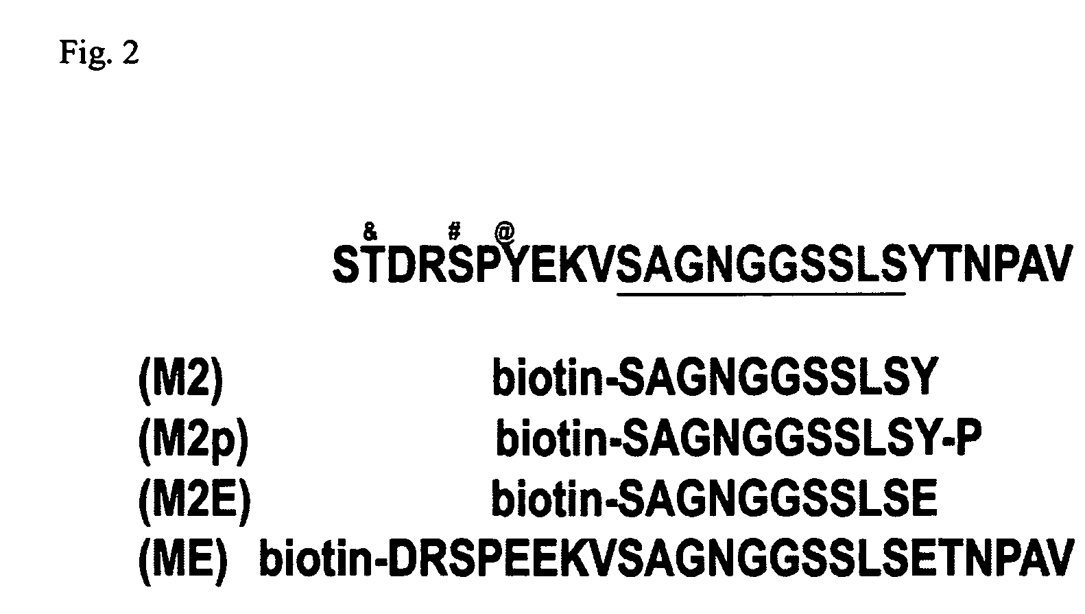
[0034]Peptide design: We have designed peptides to the MUC1 cytoplasmic domain that encompass the published binding sites β-catenin (Li et al., 1998; Li et al., 2001 a; Li et al., 2001b). Due to the tyrosine phosphorylation of MUC1 by EGFR and c-src, we have designed 25 mer peptides that are both nascent and tyrosine phosphorylated, and will determine the requirement of tyrosine phosphorylation for interaction withβ-catenin. Furthermore, we have designed peptides that have the tyrosine residues mutated to glutamic acid residues to determine if this will further inhibit cellular invasion. These peptides will all be synthesized by the American Peptide Company (Sunnyvale, Calif.). The peptides are produced at an 85% purity level (purified by HPLC, assayed by Mass Spectrometry), and biotinylated at the C-terminus for identification purposes.
[0035]Peptide treatment and invasion assay: 8 um pore size Transwell inserts (Corning) will be inverted and coated with 90 ul of Type I colla...
example 2
Effect of MUC1 Expression on Breast Cancer Invasion
[0044]To investigate the functional significance of MUC1 expression on breast cancer invasion, we have incubated invasive breast cancer cell lines with MUC1-mimetic peptides designed to the β-catenin interacting domain. We then monitored the effects of peptide treatment on invasion through a filter and into a collagen gel.
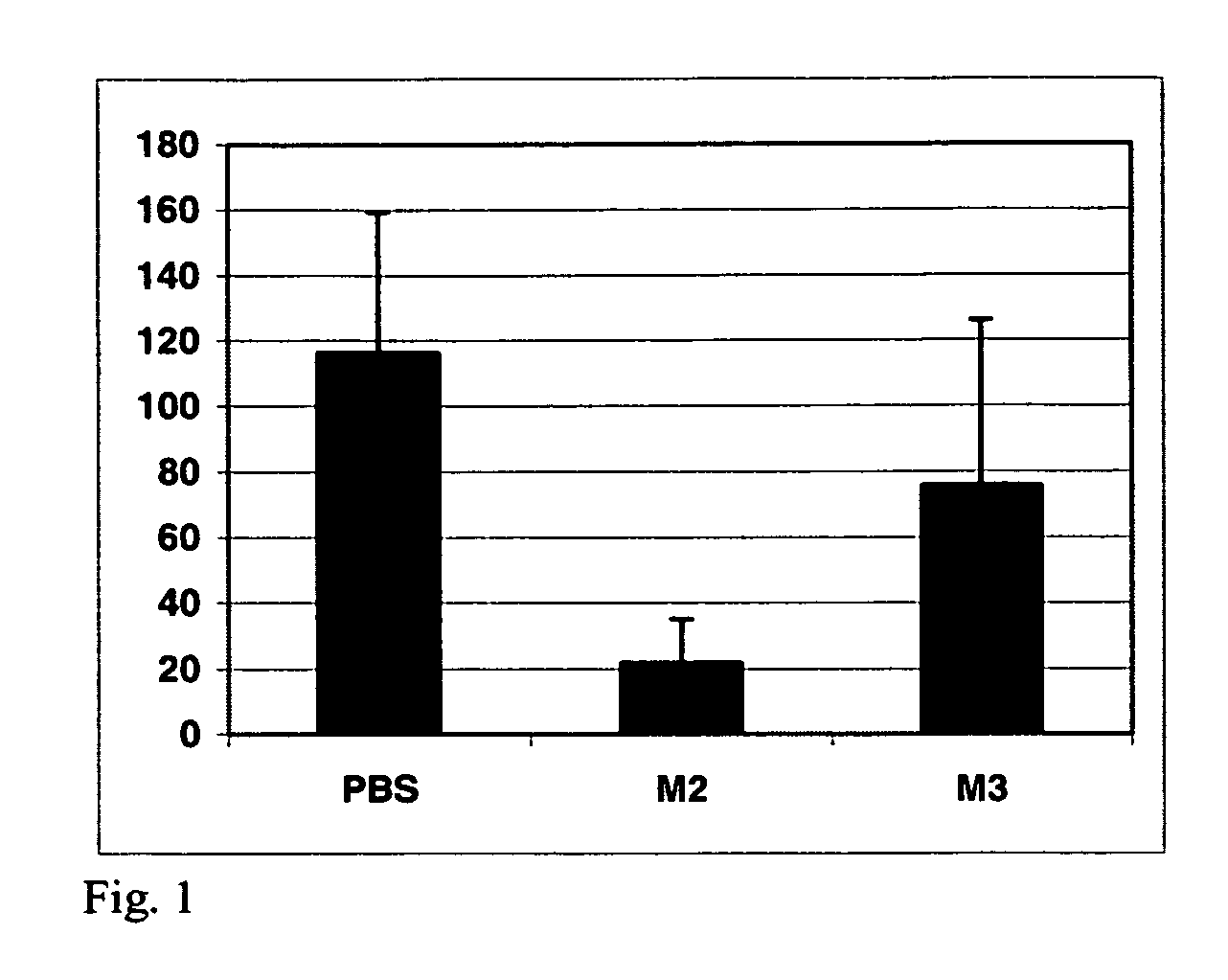
[0045]We performed initial experiments with these β-catenin-binding site (M2) peptides in MDA-MB-468 cells. Treatment with MUC1 / β-catenin (M2) peptides resulted in an approximately 8-fold inhibition of invasion of these cells into a collagen matrix (FIG. 1). We observed less than 15 M2 peptide-treated cells invaded into a collagen matrix in a 4 hour period, compared to approximately 125 cells in PBS controls. A non-specific peptide treatment (M3) gave results similar to PBS treated controls.
[0046]Analysis of the binding domains of MUC1 allows us to create a model of protein interactions from these preliminary exper...
example 3
MUC1 and β-Catenin Interact in Mouse Model
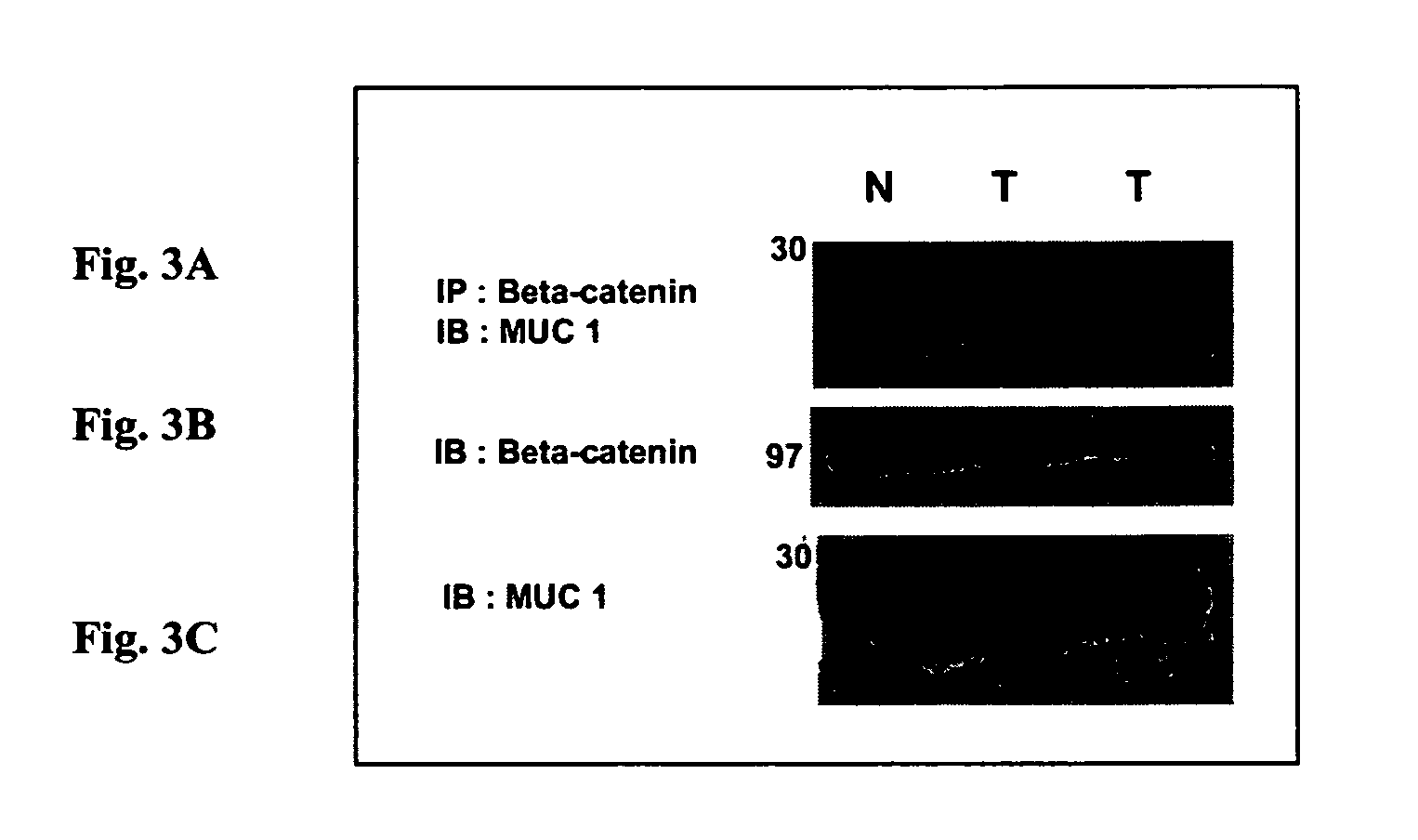
[0047]MUC1 and β-catenin interact in a spontaneous model of metastatic breast cancer. While multiple models have demonstrated a tumor-specific interaction between Muc1 and β-catenin (MMTV-Wnt-1, MMTV-MUC1, and breast cancer cell lines), they are not optimal for preclinical models for a variety of reasons (including relevance to human disease, tumor onset and latency). We therefore sought out an additional mouse model that might serve as an appropriate model in which to test our peptide-based therapies. The MMTV-pyMT (Mouse Mammary Tumor Virus—promoter driven Polyoma Middle T antigen) model of breast cancer is a transgenic mouse that develops breast cancer that is metastatic to the lung in greater than 70% of the animals analyzed (Guy et al., 1992a). This is an excellent model of breast cancer progression because it is a) metastatic, b) driven by the pyMT oncogene which interacts with a large number of cellular signaling pathways, including M...
PUM
| Property | Measurement | Unit |
|---|---|---|
| pore size | aaaaa | aaaaa |
| concentrations | aaaaa | aaaaa |
| concentrations | aaaaa | aaaaa |
Abstract
Description
Claims
Application Information
 - R&D
- Intellectual Property
- Life Sciences
- Materials
- Tech Scout
- Unparalleled Data Quality
- Higher Quality Content
- 60% Fewer Hallucinations
Browse by: Latest US Patents, China's latest patents, Technical Efficacy Thesaurus, Application Domain, Technology Topic, Popular Technical Reports.
© 2025 PatSnap. All rights reserved.Legal|Privacy policy|Modern Slavery Act Transparency Statement|Sitemap|About US| Contact US: help@patsnap.com


