
Ophthalmologic information processing apparatus and ophthalmologic examination apparatus

a technology of information processing and ophthalmologic examination, which is applied in the field of ophthalmologic information processing apparatus and ophthalmologic examination apparatus, can solve the problems of not being able to grasp the abnormality of the visual field, and not being able to grasp the morphology of the retina

Inactive Publication Date: 2010-01-05
KK TOPCON
View PDF14 Cites 8 Cited by
  • Summary
  • Abstract
  • Description
  • Claims
  • Application Information

AI Technical Summary

Benefits of technology

Enables accurate measurement of retina layer thickness at stimulation points, allowing for a clear correlation between visual field functions and retina morphology, thereby diagnosing abnormalities effectively.

Problems solved by technology

Conventionally, it has been possible to individually grasp an abnormality of the visual field and a morphological abnormality of the retina, but it has not been possible to grasp the morphology of the retina at a stimulation point in a visual-field examination.
Therefore, it has also not been possible to grasp an abnormality of the visual field and a morphological abnormality of the retina in the corresponding manner.
Incidentally, with the perimeter described in JP-A 2005-102946, it is possible to grasp, by referring to a fundus oculi image, in what site of the fundus oculi a visual field has been measured, but it is not possible to grasp a morphological abnormality of the retina, especially an abnormality inside the retina.
Therefore, even with this perimeter, it is not possible to grasp the correspondence between an abnormality of the visual field and a morphological abnormality of the retina.

Method used


Image

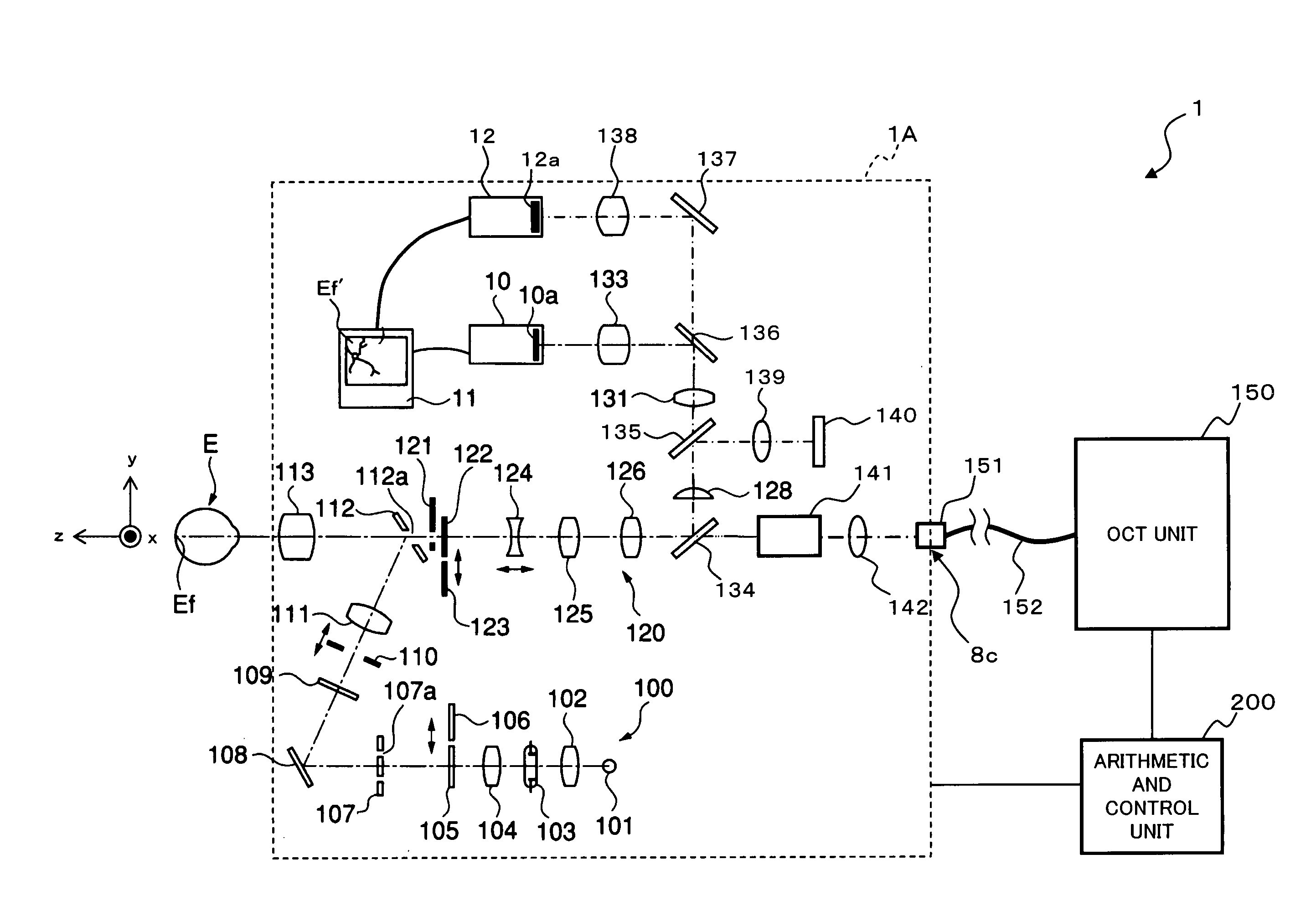
  • Ophthalmologic information processing apparatus and ophthalmologic examination apparatus
  • Ophthalmologic information processing apparatus and ophthalmologic examination apparatus
  • Ophthalmologic information processing apparatus and ophthalmologic examination apparatus

Examples


first embodiment

[0245]In a first embodiment, the program controls a computer that has a storing part for storing a 3-dimensional image representing the morphology of a retina. Examples of the storing part include the storage 232 and the storage 332 of the above embodiments.

[0246]This program makes this computer function as both a specifying part and an analyzing part. The specifying part specifies, in a 3-dimensional image, a plurality of positions that correspond to a plurality of stimulation points in a visual-field examination. Examples of the specifying part include the examination-position specifying part 231 and the examination-position specifying part 331 of the above embodiments.

[0247]The analyzing part analyzes the 3-dimensional image to find the layer thickness of the retina at each position that has been specified. Examples of the analyzing part include the layer-thickness measuring part 235 (and the examination-results comparing part 236) and the layer-thickness measuring part 335 (and ...

second embodiment

[0249]In a second embodiment, the program makes a computer, which has a storing part for storing a 3-dimensional image representing the morphology of a retina, function as a designating part, a specifying part, and an analyzing part. The designating part designates a position in the 3-dimensional image. Examples of the designating part include the user interface 240 (operation part 240B) and the user interface 340 (operation device).

[0250]The specifying part specifies a new position related to the designated position. Examples of the specifying part include the examination-position specifying part 231 and the examination-position specifying part 331 of the above embodiments.

[0251]The analyzing part analyzes the 3-dimensional image to find the layer thickness of the retina at the new position. Examples of the analyzing part include the layer-thickness measuring part 235 (and the examination-results comparing part 236) and the layer-thickness measuring part 335 (and the examination-re...


PUM


Abstract

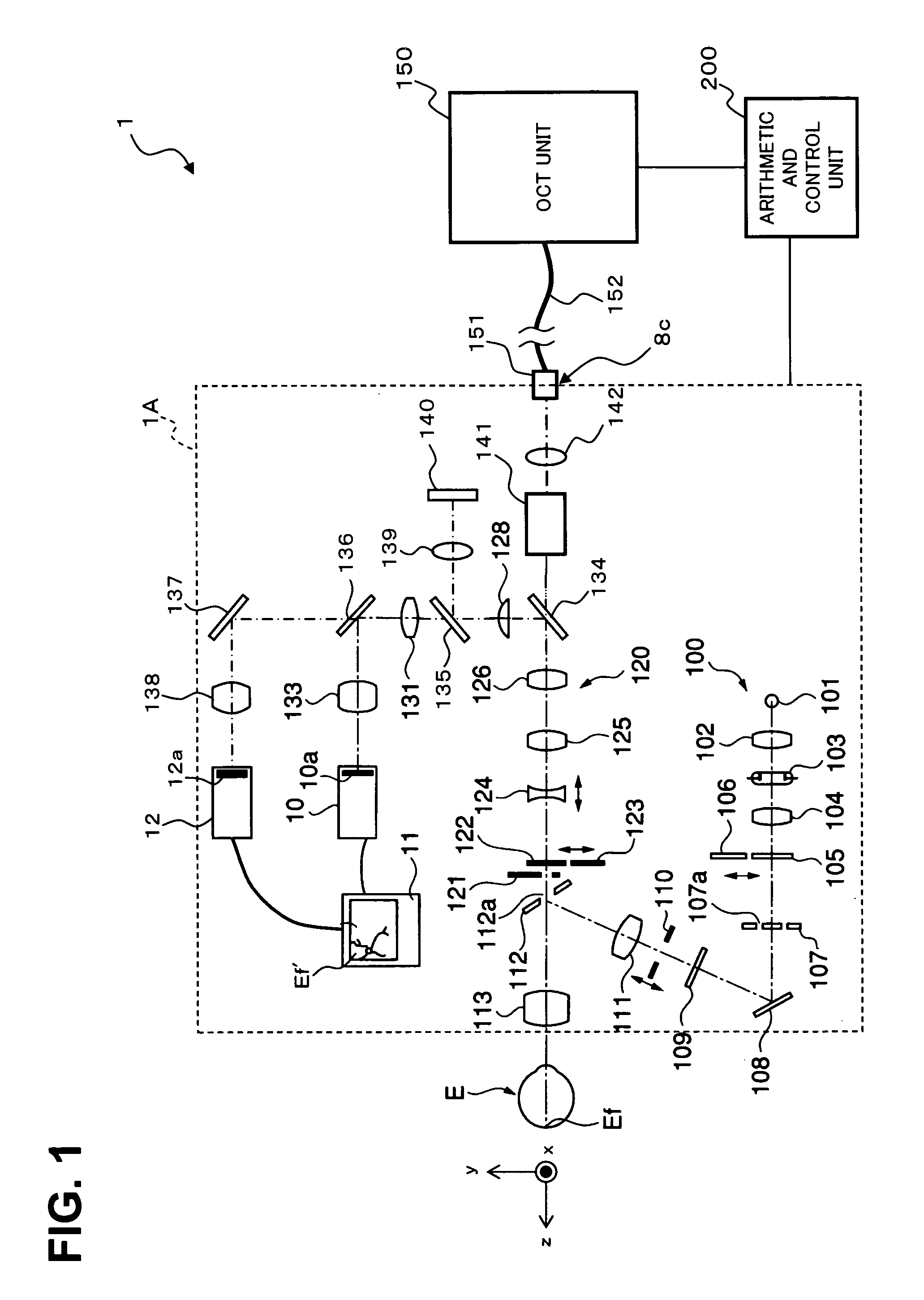
An ophthalmologic examination apparatus 1 projects a light onto a fundus oculi, detects the reflected light thereof, and forms a 3-dimensional image that represents the morphology of a retina based on the detected results. A stimulation-position specifying part 233 specifies, in the 3-dimensional image, a plurality of stimulation positions that correspond to a plurality of stimulation points Pi in a visual-field examination. A layer-thickness measuring part 235 analyzes the 3-dimensional image to find the layer thickness of the retina at each stimulation position. In addition, a displacement calculation part 234 specifies a related position of the stimulation position. A layer-thickness measuring part 235 finds the layer thickness of the retina at the related position.

Description

BACKGROUND OF THE INVENTION[0001]The present invention is related to an ophthalmologic information-processing apparatus and an ophthalmologic examination apparatus that are used for examinations in the field of ophthalmology.[0002]In the examinations used in the field of ophthalmology, there is a visual field examination for measuring the visual field of an eye. The visual-field examination is carried out for diagnosing diseases in which abnormalities of the visual field occur, such as glaucoma, retinal pigmentary degeneration, central chorioretinopathy, optic neuritis, and macular hole. Abnormalities of the visual field include stenosis, hemianopia, scotoma, and the like.[0003]For the visual-field examination, a dynamic perimeter for measuring dynamic visual field and a static perimeter for measuring static visual field are used (e.g. cf. Japanese Unexamined Patent Publications JP-A 2000-262471, JP-A 2002-253502, and JP-A 2003-164424). The dynamic perimeter is referred to as an aut...

Claims


Application Information

Patent Timeline
Patent Type & Authority Patents(United States)
IPC IPC(8): A61B3/14A61B3/00A61B3/10
CPCA61B3/102A61B3/024
Inventor HANGAI, MASANORIYOSHIMURA, NAGAHISAHUANG, YIJUNNADACHI, RYOICHI
Owner KK TOPCON
Learn More