
Method for the integration of medical imaging data and content for wireless transmission and remote viewing

a technology of medical imaging and wireless transmission, applied in the field of wireless transmission and remote viewing of medical imaging data and content, can solve the problems of obviating the comparative ease of manipulating digital images, complicating the transmission and viewing of digital images created on one machine, and doctors often being unable to view digital images captured by scanning technicians

Active Publication Date: 2008-05-27
TICSA STELIAN DORU
View PDF3 Cites 31 Cited by
  • Summary
  • Abstract
  • Description
  • Claims
  • Application Information

AI Technical Summary

Benefits of technology

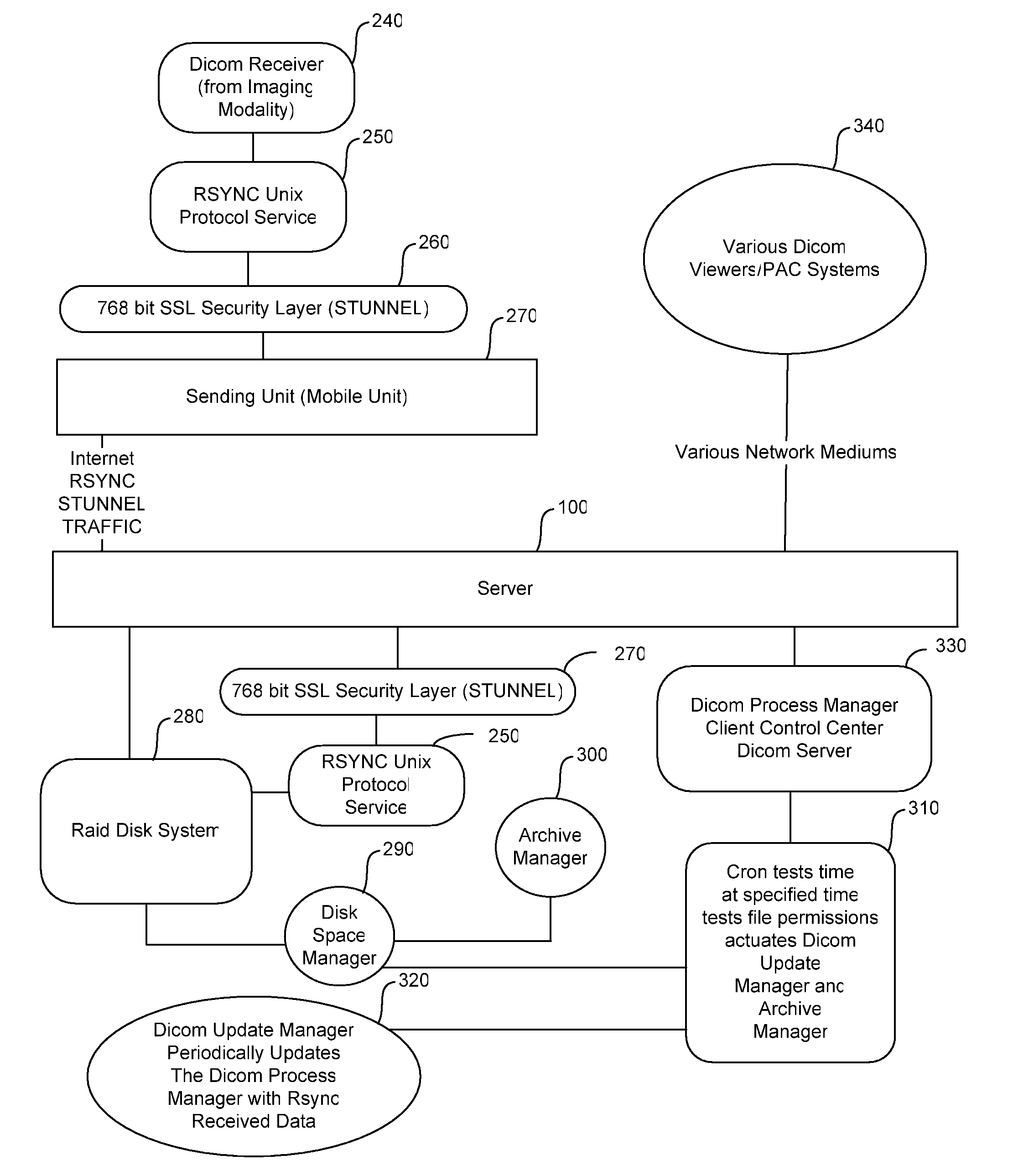
[0007]The present invention is a method of transmitting medical diagnostic imaging over low-speed wireless networks to remote locations including the steps of: accessing an imaging modality; receiving an image from the imaging modality; establishing a wireless connection with a remote primary server; establishing a security layer over the remote connection; compressing the image; encrypting the compressed image; establishing an incremental file transfer of the compressed image over the security layer to the primary server; transmitting the compressed image to the primary server; decrypting the compressed image; decompressing the image; substantially losslessly storing the image on a storage device local to the primary server; accepting authenticated remote inbound requests for viewing the image; and sending the image to an authenticated requestor. The at least one image received from the imaging modality is preferably formatted to a DICOM specification and the connection with the remote primary server is through TCP / IP. The protocol for the incremental file transfer is an RSYNC-based protocol and may include the step of archiving the at least one image to an optical storage medium for long term storage.

Problems solved by technology

The comparative ease of manipulating these digital images is obviated by problems arising from incompatible software among the various manufacturers of the imaging scanners.
Despite the creation of the DICOM standard manufacturers have developed proprietary software, and image formats, which complicate the transmission and viewing of digital images that are created on one machine but need to be viewed on another.
As a result, Doctors are often unable to view digital images captured by scanning technicians.
If the technician reduces the image to a tangible picture the Doctor cannot manipulate the image to zoom in, rotate, or change the contrast of the still image.
Furthermore, the process of reducing the images to tangible form is costly in both time and materials.

Method used


Image

  • Method for the integration of medical imaging data and content for wireless transmission and remote viewing
  • Method for the integration of medical imaging data and content for wireless transmission and remote viewing
  • Method for the integration of medical imaging data and content for wireless transmission and remote viewing

Examples


Embodiment Construction

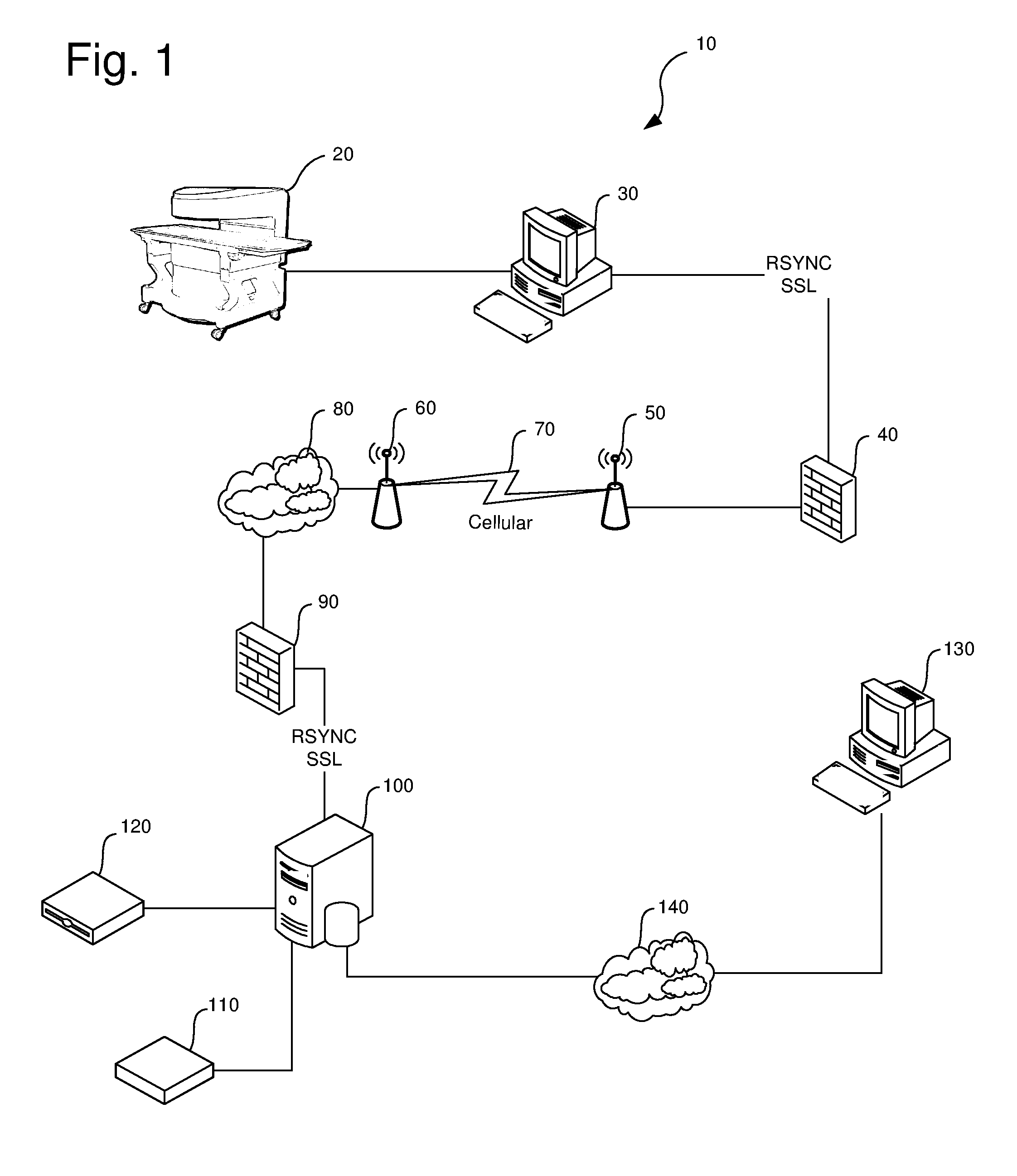
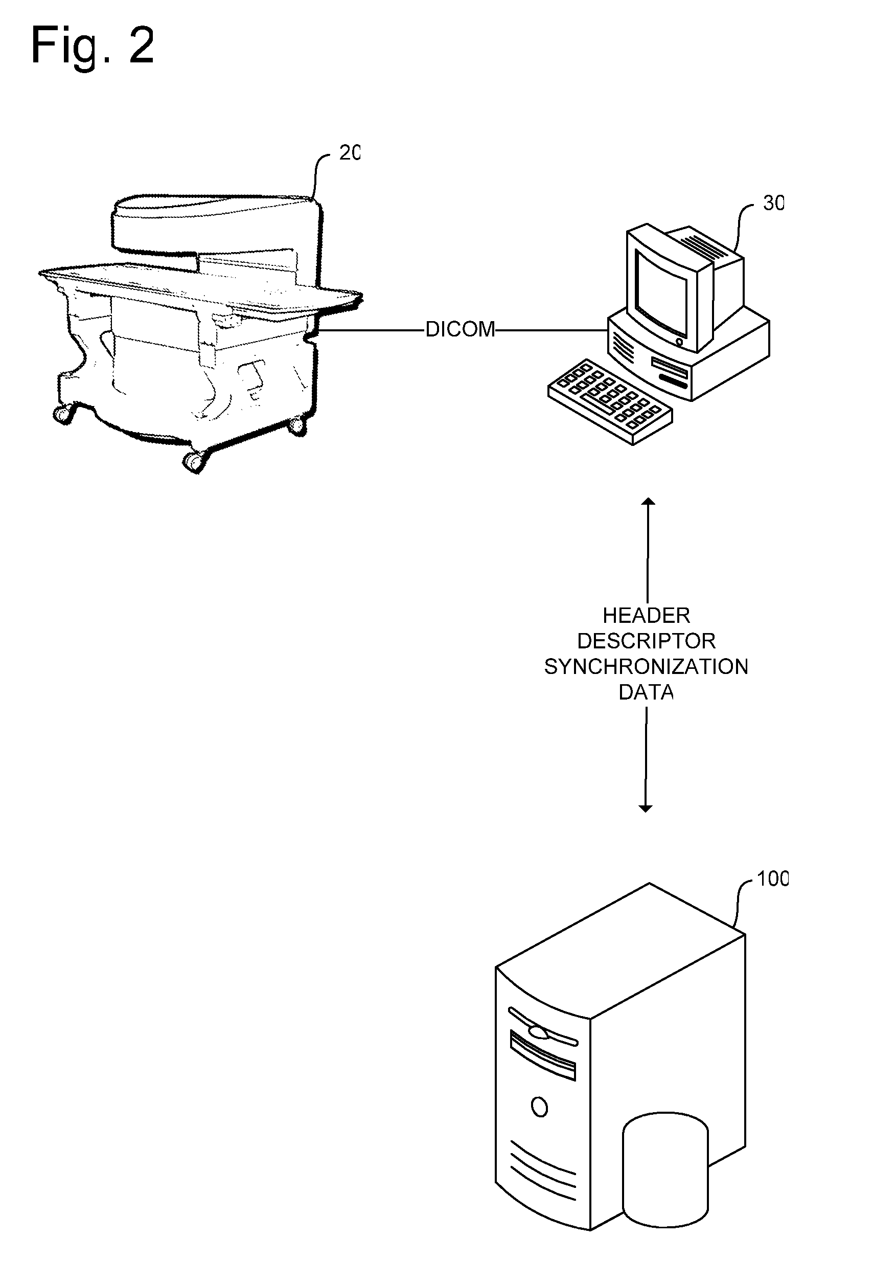
[0014]Turning to FIG. 1, the invention is denoted numeral 10 as a whole. Imaging modality 20 generates a DICOM data file received by client computer 30. Client computer 30 establish first security layer 40 including SSL-based encryption over an RSYNC incremental protocol. The RSYNC protocol allows the resumption of the stream data over high latency, low bandwidth networks that occasionally fail. Absent this incremental protocol, a network failure, even if brief, or if a single packet in a transfer times out, the sender or server point must restart the transmission from the beginning. Because the DICOM data files may be necessarily large, even when compressed, the RSYNC protocol is advantageous as it permits the transmission of the files through relatively unstable network connections.

[0015]First wireless transceiver 50 transmits to second wireless transceiver 60 through wireless network 70. Wireless network 70 may include, but is not limited to, cellular networks, WiFi, Bluetooth, o...


PUM


Abstract

A method of transmitting medical imaging data files over high latency low bandwidth networks by using an incremental RSYNC protocol over an encrypted connection.

Description

PRIORITY CLAIM[0001]This invention claims priority from U.S. Provisional Patent Ser. No. 60 / 481,345 filed Sep. 9, 2003 and titled “Method for the Integration of Medical Imaging Data and Content for Wireless Transmission and Remote Viewing.”FIELD OF INVENTION[0002]This invention relates to a system for the acquisition and verification of the integrity of medical imaging data for encrypted transmission via wireless VPN network and embedding upon a CD-ROM.BACKGROUND OF INVENTION[0003]The advent of digital imaging for the purpose of medical diagnosis has resulted in a variety of scanning systems and image formats. The comparative ease of manipulating these digital images is obviated by problems arising from incompatible software among the various manufacturers of the imaging scanners.[0004]In response to the increase in digital imaging technology, the American College of Radiology (ACR) and the National Electrical Manufacturers Association (NEMA) developed the Digital Imaging and Commun...

Claims


Application Information

Patent Timeline
Patent Type & Authority Patents(United States)
IPC IPC(8): G06K9/36G06K9/46G16H30/20G16H30/40
CPCG06F19/321H04W4/18H04L67/06H04L69/04H04L63/0428H04L63/166H04W12/02H04W80/08H04L69/16H04L67/12G16H30/20G16H30/40H04W12/033
Inventor TICSA, STELIAN DORU
Owner TICSA STELIAN DORU
Learn More