Apparatus and method for endoscopic colectomy

Active Publication Date: 2008-03-04
INTUITIVE SURGICAL OPERATIONS INC
View PDF30 Cites 821 Cited by
  • Summary
  • Abstract
  • Description
  • Claims
  • Application Information

AI Technical Summary

Benefits of technology

[0012]In keeping with the foregoing discussion, the present invention takes the form of methods and apparatus for performing endoscopic colectomy that combine the advantages of the laparoscopic and endolumenal approaches. The diseased portion of the colon to be resected is identified using either laparoscopic and / or colonoscopic techniques or using another imaging modality. A colectomy device mounted on a colonoscope grasps the colon wall at two sites adjacent to a diseased portion of the colon. Using laparoscopic techniques, the diseased portion of the colon is separated from the omentum and the blood vessels supplying it are ligated or cauterized. The colon wall is transected to remove the diseased portion and the excised tissue is removed using the laparoscope or drawn into the colectomy device for later removal upon withdrawal of the colonoscope. The colectomy device approximates the two ends of the colon and performs an end-to-end anastomosis. If the part to be resected is a tumor, prior to the resection, the edges of the segment to be resected will be stapled to seal it and prevent spillage of malignant cells to the healthy tissue.
[0013]The methods and apparatus of the present invention provide a number of benefits not realized by the prior art approaches to colectomy. As stated above, the purely endolumenal approach does not provide for separation of the colon from the omentum, which is necessary when resecting more than just a small portion of the colon wall. By combining laparoscopic techniques with a colonscope-mounted colectomy device, the present invention overcomes this deficiency in the prior art allowing a more comprehensive approach to colectomy. Unlike prior art laparoscopic techniques, however, the colon does not need to be exteriorized for excision of the diseased portion or anastomosis of the remaining colon. The colonscope-mounted colectomy device approximates the ends of the colon and performs an anastomosis from the interior of the lumen of the colon. The excised tissue can be drawn into the colectomy device for removal through the lumen of the colon along with the colonoscope or can be taken out by the laparoscope, which can be done through a very small incision in the patient's skin. The prior art approach also does not protect from leaking of malignant cells to the periphery. This idea will enable sealing of the tissue with staples at its ends to prevent such leakage. Optionally, it will be done with the help of a laparoscopic device that will serve as an anvil. Unlike the prior art procedure, the present invention will optionally use a balloon inflated in the lumen of the colon or any other resected organ before stapling, and by this assure the anastomosis will be ideal with the best possible approximation of the edges.
[0014]The use of colonoscopic techniques in the present invention provides another benefit not realized by a purely laparoscopic approach. Since colonoscopic examination is at present the most definitive diagnostic method for identifying diseases of the colon, locating the lesions through the exterior of the colon by laparoscopy or even by direct visualization can be somewhat problematic. Using the colonoscope to identify and isolate the diseased portion of the colon from within the lumen helps assure that the correct portions of the colon wall are excised and makes clean surgical margins without residual disease more assured as well.
[0015]In a preferred embodiment, the present invention utilizes a steerable colonoscope as described in U.S. patent application Ser. Nos. 09 / 790,204 (now U.S. Pat. No. 6,468,203); 09 / 969,927; and 10 / 229,577, which have been incorporated by reference. The steerable colonoscope described therein provides a number of additional benefits for performing endoscopic colectomy according to the present invention. The steerable colonoscope uses serpentine motion to facilitate rapid and safe insertion of the colonoscope into the patient's colon, which allows the endoscopic colectomy method to be performed more quickly and more safely. Beyond this however, the steerable colonoscope has the capability to create a three-dimensional mathematical model or map of the patient's colon and the location of any lesions identified during the initial examination. Lesions found during a previous examination by CT, MRI or any other imaging technology can also be mapped onto the three dimensional map of the colon. By generating a three dimensional map of the colon, the system knows where each part of the endoscope is in the colon and will be able to localize the two parts of the dissecting and stapling system exactly in the desired location. During surgery, this information can be used to quickly and accurately return the colonoscope to the location of the identified lesions where the colonoscope-mounted colectomy device will be used to complete the endoscopic colectomy procedure.

Problems solved by technology

As stated above, the purely endolumenal approach does not provide for separation of the colon from the omentum, which is necessary when resecting more than just a small portion of the colon wall.
The prior art approach also does not protect from leaking of malignant cells to the periphery.
Since colonoscopic examination is at present the most definitive diagnostic method for identifying diseases of the colon, locating the lesions through the exterior of the colon by laparoscopy or even by direct visualization can be somewhat problematic.

Method used


Image

  • Apparatus and method for endoscopic colectomy
  • Apparatus and method for endoscopic colectomy
  • Apparatus and method for endoscopic colectomy

Examples


first embodiment

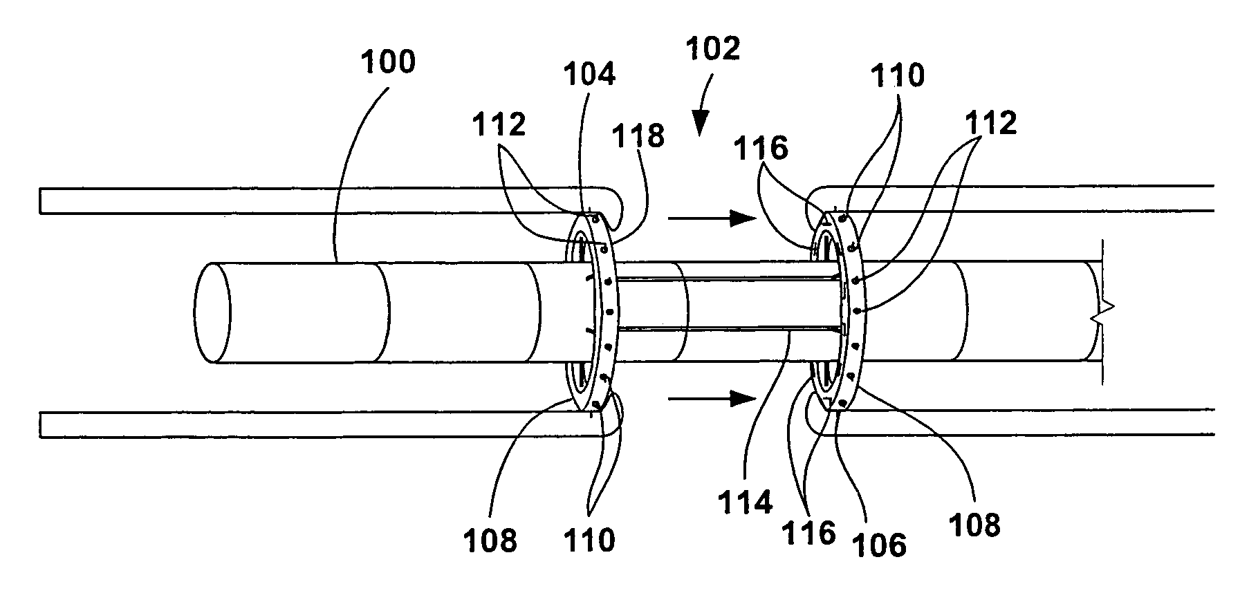
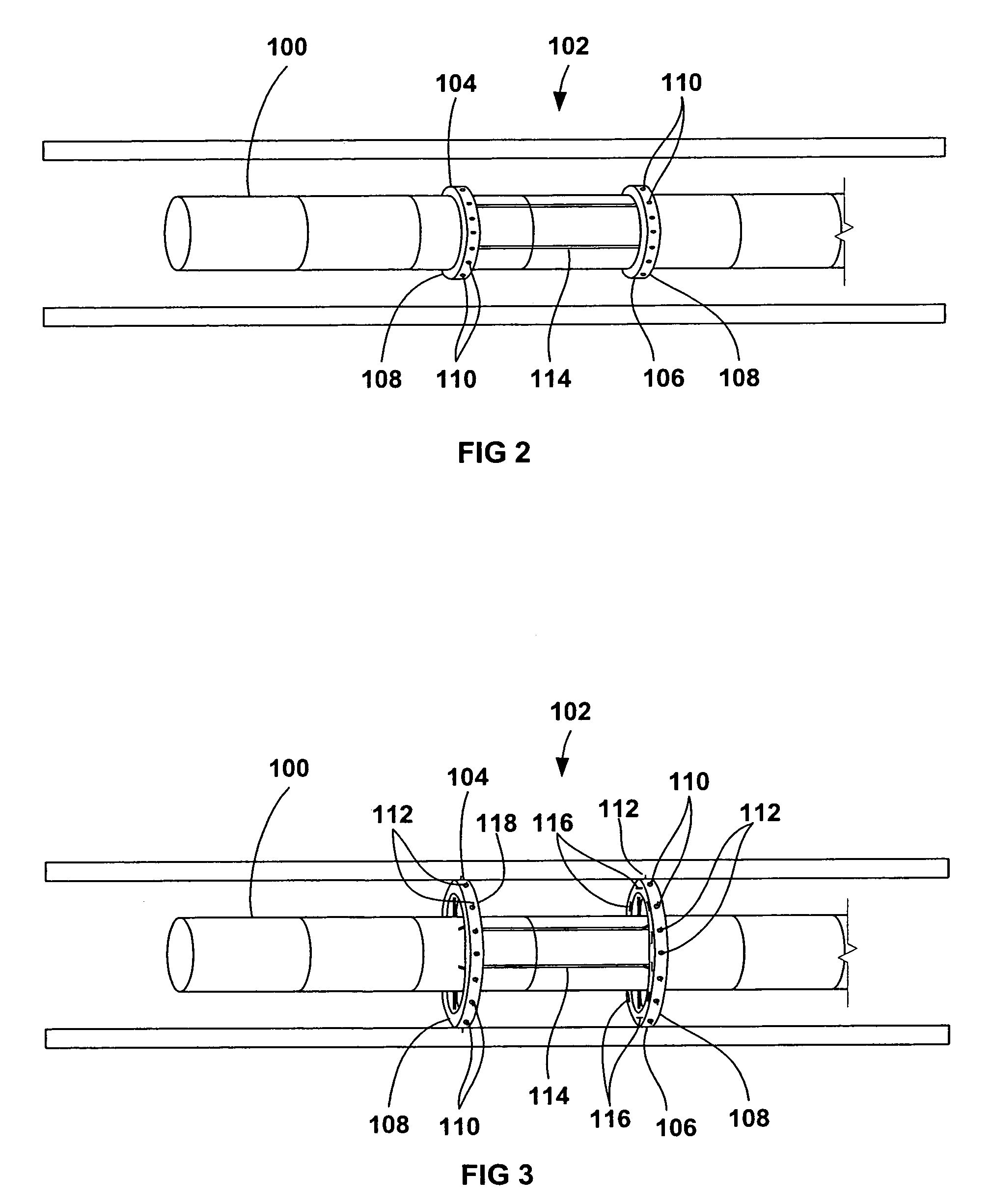
[0053]FIG. 6 shows the steerable endoscope 100 of the present invention. The endoscope 100 has an elongate body 103 with a manually or selectively steerable distal portion 105 and an automatically controlled proximal portion 107. The selectively steerable distal portion 105 can be selectively steered or bent up to a full 180 degree bend in any direction. A fiberoptic imaging bundle 113 and one or more illumination fibers 115 extend through the body 103 from the proximal end 111 to the distal end 109. Alternatively, the endoscope 100 can be configured as a video endoscope with a miniaturized video camera, such as a CCD camera, positioned at the distal end 109 of the endoscope body 103. The images from the video camera can be transmitted to a video monitor by a transmission cable or by wireless transmission. Optionally, the body 103 of the endoscope 100 may include one or two instrument channels 117, 119 that may also be used for insufflation or irrigation. The body 103 of the endosco...

second embodiment

[0057]FIG. 7 shows the endoscope 100 of the present invention. As in the embodiment of FIG. 6, the endoscope 100 has an elongate body 103 with a selectively steerable distal portion 105 and an automatically controlled proximal portion 107. The steering control 122 is integrated into proximal handle 121 in the form or one or two dials for selectively steering the selectively steerable distal portion 105 of the endoscope 100. Optionally, the electronic motion controller 140 may be miniaturized and integrated into proximal handle 121, as well. In this embodiment, the axial motion transducer 150 is configured with a base 154 that is attachable to a fixed point of reference, such as the surgical table. A first roller 156 and a second roller 158 contact the exterior of the endoscope body 103. A multi-turn potentiometer 160 or other motion transducer is connected to the first roller 156 to measure the axial motion of the endoscope body 103 and to produce a signal indicative of the axial po...

embodiment 200

[0066]As mentioned above, such a segmented body may be actuated by a variety of methods. A preferable method involves the use of electromechanical motors individually mounted on each individual segment to move the segments relative to one another. FIG. 12 shows a preferable embodiment 200 having motorized segmented joints. Each segment 192 is preferably comprised of a backbone segment 202, which also preferably defines at least one lumen running through it to provide an access channel through which wires, optical fibers, air and / or water channels, various endoscopic tools, or any variety of devices and wires may be routed through. The backbone segment may be made of a variety of materials which are preferably biocompatible and which provide sufficient strength to support the various tools and other components, e.g., stainless steel. Although much of the description is to an individual segment 192, each of the segments 192 are preferably identical, except for the segment (or first fe...


PUM


Abstract

Apparatus and methods for endoscopic colectomy are described herein. A colectomy device having a first and a second tissue approximation device is mounted on a colonoscope separated from one another. During deployment of the colectomy device, a diseased portion of the colon is positioned inbetween the tissue approximation devices. The tissue approximation devices are radially expanded such that they contact and grasp the colon wall at two sites adjacent to the diseased portion of the colon. The diseased portion is separated from the omentum and is transected using a laparoscope or is drawn into the colonoscope for later removal. The tissue approximation devices are then urged towards one another over the colonoscope to approximate the two free edges of the colon into contact together where they are fastened to one another using the tissue approximation device as a surgical stapler to create an end-to-end anastomosis.

Description

CROSS-REFERENCE TO RELATED APPLICATIONS[0001]This application claims the benefits of priority to U.S. Provisional Patent Application Ser. No. 60 / 347,674 filed Jan. 9, 2002, the entirety of which is incorporated herein by reference.FIELD OF THE INVENTION[0002]The present invention relates generally to surgical methods and apparatus. More particularly, it relates to methods and apparatus for performing endoscopic colectomy.BACKGROUND OF THE INVENTION[0003]Endoscopy studies the intralumenal aspects of hollow organs of the upper and lower intestine including the esophagus, stomach and the colon through cannulation of the lumen via the mouth or anus. Endoscopic polypectomy is presently limited to a submucosal resection. The endoscopist is often unable to completely resect a sessile polyp or lesion and therefore the patient is subjected to subsequent definitive surgery, i.e. resection of the base of the tumor. Endoscopic polypectomy can be used to debulk sessile masses but it is unable to...

Claims


Application Information

Patent Timeline
IPC IPC(8): A61B17/04A61BA61B17/32A61B1/00A61B1/31A61B17/00A61B17/08A61B17/11A61B17/115A61B17/22
CPCA61B1/00147A61B1/0055A61B1/31A61B17/1114A61B1/0058A61B2019/5259A61B2017/00557A61B2017/1103A61B2017/22034A61B2017/22054A61B2017/003A61B2034/2059A61B1/008A61B1/05A61B2017/00323A61B17/00234A61B17/1155A61B2017/00296A61B2017/00314A61B2017/00349A61B2017/00353A61B2017/1132
Inventor BELSON, AMIR
Owner INTUITIVE SURGICAL OPERATIONS INC
Why Eureka
Try Eureka