
Decellularized bone marrow extracellular matrix

a bone marrow and extracellular matrix technology, applied in the direction of prosthesis, drug composition, peptide, etc., can solve the problems of resuscitation, ineffective pharmacological therapy, scarring and loss of function,

Active Publication Date: 2008-02-05
BOSTON SCI SCIMED INC
View PDF47 Cites 42 Cited by
  • Summary
  • Abstract
  • Description
  • Claims
  • Application Information

AI Technical Summary

Benefits of technology

The invention is a composition made from decellularized bone marrow extracellular matrix that can be injected or implanted into a person to treat damaged or defective tissues or organs. The composition is made by decellularizing the bone marrow extracellular matrix in a way that preserves its structure and removes non-extracellular matrix components like cells, cell components, and proteins. The decellularized bone marrow extracellular matrix can also be combined with other therapeutically or prophylactically effective agents to repair, regenerate, or strengthen tissues or organs. The composition can be coated onto a medical device like a stent or artificial heart, and can be used alone or in combination with other materials like paclitaxel or other polymers. The invention also includes methods for preparing the decellularized bone marrow extracellular matrix by physical, chemical, and biological steps. The technical effects of this invention include improved tissue regeneration, reduced immune response, and improved biocompatibility of the composition.

Problems solved by technology

The healing of damaged or diseased tissues or organs is, in general, imperfect, resulting in scarring and loss of function.
The ineffectiveness of pharmacological therapy has led physicians to resort to reconstructive surgery that repairs or reconstitutes soft tissue or skin by introducing an injectable or implantable material.
Implantation or injection of these synthetic materials, however, can become nazardous to the health of the patient due to leakage, calcium deposit, hematoma, nodules, cellulitis, skin ulcers, and the triggering of autoimmune diseases (e.g., joint swelling and flu-like symptoms).
The main problems associated with solid implants, however, are that they must be implanted surgically and often lose flexibility due to continuing cross-linking in situ.
In contrast, while injectable collagen implant materials might have improved volume consistency and resistance to physical deformation, they are expensive and time consuming to prepare.

Method used


Image

  • Decellularized bone marrow extracellular matrix

Examples


example 1

6.1 Example 1

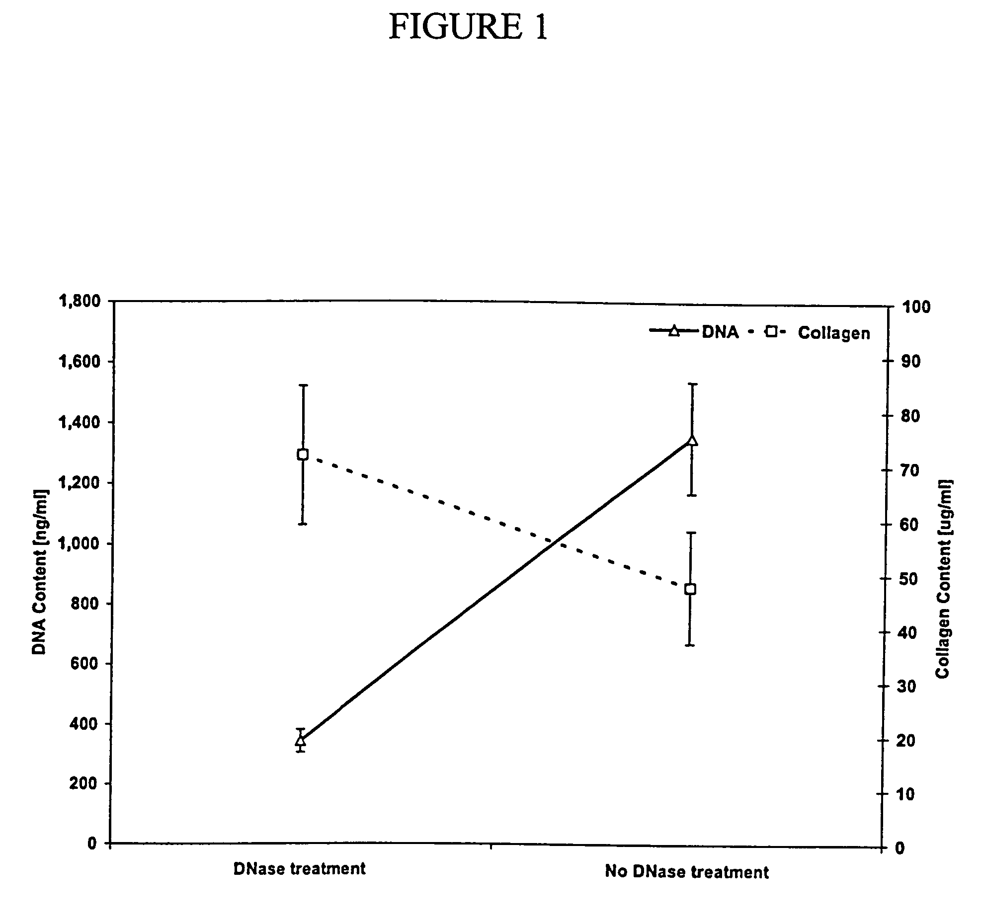
[0115]First, a bone marrow sample is isolated from a pig or cow. Second, the bone marrow sample is filtered to remove bone spicules. Third, the remaining bone marrow sample is centrifuged in saline to remove the fat. Fourth, a non-ionic detergent is added to the remaining bone marrow sample to solubilize the cell membranes and any remaining fat. The remaining bone marrow sample is then rinsed with saline and filtered to remove the remaining extracellular matrix components. The rinsing step is repeated at least one time. Further, the remaining bone marrow sample is further treated with DNase and RNase to breakdown remaining nucleic acids and rinsed with saline to remove the remaining extracellular matrix components. Finally, the resulting extracellular matrix is sterilized using standard techniques in the art, e.g., peracetic acid (0.1%).

example 2

6.2 Example 2

[0116]First, a bone marrow sample is removed from the bone marrow cavity and the cortical bone by cutting, scooping, chiseling or sawing. The marrow is then reduced to small pieces by cutting, grinding, chopping, shearing, or crushing. The blood components are removed by rinsing, one or more times, in buffered saline or water. Next, the bone is separated from the ECM and any other remaining components by vigorous agitation (e.g., vortexing, shaking). After allowing the bone fragments to settle to the bottom, the non-bone components are removed by carefully decanting or aspirating (e.g., pipet). This process is repeated by adding more saline or water to the remaining bone. The non-bone components are treated with a combination of non-ionic detergent, acidic solution, alcohol solution, and basic solution to solubilize non-ECM components. Alternatively, the non-blood components are treated with an acidic or enzymatic solution (0.5 M acetic acid with pepsin 1 mg / ml). The so...

example 3

6.3 Example 3

[0117]6.3.1 Isolation Procedure

[0118]Ribs and sternum from a freshly killed pig are collected and the cortical bone is removed. Large pieces of bone marrow are obtained by piercing, cutting and prying at the cortical bone-bone marrow interface with a sharp, stout knife. The large pieces of bone marrow are subsequently reduced in size to less than 5 mm on a side by cutting, crushing or shearing. Blood which remains in these pieces is rinsed away by multiple rinses using a buffered saline with protease inhibitor Protease-Arrest™ (Genotech, St. Louis, Mo.). For each rinse, the marrow is first placed in the solution and slowly agitated for 30 minutes. The rinsing solution is then removed from the bone marrow pieces with a pipet. Following the final rinse, more buffered saline with protease inhibitor Protease-Arrest™ is added to the bone marrow piece. The marrow is separated from the bone pieces by vigorous agitation with a standard lab vortex mixer. The resulting suspension...


PUM

PropertyMeasurementUnit
pore sizeaaaaaaaaaa
pore sizeaaaaaaaaaa
pHaaaaaaaaaa

Abstract

The invention is directed to compositions comprising decellularized bone marrow extracellular matrix and uses thereof. Methods for repairing or regenerating defective, diseased, damaged or ischemic tissues or organs in a subject, preferably a human, using the decellularized bone marrow extracellular matrix of the invention are also provided. The invention is further directed to a medical device, preferably a stent or an artificial heart, and biocompatible materials, preferably a tissue regeneration scaffold, comprising decellularized bone marrow extracellular matrix for implantation into a subject.

Description

1. FIELD OF THE INVENTION[0001]The invention relates generally to decellularized bone marrow extracellular matrix, as well as methods for the production and use thereof. In particular, the invention relates to treating defective, diseased, damaged or ischemic tissues or organs in a subject, preferably a human, by injecting or implanting decellularized bone marrow extracellular matrix into the subject. More particularly, the invention is directed to a medical device, preferably a stent or an artificial heart, comprising decellularized bone marrow extracellular matrix for implantation into a subject. Methods for manufacturing and coating a surface of the medical device with decellularized bone marrow extracellular matrix are also provided. The invention further relates to a biocompatible material, preferably a tissue regeneration scaffold, comprising decellularized bone marrow extracellular matrix.2. BACKGROUND OF THE INVENTION[0002]The healing of damaged or diseased tissues or organs...

Claims


Application Information

Patent Timeline
Patent Type & Authority Patents(United States)
IPC IPC(8): C12N5/00C12N5/08A61K35/28A61K38/18A61K38/19A61K38/30A61K38/55A61L27/28A61L27/36A61L31/08
CPCA61K35/28A61L27/28A61L27/3683A61L31/08A61L27/3633A61K38/00A61P9/00A61P19/00
Inventor FREYMAN, TOBY
Owner BOSTON SCI SCIMED INC
Learn More