Implantation of tissue bulking devices

a tissue bulking and implanted technology, applied in the field of tissue bulking, can solve the problems of undesirable mucosal tissue erosion, loss of bulking devices implanted within or directly beneath a submucosal layer, and pronounced protrusion into the lumen, so as to increase the closing pressure of the les, and reduce the likelihood of reflux fluid flow

Active Publication Date: 2007-12-11
MEDTRONIC INC
View PDF33 Cites 12 Cited by
  • Summary
  • Abstract
  • Description
  • Claims
  • Application Information

AI Technical Summary

Benefits of technology

[0010]Various embodiments of the present invention may be capable of solving at least some of the foregoing problems. For example, medical methods and systems according to some embodiments provide for implantation of bulking devices between a muscular layer of a luminal wall and an adventitial layer that at least partially covers the luminal wall, or within the adventitial layer. Bulking devices implanted in these locations may be less likely to migrate than bulking devices implanted within or directly beneath a submucosal layer. Further, bulking devices implanted in these locations may cause less pronounced protrusions into a lumen, which may reduce the likelihood of mucosal erosion and contact between mucosal tissues across the lumen relative to known implantation locations.
[0014]In exemplary embodiments, the endoscopic delivery device is sized for introduction into an esophagus of a patient, and a plurality of bulking devices are implanted within the esophageal wall of a patient proximate to the LES to treat GERD. The bulking devices cooperate with the LES to increase the closing pressure of the LES, thereby reducing the likelihood of reflux flow of fluid from the stomach into the esophagus. The bulking devices may also interact with a flap valve near the LES, or cause fibrosis within or near the LES, which may further reduce the likelihood of reflux fluid flow. The endoscopic delivery device may be steerable to allow a plurality of bulking devices to be implanted at various angles about the lumen defined by the esophageal wall, such as four bulking devices implanted at approximately 90 degree intervals about the lumen.
[0015]The bulking devices may have a predetermined form, such as a substantially cylindrical form with blunt, a traumatic edges. In some embodiments, the bulking devices are expandable after implantation, e.g., have a first, smaller size for ease of implantation, and expand to a second, larger size that provides a desired amount of tissue bulking. In exemplary embodiments, the bulking devices include a hydrogel material to provide expandability. Bulking devices including hydrogel are implanted with the hydrogel in an at least partially dehydrated state. Upon implantation, the hydrogel rehydrates to expand the size of the bulking device.

Problems solved by technology

Such problems include, by way of example, migration and loss of bulking devices implanted within or directly beneath a submucosal layer.
Additional problems include the pronounced protrusions into a lumen caused by bulking devices implanted within or below the submucosal layer, or within a muscular layer below the submucosal layer.
Such protrusions can lead to undesirable mucosal tissue erosion and contact between mucosal tissues disposed on opposite sides of the lumen.
Further, bulking devices implanted in these locations may cause less pronounced protrusions into a lumen, which may reduce the likelihood of mucosal erosion and contact between mucosal tissues across the lumen relative to known implantation locations.

Method used


Image

  • Implantation of tissue bulking devices
  • Implantation of tissue bulking devices
  • Implantation of tissue bulking devices

Examples


Embodiment Construction

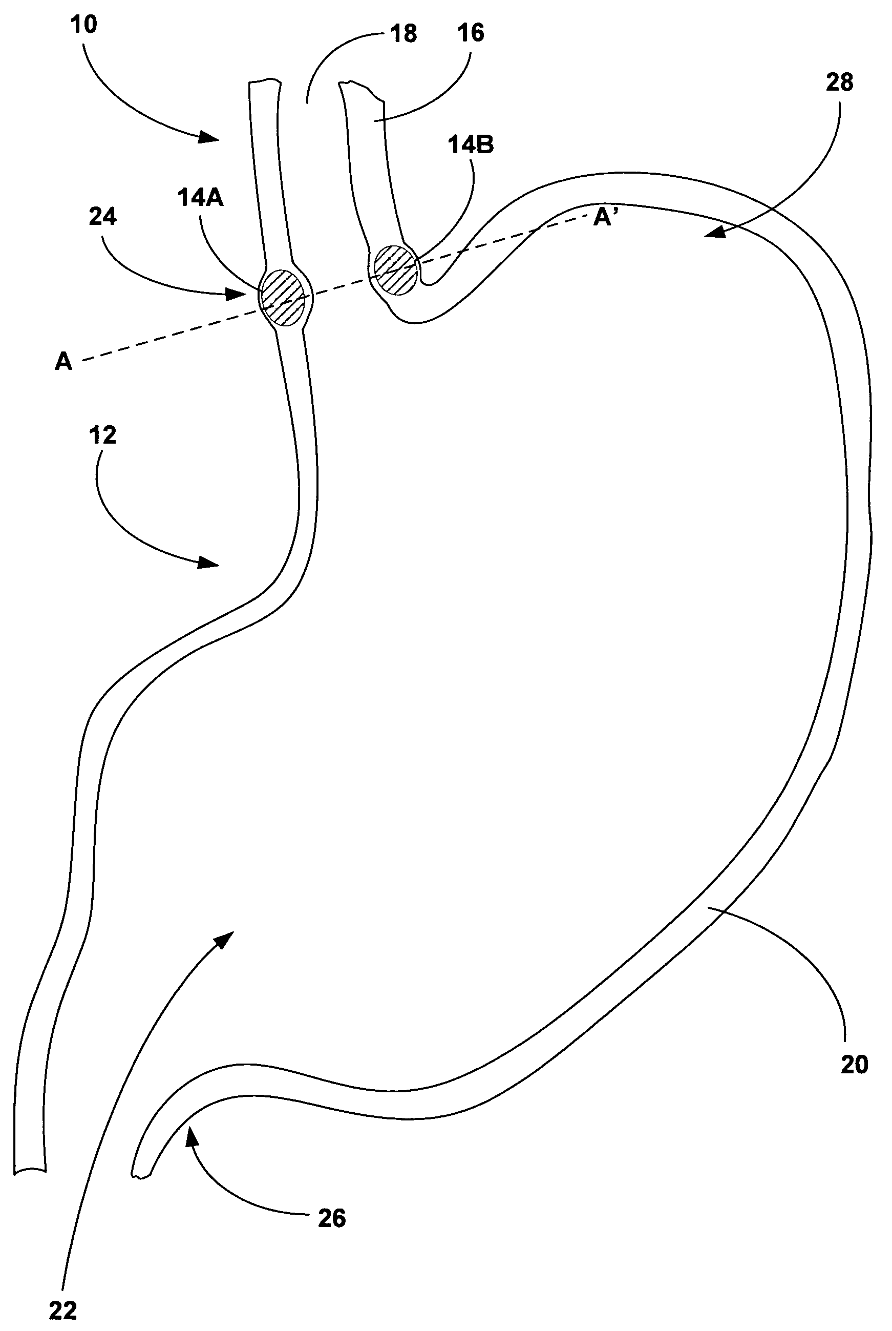
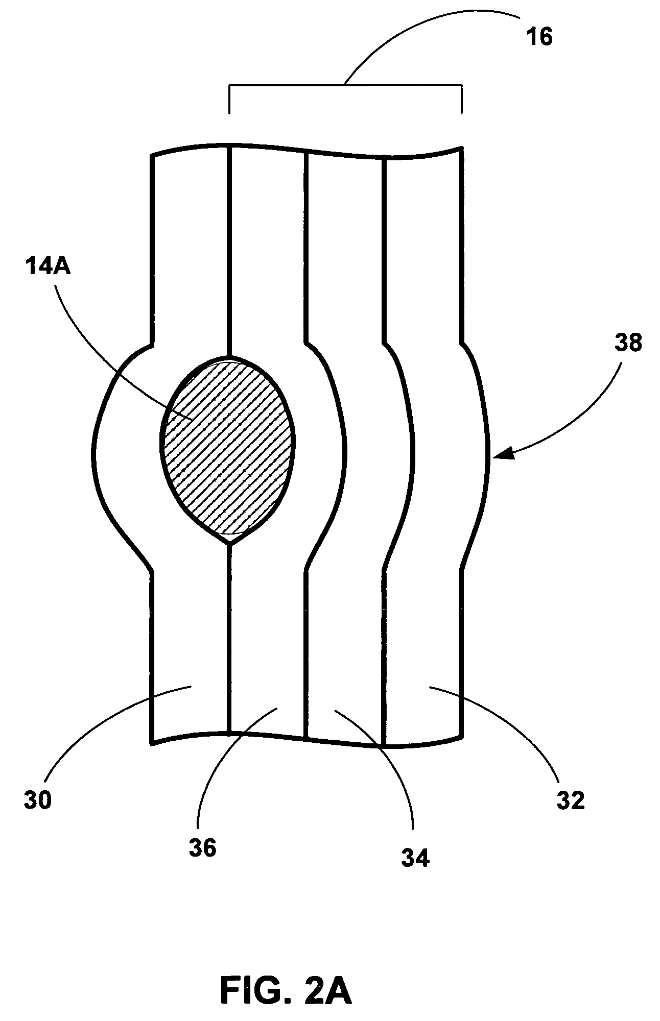
[0025]FIG. 1 is a cross-sectional diagram of an esophagus 10 and stomach 12 illustrating an example implantation of tissue bulking devices 14A and 14B (collectively “bulking devises 14”) to bulk a wall 16 of esophagus 10. In some embodiments, as will be described in greater detail below, bulking devices 14 are implanted between a luminal wall within a patient, such as esophageal wall 16, and an adventitia or adventitial layer (not shown in FIG. 1) that at least partially covers the luminal wall. In other embodiments, bulking devices 14 are implanted within the adventitial layer. Implantation of bulking devices 14 at either of these locations may provide desired bulking of a structure, while avoiding problems associated with implantation of bulking devices 14 within or between the various layers of a luminal wall. Such problems may include migration or loss of bulking devices 14, mucosal erosion, and contact between mucosal tissues across an inner lumen defined by the luminal wall.

[0...


PUM


Abstract

One or more tissue bulking devices are implanted to bulk a structure within a patient. The tissue bulking devices may be implanted between the structure and an adventitial layer that at least partially covers the structure, or within the adventitial layer. In some embodiments, the structure is a luminal wall that defines an inner lumen, and the bulking devices are implanted endoscopically via the lumen. In such embodiments, the tissue bulking devices may be implanted between a muscular layer of the luminal wall and an adventitial layer that at least partially covers the luminal wall, or within the adventitial layer. In exemplary embodiments, the luminal wall is the wall of the esophagus of the patient, and the tissue bulking devices are implanted proximate to the lower esophageal sphincter (LES) of the patient to treat gastroesophageal reflux disease (GERD).

Description

FIELD OF THE INVENTION[0001]The invention relates to tissue bulking and, more particularly, to medical methods and systems for implantation of bulking devices.BACKGROUND[0002]Tissue bulking involves the implantation of bulking devices within tissue of a selected structure within a patient. By bulking the tissue of the structure, the bulking devices can alter the function of the structure. For example, bulking devices may be implanted proximate to the lower esophageal sphincter (LES) of a patient to treat gastroesophageal reflux disease (GERD). When implanted proximate to the LES, the bulking devices treat GERD by cooperating with the LES to increase the closing pressure of the LES, thereby reducing the likelihood of reflux flow of fluid from the stomach into the esophagus. As other examples, bulking devices may be implanted near the pyloric sphincter or within the fundus of the stomach to treat obesity, or proximate to a urethral or anal sphincter to treat incontinence.[0003]Luminal...

Claims


Application Information

Patent Timeline
Patent Type & Authority Patents(United States)
IPC IPC(8): A61B19/00A61F2/04
CPCA61B17/3468A61B2017/306A61B2017/00292
Inventor TROPSHA, YELENA G.DEEGAN, CHRIS A.TRONNES, CAROLE A.
Owner MEDTRONIC INC
Why Eureka
Try Eureka