High sensitivity immunoassay method
a high-sensitivity, immunoassay technology, applied in the direction of assay labels, instruments, peptides, etc., can solve the problems of inability to accurately measure the concentration of such cytokines in plasma, and no instances are known in which such a eusup>3+/sup> complex has been applied, and achieves the effect of ease and high sensitivity
- Summary
- Abstract
- Description
- Claims
- Application Information
AI Technical Summary
Benefits of technology
Problems solved by technology
Method used
Image
Examples
example 1
Preliminary Study for TR-FIA
[0114]Initially, efforts were made to identify good combinations of solid-phase-bound capture antibodies and detection antibodies which are appropriate for an ELISA-based immunoassay system for SDF-1 measurement. For this purpose, various combinations were studied from a total of five kinds including polyclonal rabbit anti-SDF-1 antibodies and polyclonal goat anti-SDF-1 antibodies. Specific detection of reference SDF-1 was observed in three combinations. However, the detection limit for SDF-1 in the ELISA assay never exceeded about 10 to 20 ng / ml. Usually, the level of SDF-1 present in plasma is much lower than such a detection limit. Thus, it was confirmed that it is virtually impossible to detect SDF-1 in plasma samples with an ELISA assay.
[0115]By employing the most preferable combinations of polyclonal antibodies that were found in the aforementioned manner, SDF-1 detection was carried out by modifying the usual TR-FIA conditions as described below.
example 2
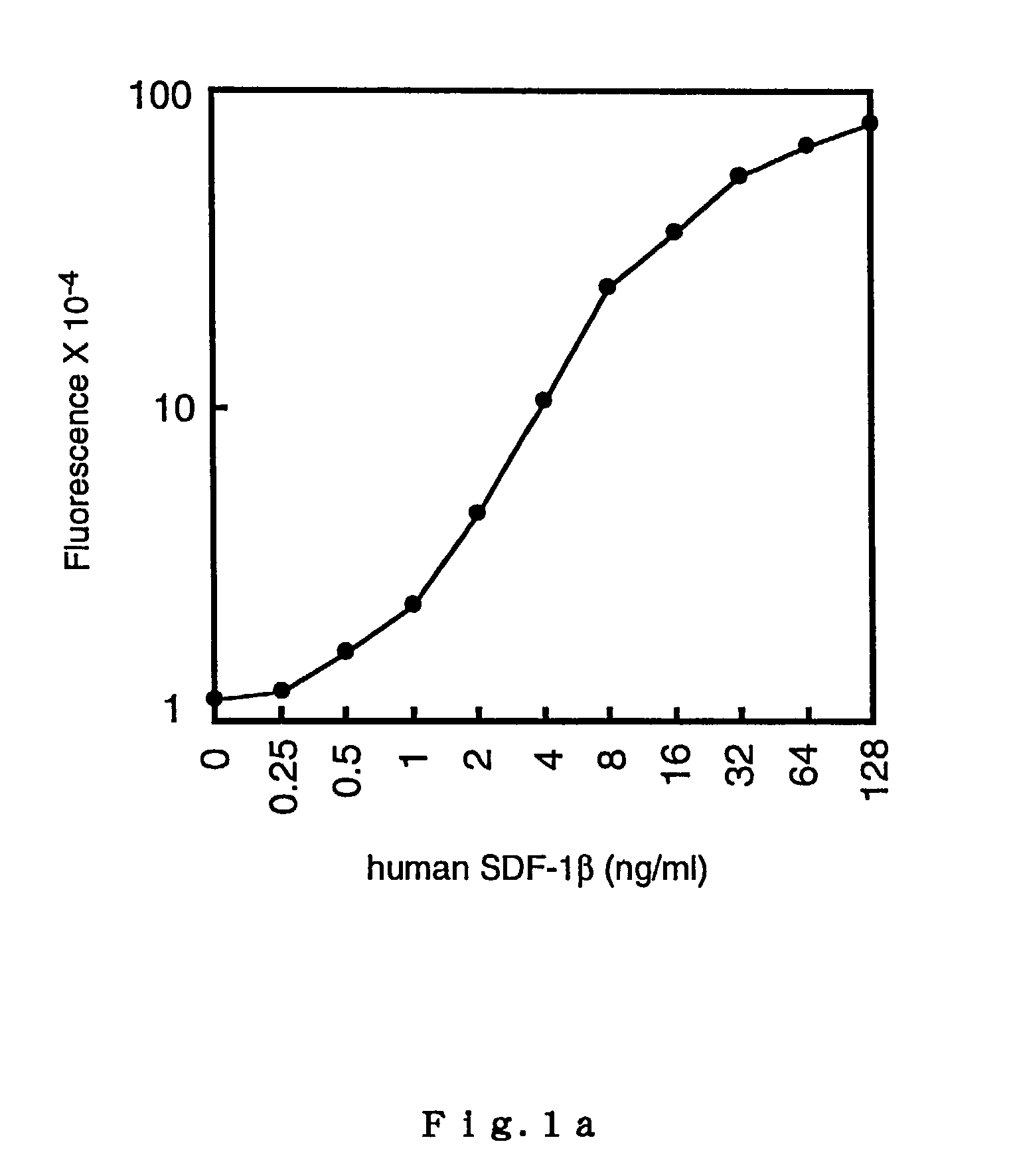
TR-FIA for Reference SDF-1
[0116]Four kinds of assay buffer solutions were prepared for TR-FIA: Buffer Solution 1 for coating a 96-well microtiter plate (0.15 M phosphate buffer (PBS) containing 0.14 M NaCl); Buffer Solution 2 for washing plates (0.05 M Tris-HCl containing 0.05% Tween20, pH 7.8); Buffer Solution 3 for washing plates (0.05 M Tris-HCl, pH 7.8); and Buffer Solution 4 for diluting protein solutions (0.05 M Tris-HCl containing 0.2% BSA, 0.1% NaN3, and 0.9% NaCl, pH 7.8).
[0117]The synthesis of BHHCT was performed following a method described in Yuan et al. ('98)(Document 5); and the preparation of a streptoavidin-bovine serum albumin (SA-BSA) conjugate and the labeling of the conjugate with BHHCT were performed following a method described in Yuan et al. ('97)(Document 4). A solution of the labeled conjugate was preserved at −20° C., and diluted 100× with the buffer solution below (Buffer Solution 4) immediately before use.
[0118]Rabbit polyclonal anti-human SDF-1β antibody...
example 3
Down Modulation of CXCR4 by Human SDF-1β
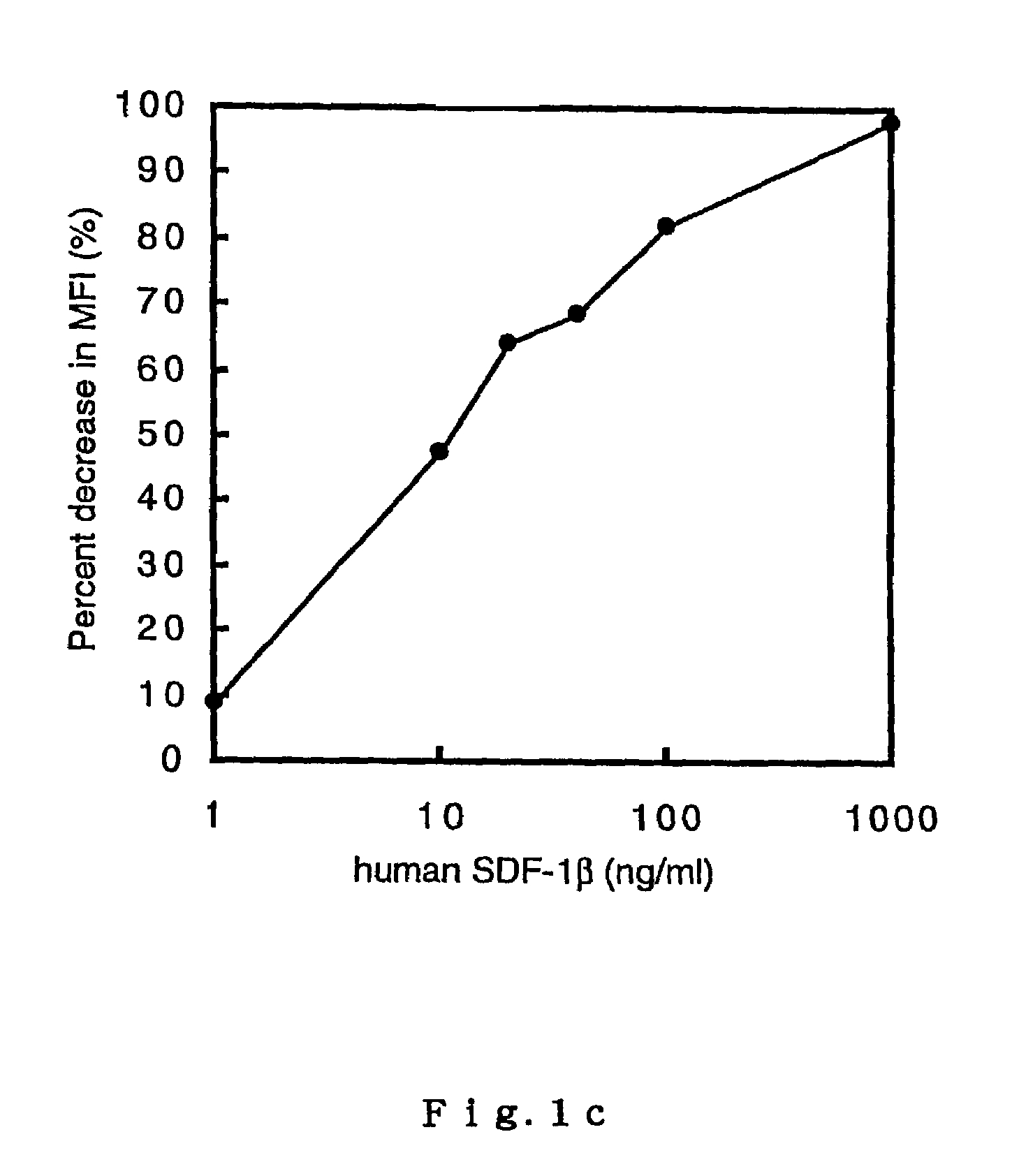
[0128]In order to confirm the interrelationship between SDF-1 measurement values by TR-FIA according to Example 2 and the biological activity of the reference SDF-1 protein, an in-vitro down modulation of a SDF-1 receptor (CXCR4) which is induced in EL-4 cells upon binding of SDF-1 was measured.
[0129]EL-4 cells were cultured in Dulbecco-modified Eagle's medium (D′ MEM), to which 10% fetal calf serum (FCS) was supplemented, under the presence or absence of human SDF-1β (1, 10, 20, 40, 100, and 1000 ng / ml). After 6 hours of incubation at 37° C., the CXCR4 on the cell surface was dyed with Fc-human SDF-1α chimeric protein and FITC-bound goat F(ab′)2 anti-human IgG (Southern Biotechnology Associates, Alabama, U.S.). A fluorescence intensity measurement was performed by fluorocytometry (FACSCalibur, BECTON DICKINSON, California, U.S.). The down modulation of CXCR4 was evaluated by calculating the percentage reduction in the mean fluorescence intens...
PUM
| Property | Measurement | Unit |
|---|---|---|
| Temperature | aaaaa | aaaaa |
| Fraction | aaaaa | aaaaa |
| Fraction | aaaaa | aaaaa |
Abstract
Description
Claims
Application Information
 - R&D
- Intellectual Property
- Life Sciences
- Materials
- Tech Scout
- Unparalleled Data Quality
- Higher Quality Content
- 60% Fewer Hallucinations
Browse by: Latest US Patents, China's latest patents, Technical Efficacy Thesaurus, Application Domain, Technology Topic, Popular Technical Reports.
© 2025 PatSnap. All rights reserved.Legal|Privacy policy|Modern Slavery Act Transparency Statement|Sitemap|About US| Contact US: help@patsnap.com


