
Method and apparatus for the detection and ligation of uterine arteries

a uterine artery and ligation technology, applied in the field of diseases and conditions, can solve the problems of 0.5 deaths, uterine fibroid disorders, and many women suffering from painful dysfunctional uterine bleeding or endometritis, and achieve the effects of reducing mortality, and reducing the risk of uterine fibroid disorders

Inactive Publication Date: 2007-06-12
VASCULAR CONTROL SYST
View PDF126 Cites 39 Cited by
  • Summary
  • Abstract
  • Description
  • Claims
  • Application Information

AI Technical Summary

Benefits of technology

Provides a simpler, less invasive means to treat uterine disorders by occluding uterine arteries externally, reducing the need for specialized radiographic equipment and skills, offering improved treatment options for conditions like uterine fibroids and dysfunctional uterine bleeding.

Problems solved by technology

Hysterectomy for treating uterine fibroid disorders, though effective, has many undesirable characteristics.
The undesirable characteristics of hysterectomy include a known mortality rate of 0.5 deaths per 1000 hysterectomies.
It is normal for portions of the lining to slough off and bleed during menstruation, but many women suffer from painful dysfunctional uterine bleeding or endometritis.
However, visualization of all of the endometrium is difficult.
In this distension process, some women become water intoxicated and hyponatremic.
Furthermore, the uterine cavity is an awkward shape, somewhat triangular and often angulated.
Endometrial destruction can also be brought about with chemical damage, photochemical injury, or thermal damage (heat or cold).
Energy that reaches and destroys the cells of the endometrial lining of the uterus potentially destroys the uterine lining and thereby treats dysfunctional uterine bleeding.
Consequently, it is difficult to know which fibroid is causing symptoms to the patient (bleeding, pain, and bulk effects on adjacent organs).
Clearly, the strategy of identifying which individual fibroid is causing symptoms (when there are often many), finding that fibroid, and then either removing or destroying that individual fibroid is a rather complex strategy.
Second, fibroids live a tenuous vascular life with very little ability to recruit a new blood supply from the host when the primary blood supply is compromised.
One problem with present methods of uterine artery embolization is that many physicians do not possess the skill or equipment necessary to perform catheter-based uterine artery embolization under radiologic direction.

Method used


Image

  • Method and apparatus for the detection and ligation of uterine arteries
  • Method and apparatus for the detection and ligation of uterine arteries
  • Method and apparatus for the detection and ligation of uterine arteries

Examples


Embodiment Construction

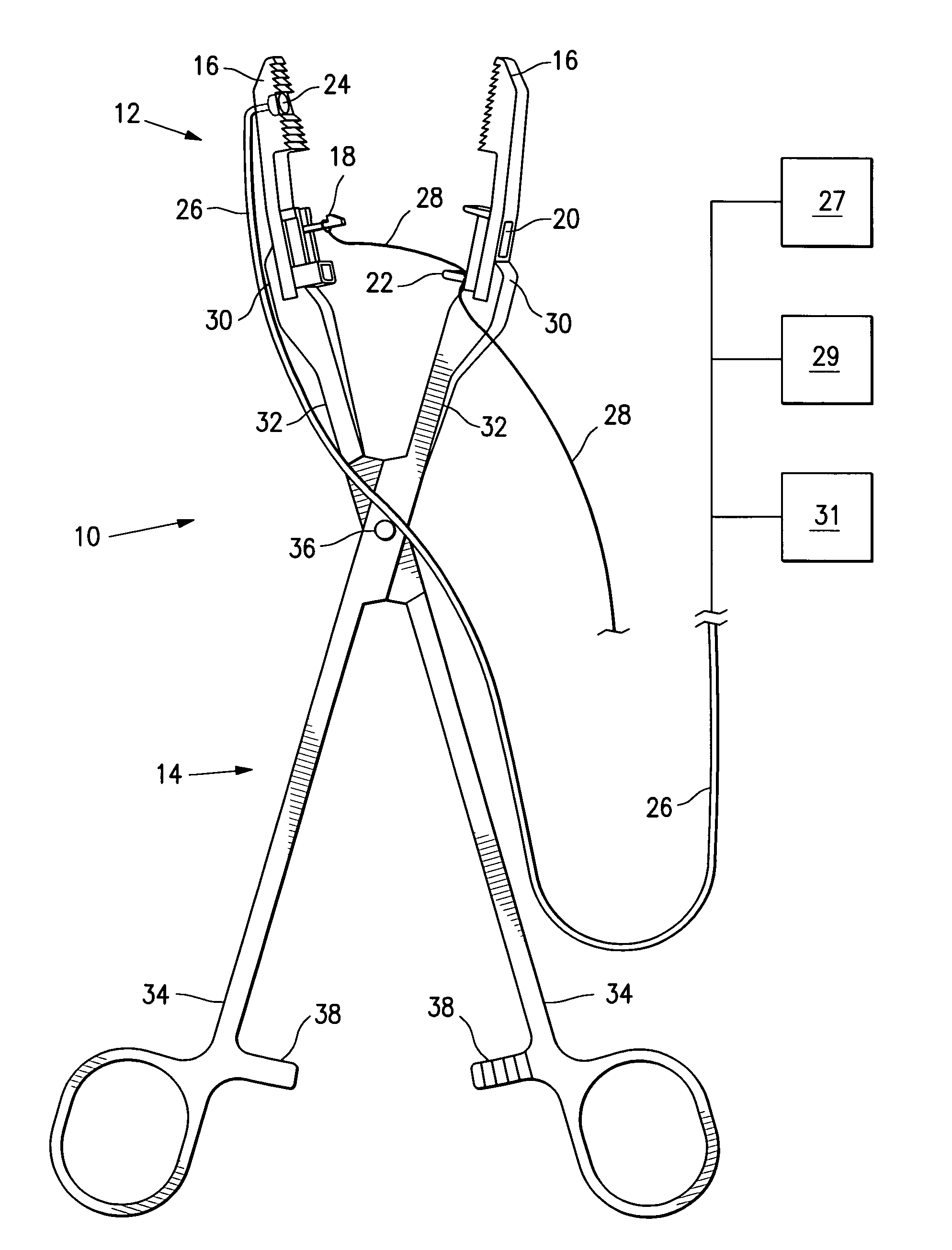
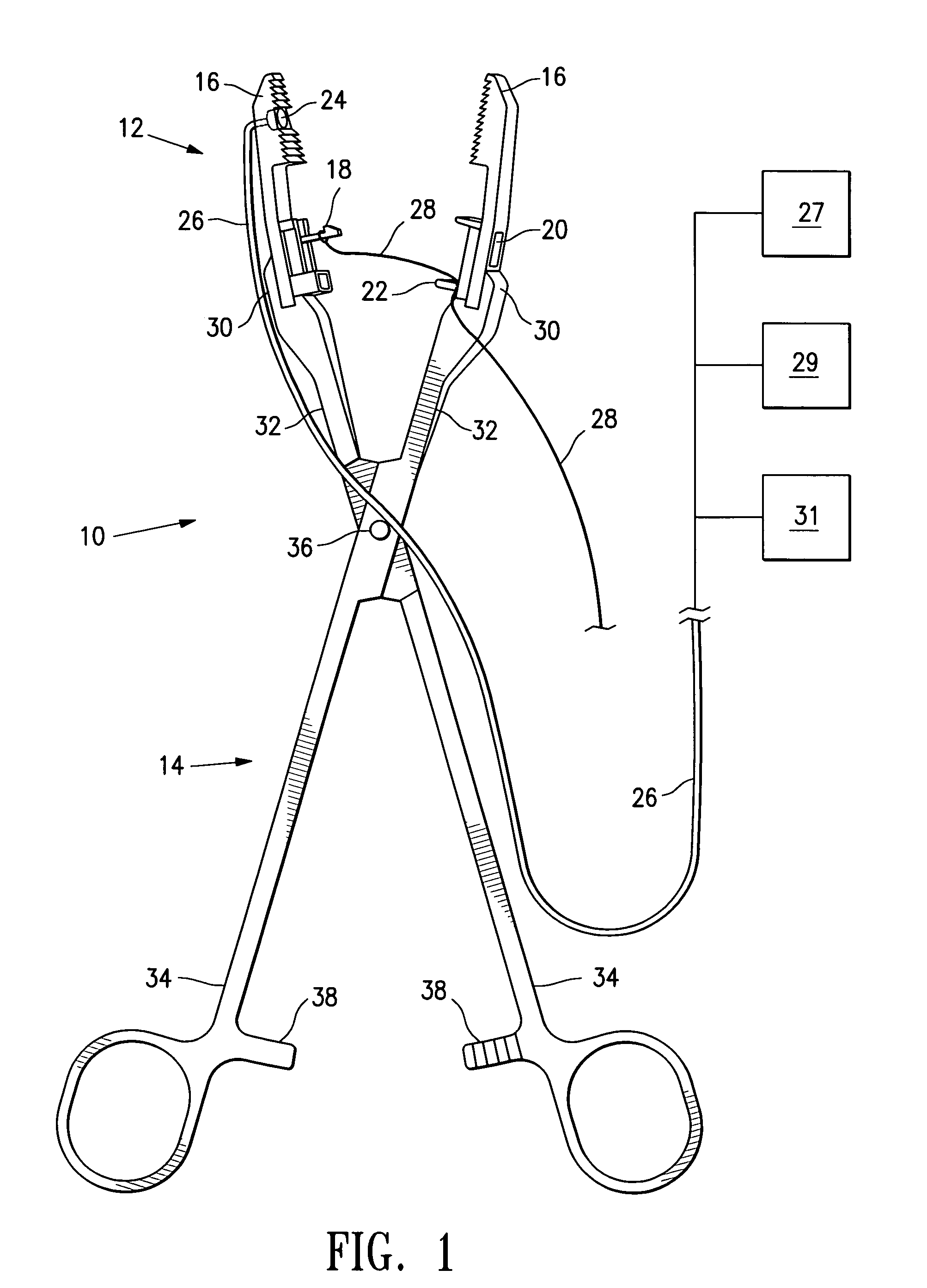
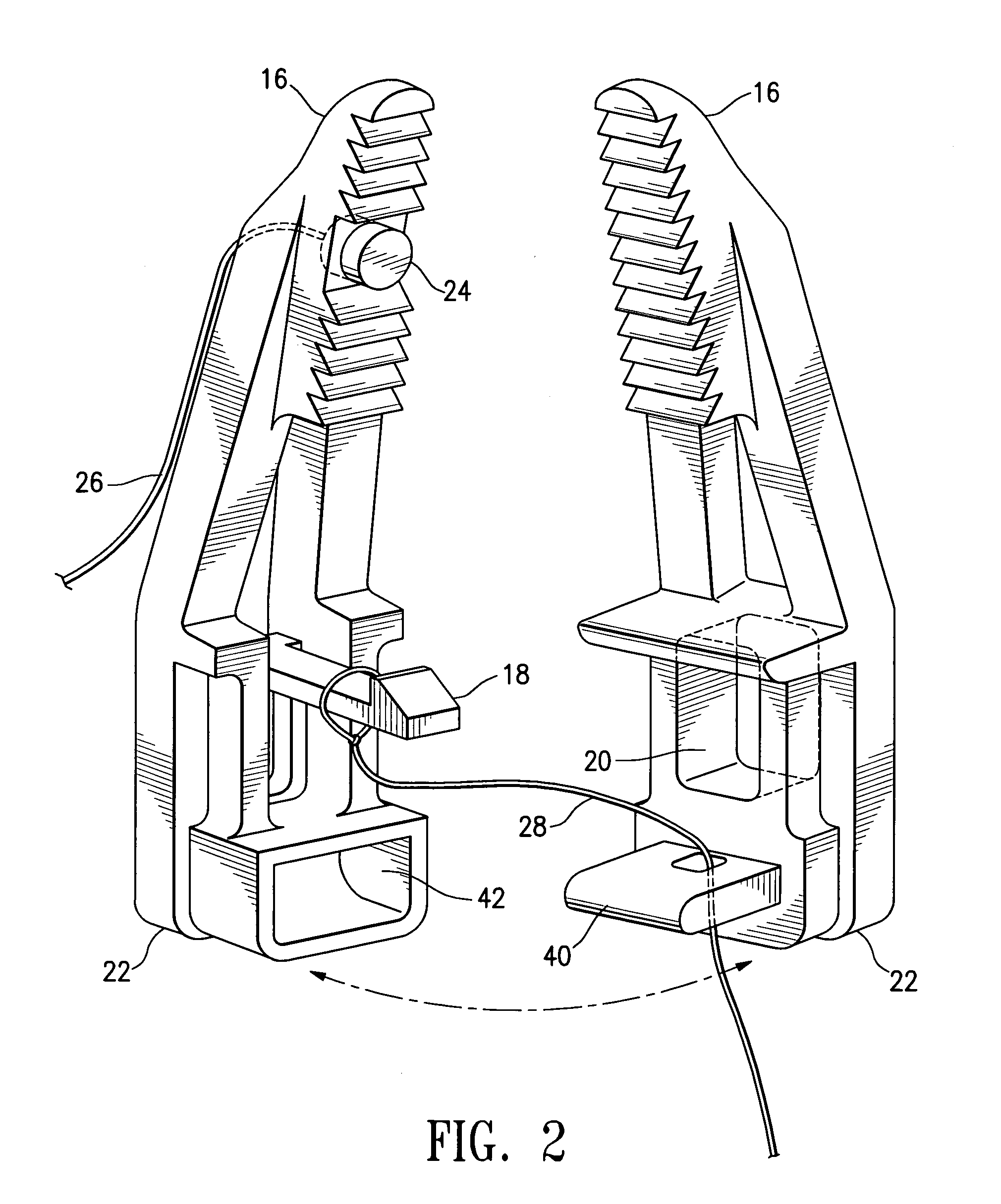
[0040]FIG. 1 shows a system 10 embodying features of the invention including a clamping device 12 and a clamping device applicator 14 embodying features of the invention. The clamping device shown in FIG. 1 is a clamp 12 having clamp jaws 16 which act as clamping members. The clamp jaws 16 are shown separated, but may be locked together by action of the clamp device applicator 14 bringing together locking mechanism components detent 18 and catch 20 effective to lock jaws 16 together. Placement of jaws 16 against tissue, such as vaginal wall tissue, surrounding an artery, such as a uterine artery, is effective to compress an artery between jaws 16 and to clamp and lock together jaws 16 to maintain such compression for a desired length of time. Clamp 12 also has a mounting portion 22 configured to engage a clamp device applicator.

[0041]FIG. 1 further illustrates blood flow sensor 24 located on a jaw 16 where it may sense blood flow in an artery within tissue near a clamp 12. Blood flo...


PUM


Abstract

The invention provides devices, systems and methods for occluding arteries without puncturing skin or vessel walls. The devices, systems and methods for occluding arteries are configured to be applied to arteries externally of the arteries. Occlusion may be temporary or permanent, and may be partial or complete. Clamping a device to tissue near to an artery is effective to compress tissue around the artery and to indirectly compress the artery. The methods, devices and systems of the invention find use in, for example, treatment of uterine disorders and conditions which may be treated by occlusion of the uterine arteries. A uterine artery may be accessed via a patient's vagina by compressing a portion of the vaginal wall around a portion of a uterine artery to occlude a uterine artery. Clamping of an artery may also be performed by clamping a device directly onto an artery.

Description

CROSS-REFERENCE TO RELATED APPLICATIONS[0001]This application is related to U.S. Provisional Application No. 60 / 279,477 filed Mar. 28, 2001, hereby incorporated by reference in its entirety and from which benefit is hereby claimed under 35 U.S.C. 119(e).FIELD OF THE INVENTION[0002]The invention relates generally to the field of treatment of diseases and conditions by regulation of blood flow; in particular, the treatment of a uterus by regulating blood flow thereto.BACKGROUND OF THE INVENTION[0003]Hysterectomy (surgical removal of the uterus) is performed on approximately 600,000 women annually in the United States. For approximately 340,000 women, hysterectomy is probably the best current therapeutic choice for the treatment of their diseases (uterine cancer, endometriosis, menorrhagia, and prolapse). For approximately 60,000 women with dysfunctional uterine bleeding (abnormal menstrual bleeding that has no discrete anatomic explanation such as a tumor or growth), newer endometrial...

Claims


Application Information

Patent Timeline
Patent Type & Authority Patents(United States)
IPC IPC(8): A61B17/00A61B17/08A61B17/42A61B5/02A61B17/12A61B5/026A61B8/06A61B17/122A61B17/128A61B18/00A61M25/00
CPCA61B5/415A61B5/418A61B8/06A61B8/12A61B17/12A61B17/122A61B17/1285A61B17/2812A61B5/026A61B17/00234A61B17/128A61B17/42A61B2017/0046A61B2017/4216
Inventor BURBANK, FRED H.JONES, MICHAEL L.SERRA, R. J.ALTIERI, GREIGUYENO, JILLWONG, YU-TUNGWERNETH, RANDY
Owner VASCULAR CONTROL SYST
Learn More