Eureka AIR delivers breakthrough ideas for toughest innovation challenges, trusted by R&D personnel around the world.

Implantable medical device having optical fiber for sensing electrical activity

a technology of optical fiber and electrical activity, applied in the field of implantable medical devices, can solve the problems of electrical noise, interference with detection, and number of limitations

Active Publication Date: 2007-03-13
MEDTRONIC INC
View PDF32 Cites 59 Cited by
  • Summary
  • Abstract
  • Description
  • Claims
  • Application Information

AI Technical Summary

Problems solved by technology

A number of limitations exist, however, in using medical electrical leads that rely on metallic wire conductors for carrying sensed electrical signals from excitable body tissue to an implanted or external device.
One limitation is the presence of electrical noise, which may be in the form of electromagnetic interference or electrical potential signals arising from other nearby excitable tissue, sometimes referred to as “far-field signals”.
Such noise or far-field signals contaminate the sensed signal, interfering with the detection of electrical signals of interest.
Oversensing or undersensing of these intrinsic events by a cardiac rhythm management device can result in incorrect detection and classification of a rhythm (normal versus pathological), potentially triggering the delivery of unnecessary cardiac stimulation therapy or inappropriately withholding stimulation when it is actually needed.
Inappropriate withholding of cardiac stimulation is undesirable when the patient is pacemaker dependent or the stimulation therapy is life saving.
Inappropriate delivery of cardiac stimulation is undesirable because it may cause unnecessary pain to the patient if the therapy delivered is a shock, and can also lead to premature device battery depletion.
Moreover, delivery of stimulation therapies in the presence of normal intrinsic cardiac activity can result in stimulation pulses being delivered during the so-called “vulnerable period” of the cardiac cycle, during which cardiac arrhythmias are easily induced in some patients, creating a potentially life-threatening situation.
The repolarization time of cells located at the stimulation site is difficult to ascertain from EGM signals sensed using metallic wire based leads because EGM signals reflect the summation of many cellular action potential signals as a depolarization wavefront moves through the myocardium.
The EGM signal does not resemble an action potential signal and therefore the recovery time of local cells cannot be accurately estimated from an EGM.
Moreover, the T-wave, which contains the repolarization information in a far-field EGM, can have complex morphology making it difficult to ascertain exact repolarization time and characteristics using signal processing techniques.
However, despite these improvements, noise and far-field signals remain an infrequent but serious problem that undermines the accuracy of cardiac event sensing using metallic wire based medical electrical leads.
Another limitation encountered with metallic wire based lead systems relates to sensing of an evoked response following a cardiac pacing pulse.
False capture detections due to oversensing of noise or far-field signals may result in prolonged episodes of subthreshold cardiac pacing that is ineffective in maintaining a base heart rate.
False loss of capture detections can result from undersensing of the evoked response and can trigger the delivery of unnecessary backup pacing pulses and pacing threshold searches.
Increases in pacing pulse energy due to false loss of capture detections can lead to premature pacemaker battery depletion.
Evoked response sensing using metallic wire based leads is difficult, however, for a number of reasons.
A major challenge in evoked response sensing arises due to the post-pace polarization artifact at the electrode-tissue interface.
The polarization artifact may diminish during the blanking interval, however, it may still interfere with evoked response sensing.
Sensing in other areas of the heart could lead to erroneous evoked response detection due to noise or other myopotentials being sensed as an evoked response.
Furthermore, sensing for an evoked response conducted to another area of the heart may not be possible in patients having conduction disorders.
Because the extrasystole is most effective just after repolarization, a perceived risk in delivering PESP stimuli is that the extrasystole may fall in the vulnerable period.
As indicated above, an electrical signal sensed by a sensing lead or catheter utilizing an electrical wire conductor will generally be subject to electrical noise and artifacts.
Another limitation of metallic wire based leads is that they are generally incompatible with magnetic resonance imaging because the magnetic field can induce unwanted current in metallic wire conductors.

Method used


Image

  • Implantable medical device having optical fiber for sensing electrical activity
  • Implantable medical device having optical fiber for sensing electrical activity
  • Implantable medical device having optical fiber for sensing electrical activity

Examples


Embodiment Construction

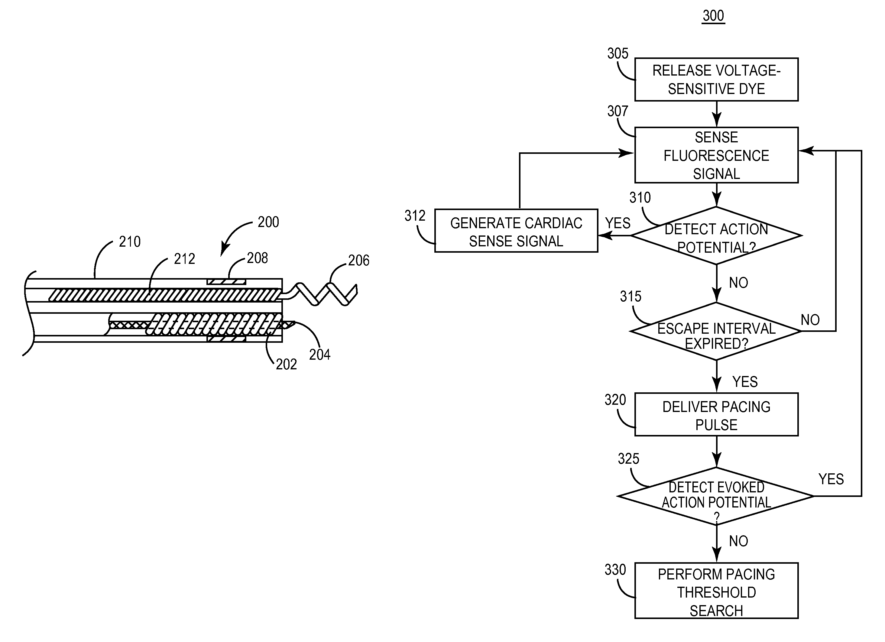
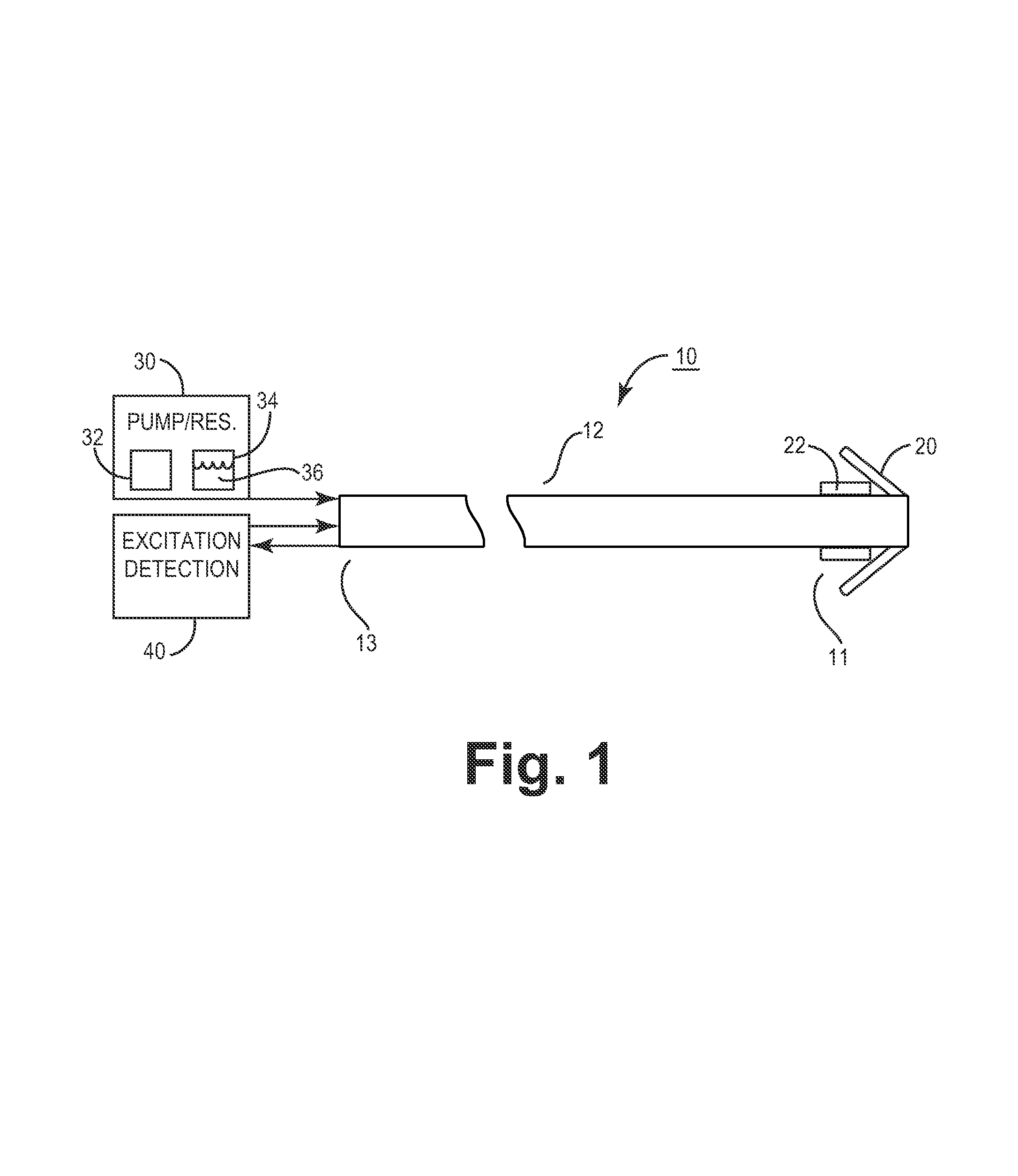
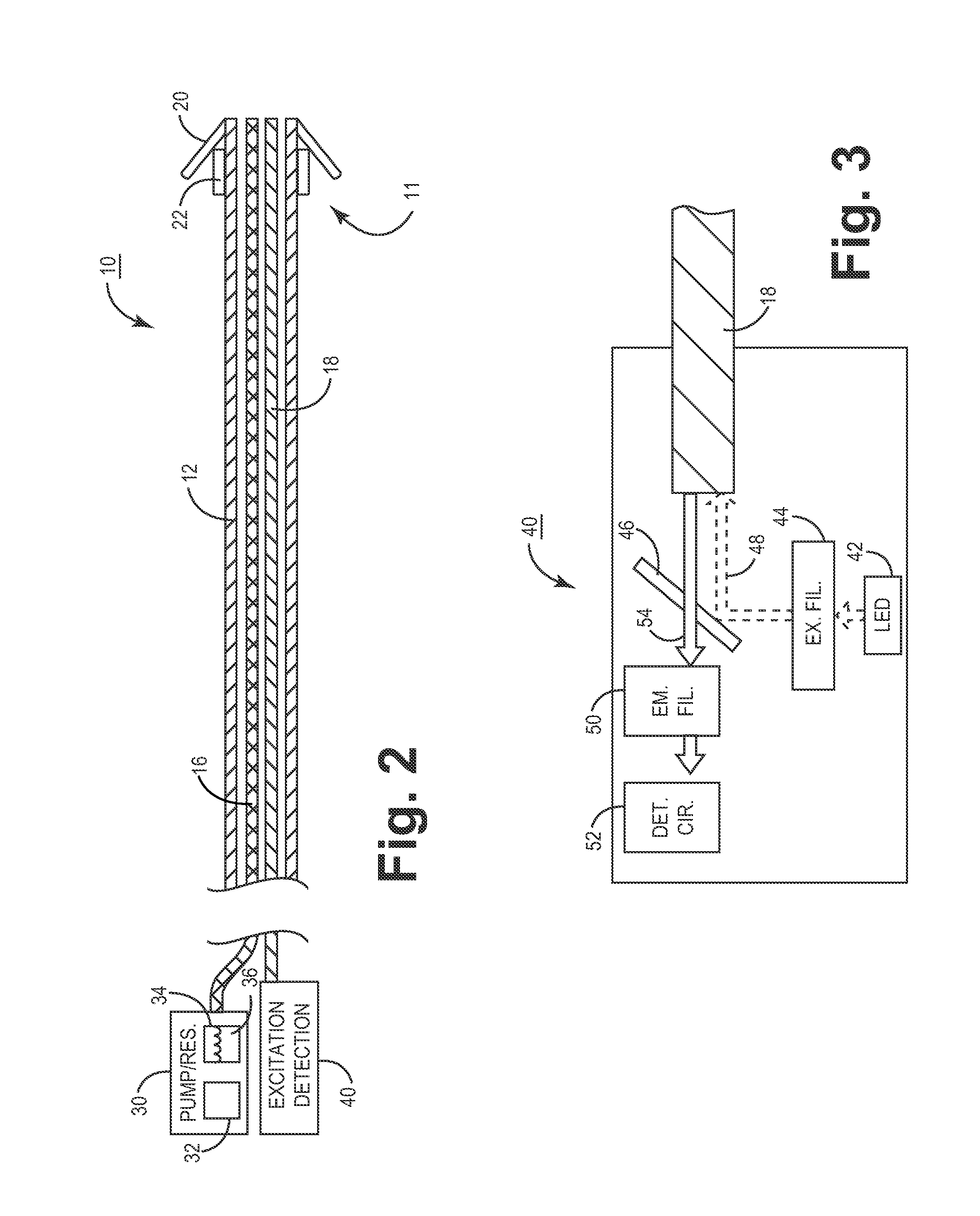
[0046]The present invention provides an implantable medical lead for sensing electrical activity of excitable body tissue using an optical fiber for transmitting fluorescence signals associated with the local transmembrane cellular potential changes. The lead is provided with an elongated body carrying at least a first optical fiber extending between a proximal lead end and a distal lead end that is positioned in or against excitable body tissue for monitoring local electrical activity. At the proximal lead end a connector is provided for connecting the lead to an implantable or external monitoring device for acute or chronic monitoring of electrical activity and optionally delivering electrical stimulation or other therapy. Optical excitation and photodetection circuitry are included in a lead connector assembly and / or in the associated medical device. Excitation circuitry includes a light emitting diode positioned relative to the proximal end of the optical fiber to permit emitted...


PUM


Abstract

An implantable medical device for optically sensing action potential signals in excitable body tissue. The device includes an elongated tubular lead body carrying an optical fiber extending from a proximal lead end to a distal lead end to position the optical fiber at a target site. The lead body additionally carries a conduit for dispensing a voltage-sensitive fluorescent dye into tissue surrounding the target site. The optical fiber transmits excitation light to the fluorescent dye to cause the dye to fluoresce with varying intensity as the transmembrane potentials of local tissue cells vary due to passing depolarization wavefronts. The optical fiber transmits the fluorescence signal to the device to generate an action potential signal or fiducial points of an action potential signal for use in accurately measuring and characterizing electrical activity of excitable tissue.

Description

FIELD OF THE INVENTION[0001]The present invention relates generally to implantable medical devices and, more particularly, the present invention relates to a method and apparatus for sensing an action potential signal of excitable tissue.BACKGROUND OF THE INVENTION[0002]Medical electrical leads are used in conjunction with numerous types of medical devices for monitoring the electrical activity of and / or stimulating excitable body tissue. Such devices include cardiac pacemakers, cardiac defibrillators, cardioverters, myostimulators, neurostimulators, and other devices for delivering electrical signals to excitable body tissue and / or receiving electrical signals from the tissue. Cardiac rhythm management devices, for example, including pacemakers, cardioverters and defibrillators, are designed to operate so as to sense the intrinsic cardiac electrical activity and deliver appropriately timed electrical stimulation signals when needed, in order to maintain a normal sinus rhythm at a p...

Claims


Application Information

Patent Timeline
Patent Type & Authority Patents(United States)
IPC IPC(8): A61B5/04A61B5/042A61N1/05A61N1/362A61N1/37
CPCA61B5/042A61N1/371A61N1/056A61B5/283A61B5/29
Inventor SHARMA, VINODXIAOHONG, ZHOU
Owner MEDTRONIC INC
Why Eureka
Eureka Blog
Learn More