
Multiportal device with linked segmented cannulae and method for percutaneous surgery

a multi-portal device and segmented technology, applied in the field of medicine, can solve the problems of long recovery time, postoperative scarring, and large scarring, and achieve the effects of short incisions, simple construction, and simple us

Inactive Publication Date: 2006-10-10
NEVMET CORP
View PDF11 Cites 2 Cited by
  • Summary
  • Abstract
  • Description
  • Claims
  • Application Information

AI Technical Summary

Benefits of technology

[0025]It is an object of the present invention to provide a multiportal device with linked segmented cannulae for percutaneous surgery, which is very simple in construction, reliable and simple in use, allows insertion of cannulae and permanently maintaining them in controlled positions without resorting to additional X-ray for the duration of the procedure. It is another object to provide a device and method that allow performing a surgery with short incisions. It is a further object of the invention to provide a device and method, which ensure angular arrangement of cannulae inserted into the patient's body even on relatively flat portions of the body.

Problems solved by technology

Low back pain and radiculopathy as a result of herniated intervertebral disc represents a major health problem in the United States and all over the world.
A laminectomy is a somewhat destructive procedure, which might cause extensive scarring and long (up to 9 days) hospitalization, with an up to 3-month postoperative recovery period.
This is less invasive compared to the laminectomy, though the existing microdiscectomy may still cause some complications similar to those associated with laminectomy, for example, possible injury to the nerve root and dural sac, postoperative scarring and a relatively long recovery time.
The method of U.S. Pat. No. 4,545,374 has the following drawbacks: 1) the material is removed from the center of the disc only, thus preventing a surgeon from excising the fragments from the actual herniation site, which may or may not cause recurrent symptoms; 2) this technique is unsuitable for noncontained (or sequestered) herniations, since it does not give a surgeon access to the epidural space.
Like in the percutaneous approach described above, one of the disadvantages inherent in this procedure is that it deals with the disc nucleus, rather than with the herniation itself.
Lasers are both expensive and somewhat tedious to use in these procedures.
Another disadvantage with lasers is the difficulty in judging the depth of tissue ablation.
Both methods described in U.S. Pat. No. 6,264,650 and in U.S. Pat. No. 6,254,553 make it complicated for a surgeon to focus treatment on the symptomatic site, without affecting the surrounding tissues.
Single-portal MISS methods are limited to the use of one channel at a time.
However, this biportal procedure assumes the second portal to be created from the opposite side to the first portal (bilateral), hence increasing the operating time, post-operative morbidity, and surgeon exposure to radiation.
It may also cause excessive removal of disc nuclear tissue, therefore increasing the possibility of post-operative stenosis (narrowing) of the foramen.
These approaches do not provide a comprehensive solution for percutaneous disc surgery.
The method described in U.S. Pat. No. 5,730,754 still needs accurate targeting and it not sufficiently universal.
U.S. Pat. No. 5,762,629 allows inserting a second cannula using a special targeting device, but has the following disadvantages: 1) the targeting device is rigid and does not allow the flexibility required by a surgeon for the formation of an angle between inserted cannulae; 2) after cannulae are inserted, the targeting device is removed, leaving the cannulae completely unlinked, so that a surgeon cannot keep them interrelated.
In case the position of one of the inserted cannulae should be temporarily changed, it becomes a problem to reorient them, especially when more than two cannulae are used for the surgery.
Another common disadvantage of the existing devices for the percutaneous surgery is that they require the operation to be carried out under X-ray monitoring at all steps of the surgery, i.e., during insertion of each additional cannula and occasionally during the procedure itself.
Although the device described above is efficient in eliminating extraneous X-raying and in facilitating guiding and manipulation of the cannulae during multiportal approach to the symptomatic site, nevertheless this device has many parts and is expensive to manufacture.
Therefore it cannot be made entirely of plastics and cannot be made disposable.
This may cause more extensive scarring and longer healing.

Method used


Image

  • Multiportal device with linked segmented cannulae and method for percutaneous surgery
  • Multiportal device with linked segmented cannulae and method for percutaneous surgery
  • Multiportal device with linked segmented cannulae and method for percutaneous surgery

Examples


Embodiment Construction

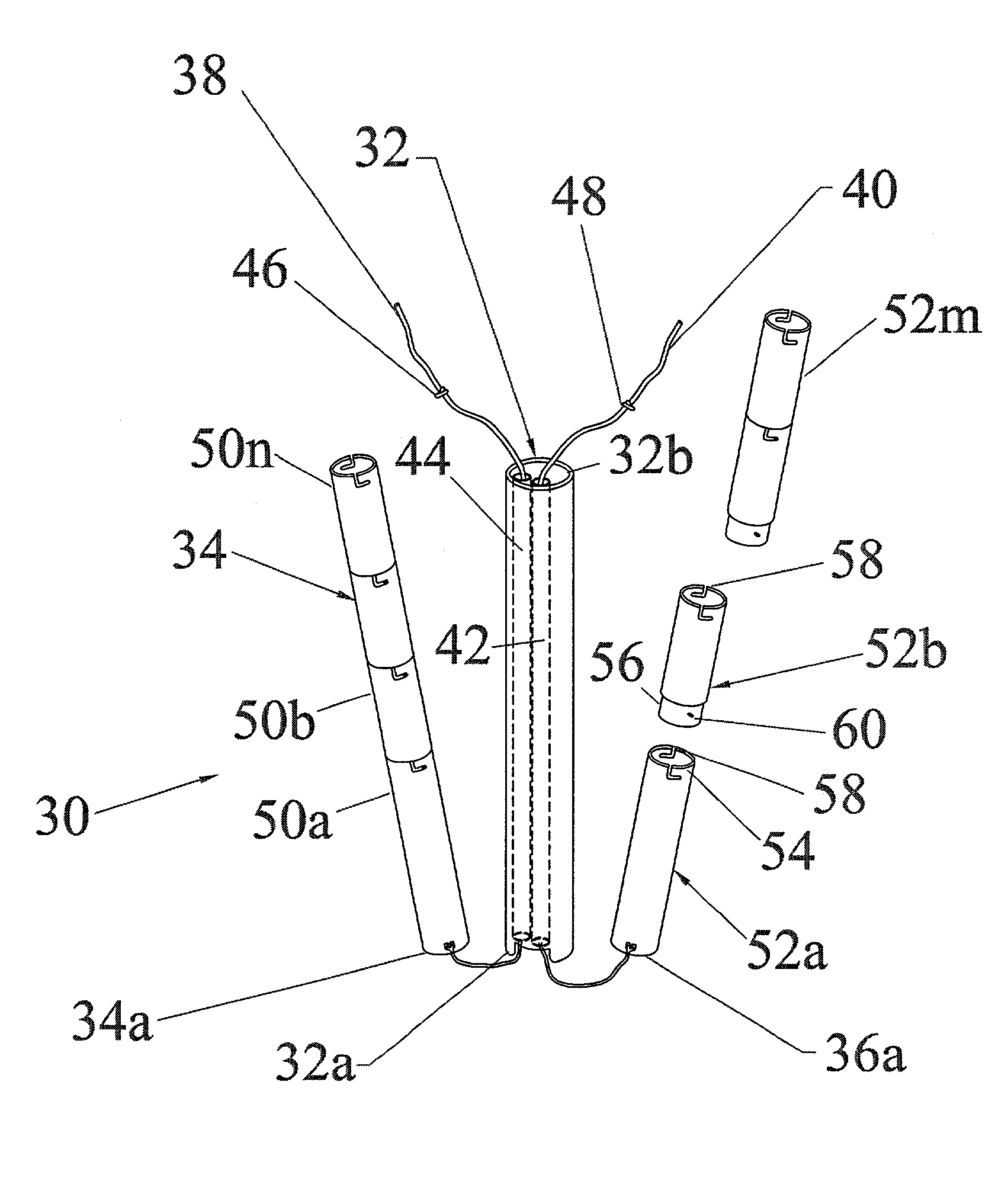
[0048]A general three-dimensional view of the multiportal device with linked segmented cannulae of the invention for percutaneous surgery (hereinafter referred to as “multiportal device”) is shown in FIG. 5. In the embodiment shown in this drawing, the multiportal device, designated in general by reference numeral 30, consists of three linked cannulae 32, 34, and 36. It is understood that these three cannulae are shown only as an example and that the principle of the invention is equally applicable to the embodiments with two or more than three cannulae. The device per se is very simple and consists of a required number of cannulae 32, 34, and 36, in the illustrated case, pre-linked at their distal ends 32a, 34a, and 36a, respectively, with flexible elements such as wires or threads 38 and 40. More specifically, both threads 38 and 40 are passed through the central cannula 32 and their ends that project through the distal end 32a, are secured to the walls of the neighboring cannulae...


PUM


Abstract

The device of the invention consists of a plurality of tubular cannulae, consisting of stackable segments, which prior to the surgery, are linked together at their distal ends by flexible elements, such as wires or threads. All the threads, the number of which is one less than the number of the used cannulae, are passed through one of the cannulae and the distal ends of the threads are either attached to the walls of other cannulae, at their distal ends, or pulled back through one of the cannula forming the loop at the distal end and fixed at the proximal end of the cannula. During surgery, the cannulae with their distal ends being linked are inserted into the patient's body through an incision, then the surgeon makes additional incisions, disconnects some segments from selected cannulae, subcutaneously pulls them to the additional incisions, and reconnects previously disconnected segments. The surgery is then performed using the cannulae for guiding various surgical tools. Due to flexible linking of the cannulae at their distal ends, the cannulae can be easily manipulated without disconnection and without a need for use of extraneous X-raying for reorientation of the cannulae.

Description

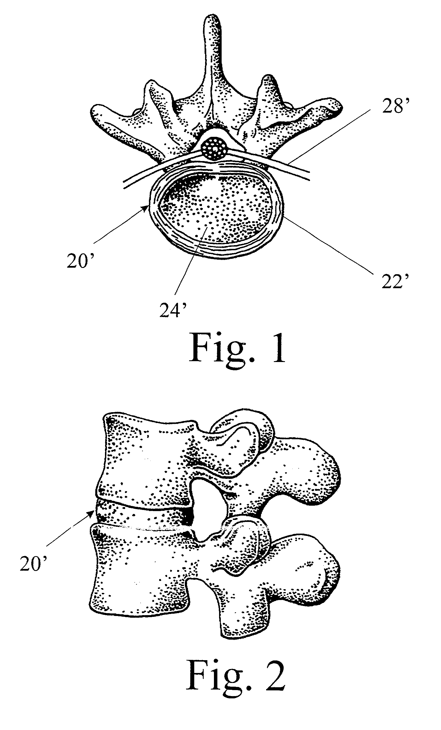
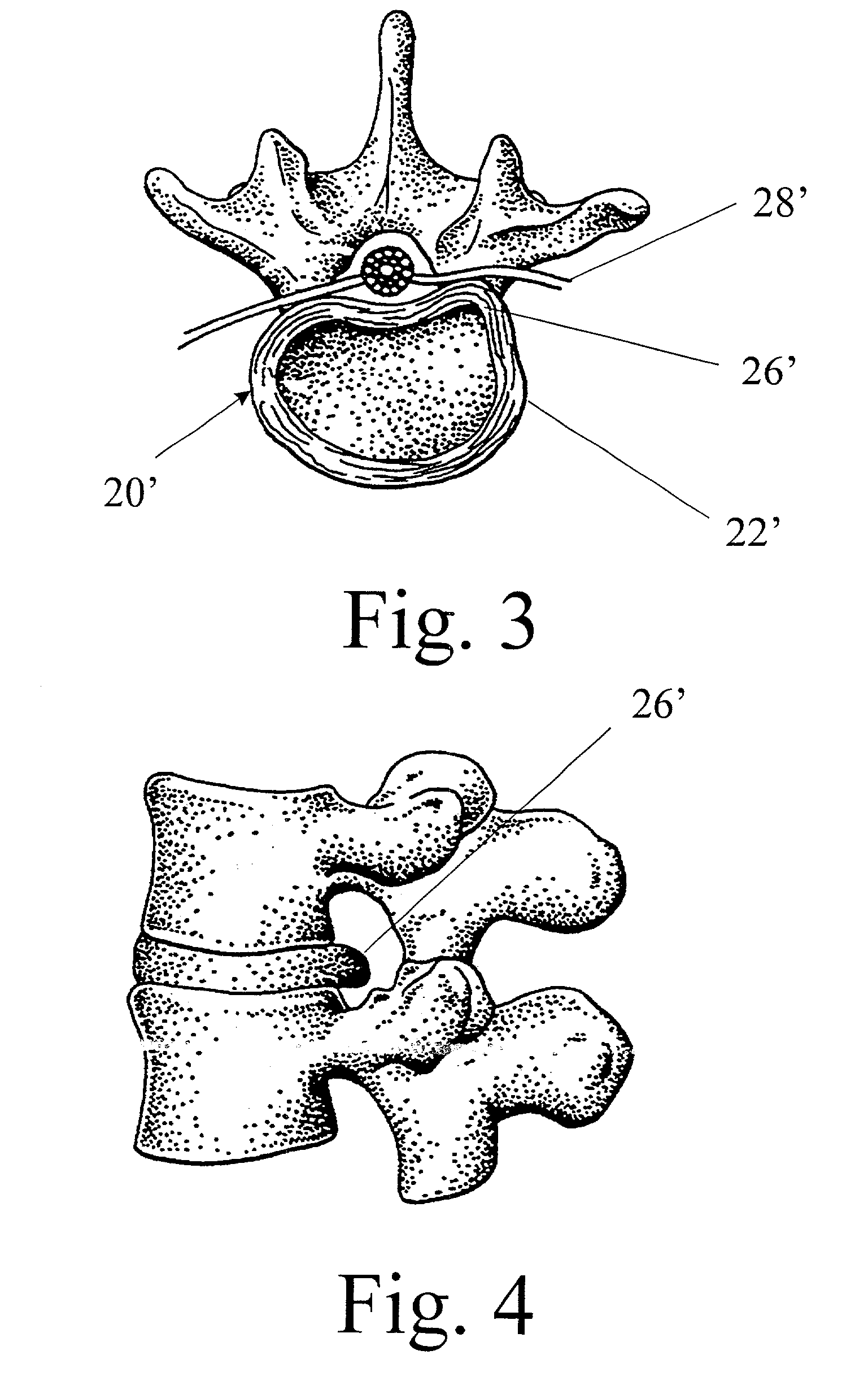
FIELD OF THE INVENTION[0001]This invention relates to the field of medicine, in particular to surgery, and specifically to a method and a device for access and removal of prolapsed nucleus pulposus material of a herniated intervertebral disc through the spinal foramen.BACKGROUND OF THE INVENTION[0002]Low back pain and radiculopathy as a result of herniated intervertebral disc represents a major health problem in the United States and all over the world. About 300,000 Americans and about the same number of people throughout the world outside the USA are operated upon each year due to this problem. Even many more people might benefit from surgical help, as those that undergo such surgery represent only about 20% of those with pathology referable to the intervertebral disc.[0003]An intervertebral disc is a structure that occupies the space between the vertebrae. It serves, in particular, as a load-absorbing cushion.[0004]FIG. 1 is a cross-sectional view of a vertebral column through a ...

Claims


Application Information

Patent Timeline
Patent Type & Authority Patents(United States)
IPC IPC(8): A61B17/56A61M39/10A61B17/00
CPCA61B17/00234A61B2017/00261
Inventor GITIS, NORMALAMIN, TODD F.MEYMAN, ALEKSANDRSHULEPOV, OLEGFAYNBERG, MIKHAIL
Owner NEVMET CORP
Learn More