
Apparatus and method of use for identifying and monitoring women at risk of developing ovarian surface epithelium derived carcinomas

a technology of ovarian epithelium and applicator, which is applied in the field of applicator and monitoring method for identifying and monitoring women at risk of developing ovarian epithelium derived carcinoma, can solve the problems of insufficient methodology for ovarian cancer screening, inability to provide a screening test as a predictive marker for identifying healthy women at increased risk of ovarian cancer, and inability to detect very early stages of the disease. to achieve the effect of improving the screening tes

Inactive Publication Date: 2006-06-06
LIFELINE BIOTECH
View PDF16 Cites 6 Cited by
  • Summary
  • Abstract
  • Description
  • Claims
  • Application Information

AI Technical Summary

Benefits of technology

[0013]It is an object of the present invention to improve screening tests for identifying and monitoring women at risk of developing OSE-derived carcinomas.
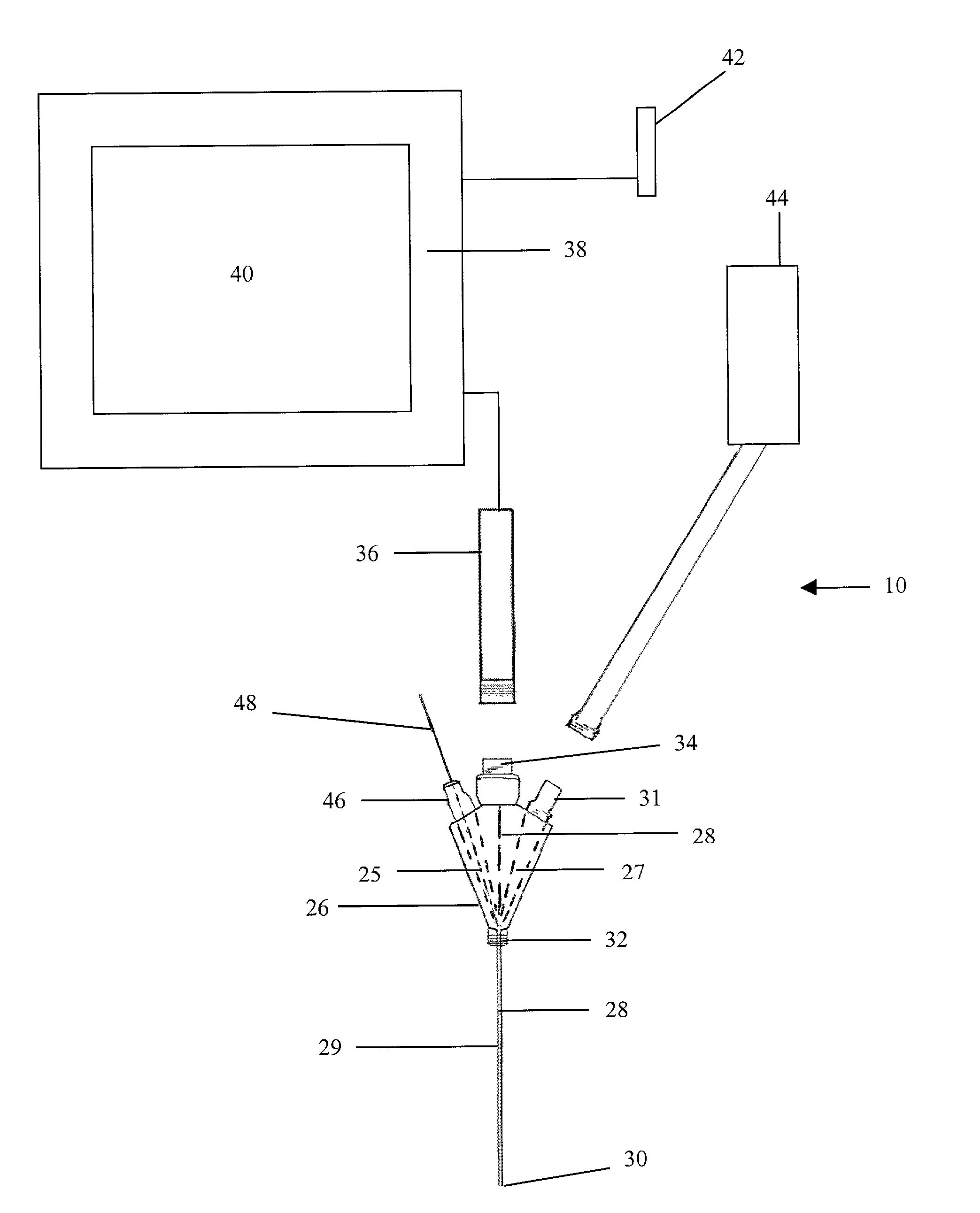
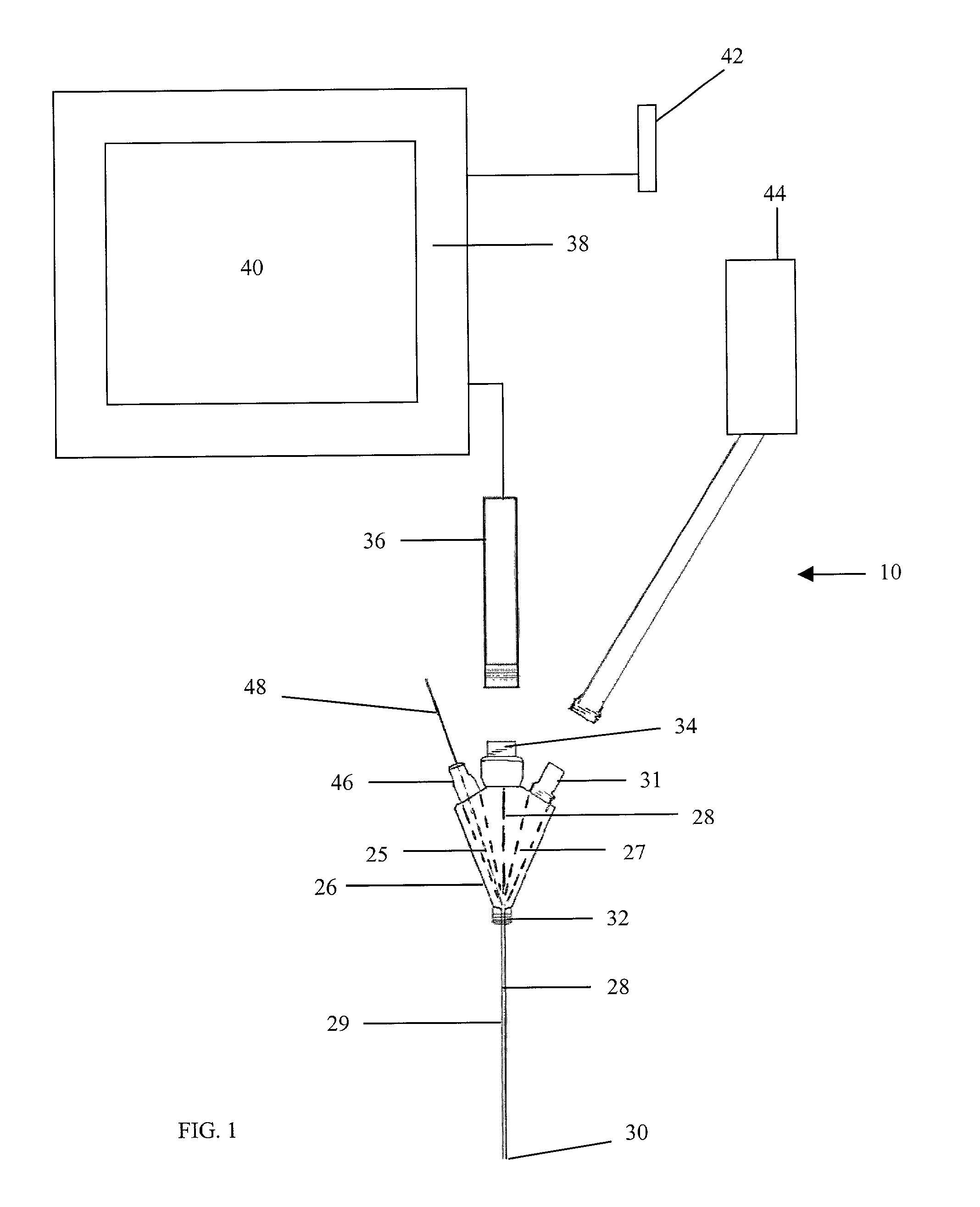
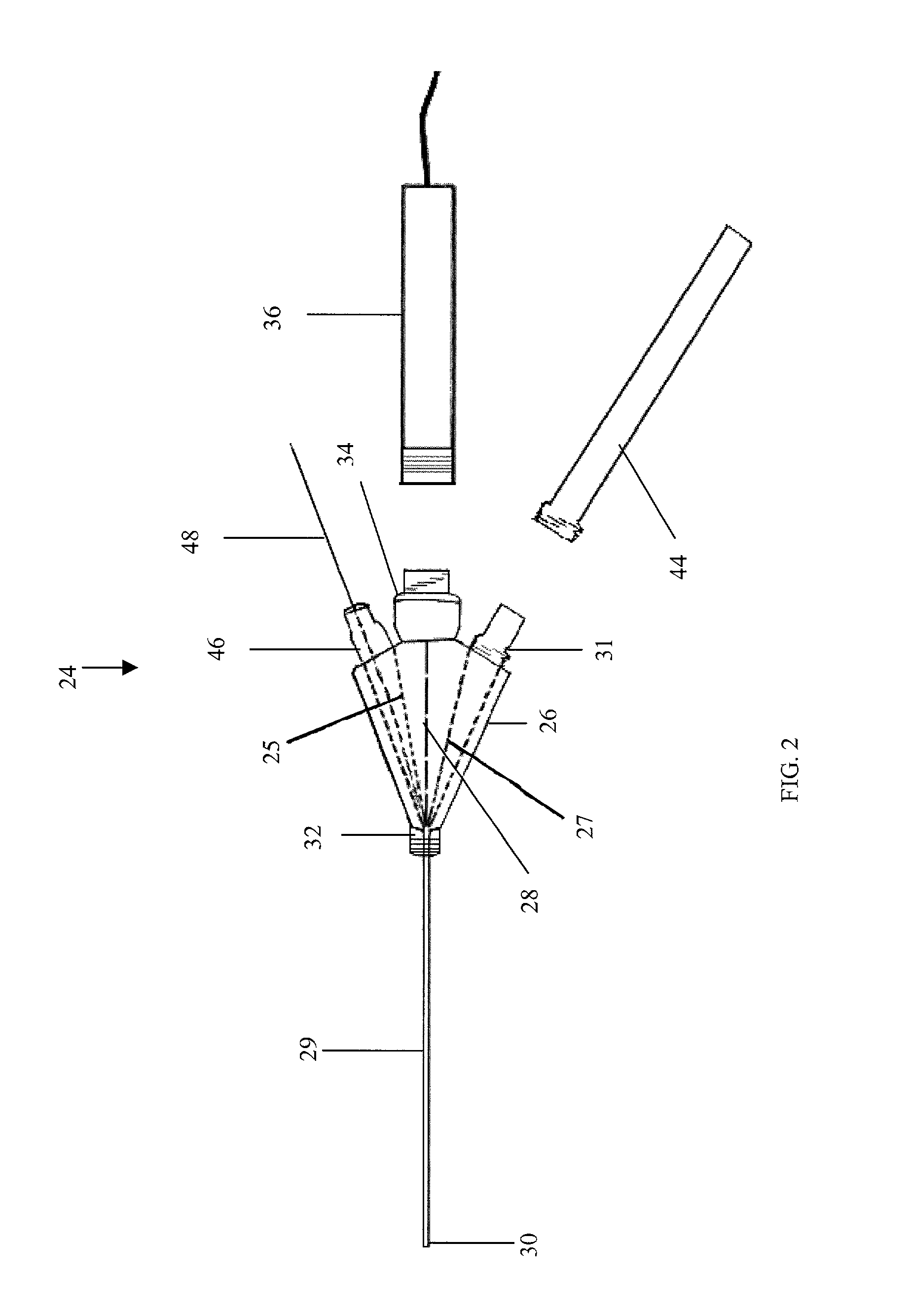
[0015]According to the present invention, there is disclosed an apparatus and method of use thereof for identifying and monitoring women at risk of developing OSE-derived cacrinomas. The apparatus includes an introducer needle configured to be capable of insertion into a female such that a terminal end of the needle is positioned adjacent an ovary of the female, a microendoscope having an optic fiber which is operably insertable into the needle in a manner to enable an image of the ovary to be obtained therethrough, and a tissue removing member operably co-insertable into the needle with the optic fiber therein to enable removal of ovarian tissue cells with minimal deleterious effect to the ovary. The needle preferably is equipped with a stylet.
[0017]The method of the invention includes the steps inserting an introducer needle having a stylet therein into a female such that a terminal such of the needle is positioned adjacent an ovary of the female, removing the stylet, operably inserting an optic fiber of a microendoscope into the needle in a manner to enable an image of the ovary to be obtained therethrough, and viewing an image through the use of the microendoscope. The method further includes operably inserting a tissue removing member into the needle and removing ovarian tissue cells with minimal deleterious effect to the ovary. The method is further characterized such that the optic fiber and the tissue removing member are co-inserted within the needle. The method further includes employing a viewing monitor with an operably associated computer to aid in viewing the image and ovarian tissue removal. Further, the method calls for employing software residing on the computer to record the image and physician notes in a data file on the computer.

Problems solved by technology

Ovarian cancer remains notoriously difficult to detect in its early stages.
Prior hereto, inadequate methodology exists for ovarian cancer screening.
Although the measurement of serum CA125 levels is an effective tool for monitoring the progression of ovarian cancers, it does not aid in providing a screening test as a predictive marker for identifying healthy women at increased risk for ovarian cancer, or for detection of very early stages of the disease.
This alone, however, is insufficient and additional contributing factors are poorly defined.
To date, however, little progress has been made in the early detection of such preneoplastic tissue changes and no reliable tumor-associated tissue markers have yet been identified.
This lack of information about early changes in the OSE poses a particular problem for women with hereditary ovarian cancer syndromes, in which there is an urgent need to define more reliable criteria for prevention and surveillance.
This has been effective for determining of existing abnormalities with the tissue, but trans-vaginal ultrasound cannot distinguish between early stage cancers and other abnormalities such as benign cysts.
However, the pap smear test is presently not used in the area of ovarian cancer detection for the lack of a suitable tissue extraction method.

Method used


Image

  • Apparatus and method of use for identifying and monitoring women at risk of developing ovarian surface epithelium derived carcinomas
  • Apparatus and method of use for identifying and monitoring women at risk of developing ovarian surface epithelium derived carcinomas
  • Apparatus and method of use for identifying and monitoring women at risk of developing ovarian surface epithelium derived carcinomas

Examples


Embodiment Construction

[0022]Referring now to the drawings, the present invention is directed to an apparatus and method of use thereof for identifying and monitoring women at risk of developing OSE-derived carcinomas and is generally designated by the numeral 10. The apparatus 10 includes an introducer needle 12 configured to be capable of insertion into a female such that a terminal end 14 of the needle is positioned adjacent an ovary of the female. The needle preferably is equipped with a stylet 16 which has a tip 18 which extends through the needle 12 and a handle 20. The style 16 prevents unwanted material from entering the needle 12 until the end 14 of the needle 12 is in its usable position. Further, the needle has a neck 22 which is formed in a manner to permit sealable connection thereto.

[0023]The invention includes a microendoscope 24 which includes a housing 26 preferably has three channels formed therein which are separated by inner partitions 25 and 27 and communicate with an open surface end...


PUM


Abstract

An apparatus and method of use thereof for identifying and monitoring women at risk of developing OSE-derived carcinomas is provided. The apparatus includes an introducer needle configured to be capable of insertion into a female such that a terminal end of the needle is positioned adjacent an ovary of the female, a microendoscope having an optic fiber which is operably insertable into the needle in a manner to enable an image of the ovary to be obtained therethrough, and a tissue removing member operably co-insertable into the needle with the optic fiber therein to enable removal of ovarian tissue cells with minimal deleterious effect to the ovary.

Description

BACKGROUND OF THE INVENTION[0001]1. Field of the Invention[0002]This application relates to a screening methodology for identifying individuals having a predisposition to develop or have ovarian cancer.[0003]2. Prior Art[0004]Ovarian cancer remains notoriously difficult to detect in its early stages. Over 80% of female ovarian carcinomas are thought to arise from the OSE. OSE-derived carcinomas are the fifth or sixth most frequent malignancy in American women and the five year survival rate of patients with ovarian cancer is less than 40%. A significant need therefore remains for a new screening test for identifying and monitoring women at risk of developing OSE-derived carcinomas.[0005]There exists various tests for the presence of female specific cancers including, for example, mammography for breast cancer and pap smears for cervical cancer. Prior hereto, inadequate methodology exists for ovarian cancer screening.[0006]One commonly used test for ovarian cancer involves measuring ...

Claims


Application Information

Patent Timeline
IPC IPC(8): A61B10/00A61B1/00
CPCA61B10/0045A61B2010/0074
Inventor REEVES, WILLIAM H.REEVES, JONATHANKEITH, LOUIS
Owner LIFELINE BIOTECH
Learn More