
Method of assaying shape and structural features in cells

a cell and structure technology, applied in image data processing, instruments, image enhancement, etc., can solve the problems of small variations in contrast introduced by cell structure, unpredictable places where such artifacts contribute to image intensity, and phase microscopy has drawbacks

Inactive Publication Date: 2006-05-23
BOWLING GREEN STATE UNIV
View PDF4 Cites 111 Cited by
  • Summary
  • Abstract
  • Description
  • Claims
  • Application Information

AI Technical Summary

Benefits of technology

[0075]In certain aspects, the present invention includes a method, in which execution of the steps serves as a rapid assay for properties or characteristics of a cell significantly related to carcinogenesis or cancer.
[0081]In certain aspects, the present invention includes a method, in which execution of the steps serves as a rapid assay for the chemotherapeutic or chemopreventive effect of an individual drug or a mixture of drugs inhibiting development of cancer.
[0083]In certain aspects, the present invention includes a method, in which execution of the steps serves as a rapid assay for properties or characteristics of a cell significantly related to carcinogenesis or cancer.
[0084]In certain aspects, the present invention includes a method, in which execution of the steps serves as a rapid assay for the chemotherapeutic or chemopreventive effect of an individual drug or a mixture of drugs inhibiting development of cancer.

Problems solved by technology

However, phase microscopy has the drawback that only small variations in contrast are introduced by cell structure.
Furthermore, the places where such artifacts contribute to image intensity are unpredictable.
This limits the value of the phase technique for the purpose of acquiring images and quantifying variations in their intensity.
Thick samples give rise to images that are too complex to interpret due to their overlapping planes of imaged structure, which planes are all projected onto the two-dimensional plane of an image.
Therefore, the usefulness of analog, video-based methods for amplifying phase shift-induced contrast is limited to specimens thinner than a cell.
Except for those few examples where software has been developed to address specific problems, the availability of such algorithms is very restricted.
Practitioners of the art in this field have not developed or refined algorithms for specification and relation of the content of multiple planes in such datasets to each other.
While the numerous advances in imaging referred to above, have been reduced to practice, they still fall short of being able to represent the complex matter composing a cell in a form that is useful for assaying the shape of cultured cells.
The arrangement of any single ingredient of a cell may now be presented in the full 3D coordinate space representing the cell in a high-speed digital computer, but there are hundreds or thousands such molecules mediating the shape of a cell, and so this increases the complexity of the analysis rather than reducing it.
Measurements based on the absorbance or optical density of a cell following staining, as disclosed in the above-claimed inventions, had the major drawback that the colors and intensity conferred by a stain could vary due to the irreproducibility in reagents.
An additional drawback of such methods is that they project the 3D image of a cell into a two-dimensional plane, which means that it is impossible to tell whether any given portion of a cell is thicker than usual or molecules are merely more dense in that portion of the cytoplasm.
Whereas it is apparent from the prior art that the shapes of cells or portions of cells can be detected and quantified, the prior work did not develop an analysis of cell shape in detail or at high resolution.
Upon reflection off the metal, this wave combines with incoming waves to introduce partial destructive interference.
A shorter wave, however, whose path through the thickness of oxide layer corresponds to the distance of one wavelength, shows a completely destructive interference effect.
One of the drawbacks of the classification method was that, although the shape variables were precise in the mathematical sense, they had little intuitive relationship to the shape features of a cell on a one-to-one basis.
This meant that the investigators might recognize a sample of cells as having the overall signature of a transformed or a nontransformed population, but not be able to distinguish among their morphological features those ones that conferred the signature values.
The drawback of such methods is that they mainly analyze a representation which is only one portion of a cell.

Method used


Image

  • Method of assaying shape and structural features in cells
  • Method of assaying shape and structural features in cells
  • Method of assaying shape and structural features in cells

Examples


example i

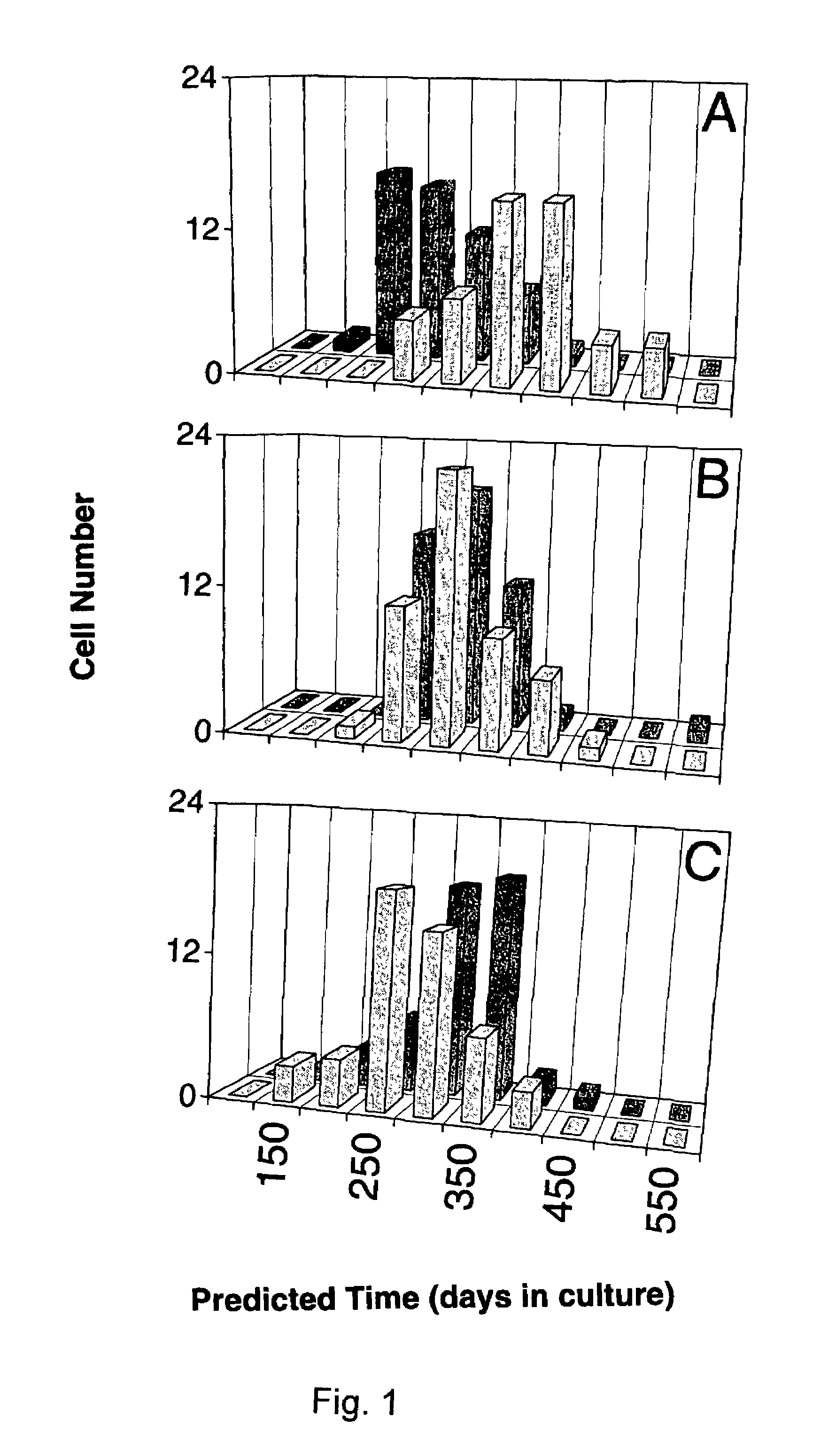
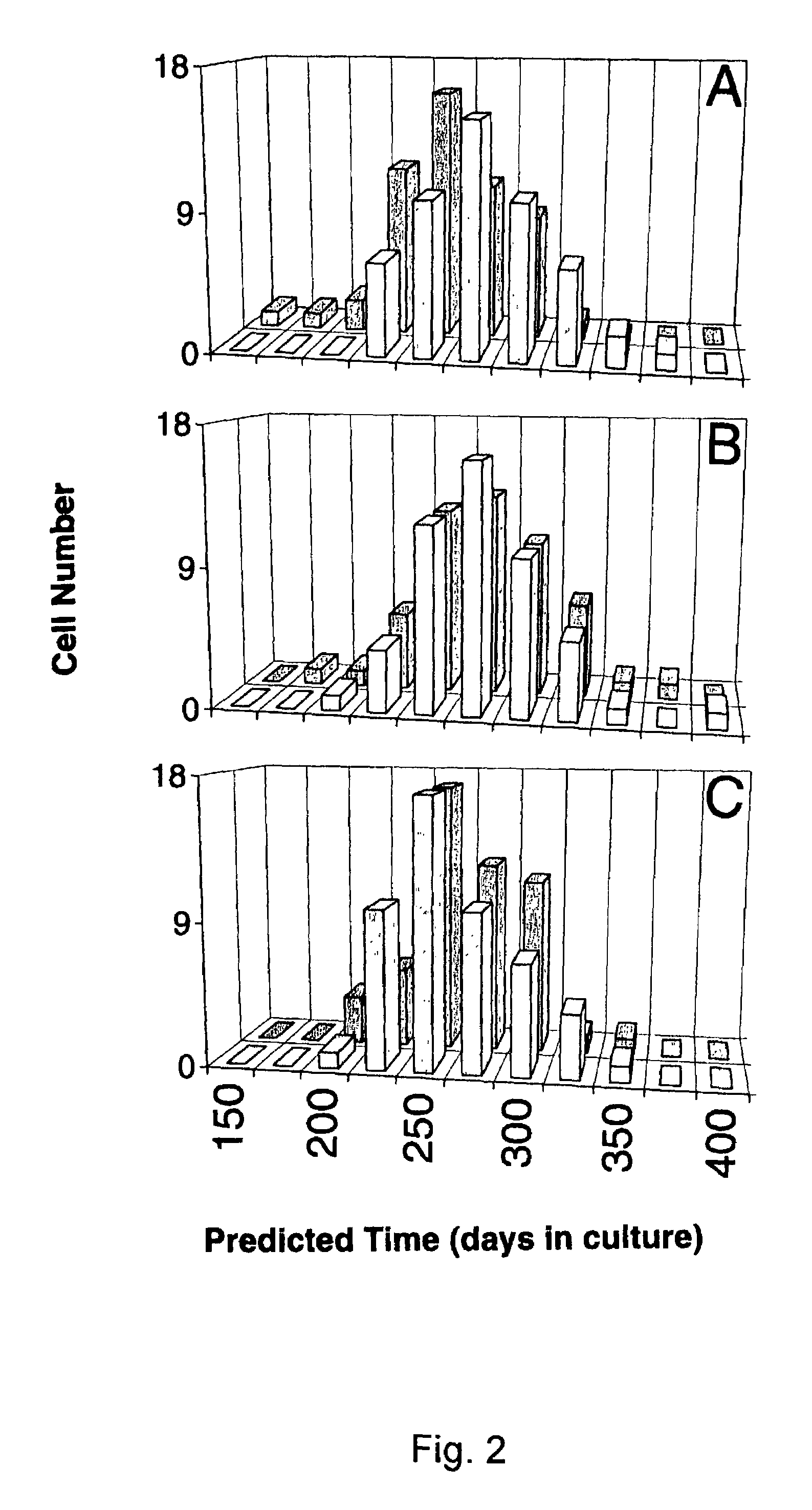
[0137]Cell lines and interference preparations. Equations were developed to define changes in the signature-type for two epithelial cell lines that underwent transformation over a prolonged time of in vivo culture. Although a different equation was developed in each case, there was considerable overlap in the factors employed as variables (3). IAR20 PC1 was a clonal derivative of a line called IAR20, which was cultured from liver cells of a BD-VI rat. The other line, 1000 W, was generated from the tracheal epithelium of a Fisher 344 rat after topical treatment with 7,12-dimethylbenz(a)anthracene. The lines become tumorigenic after 11 and 16 months of being maintained in culture, respectively (3). Liver cells were grown in William's E medium supplemented with 10% fetal bovine serum, whereas the tracheal cells were grown in Waymouth's medium containing fetal bovine serum plus 0.1 μg / ml insulin and 0.1 μg / ml hydrocortisone. The cells were maintained and subcultured as described elsewh...

example ii

[0161]The mechanism of action of the tumor promoter depends on its ability to substitute for an endogenous second messenger, diacylglycerol, and thereby activate certain members of an enzyme family known as protein kinase C. The quantitative shape phenotype of cells treated with PMA resembled the phenotype of bona fide cancer cells. The effect of PMA on this phenotype was transient as explained above. When the shape phenotype was dissected into components by relating different variable's values to shape features, several of the altered values appeared to rely upon a declining number of sharp features, such as filopodia and microspikes, at the cell edge.

[0162]Filopodia and microspikes are in turn regulated by a GTPase (79, 80) of the Rho family, Cdc42, which modulates actin architecture. Although data suggest that PMA counteracts the rearrangement of actin into filopodia and microspikes, there is no known link of Cdc42 to any PMA-responsive isozyme of protein kinase C. Further, over ...

example iii

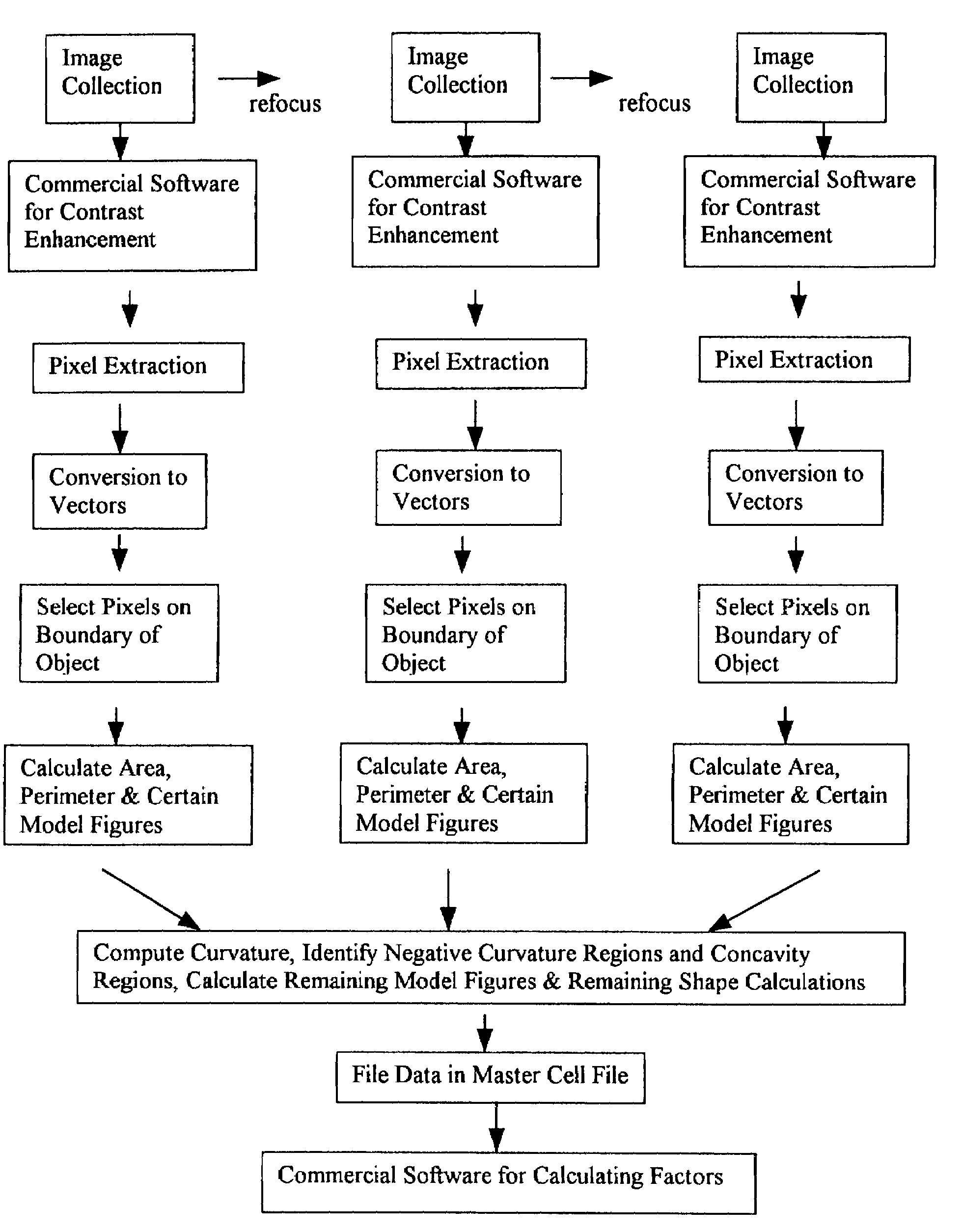
[0168]A flowchart showing the preferred method of image processing and segmentation is provided (FIG. 10). The extraction of latent factors from shape data is done in such a way that the new variables are qualitatively related to features which can be perceived by viewing the cells. The use of a large database of values for shape variables, as done herein, is desirable if factors are to be generated which approach this ideal. The definitions and names of highly correlated shape variables that contributed to a data set of exemplary factors are provided below. Here, the first 13 out of 20 factors could be given intuitive descriptions based on the variables they incorporated. Although certain factors beyond the 13th factor could also be given descriptive names, they became increasingly more contour-specific and more specific to a single kind of shape feature. Such intuitive factors can be used in interpreting alterations in cell shape characteristics that may be caused by exposing cell...


PUM


Abstract

The method for repetitively determining a profile of quantitative features for different cells in a sample by collecting and analyzing information on the mass distribution in cells or portions of cells and comparing the collected information to values from a relational database is disclosed.

Description

[0001]This application claims benefit of U.S. Provisional Application No. 60 / 280,053, filed Mar. 30, 2001.TECHNICAL FIELD[0002]This invention relates to a process for assaying the shape features of cells in order to determine whether the shape features are altered by exposure of the cells to a pharmaceutical or biological agent.BACKGROUND OF THE INVENTION[0003]There are three types of structural components of cells that become organized in certain ways to regulate cell architecture. These are actin filaments of approximately 8-nm diameter, intermediate filaments of approximately 10-nm diameter, and microtubules (MTs) of approximately 24-nm diameter. Larger macromolecular assemblages are composed from combinations of these smaller structures, which can either aggregate with components like themselves or with adaptor molecules or with other components which causes them to form an assemblage of cytoskeletal molecules. Such processes cause the assemblages to form a metastable cell struc...

Claims


Application Information

Patent Timeline
IPC IPC(8): G06K9/00G01N15/14G06T7/00G06T7/60
CPCG01N15/1475G06T7/602G06T7/604G06T7/0012G01N2015/1454G01N2015/1497G06T2207/30024G06T7/62G06T7/64G06V20/698G01N15/1433
Inventor HECKMAN, CAROL A.
Owner BOWLING GREEN STATE UNIV
Learn More