X-ray imaging method and a 3D-rotational X-ray apparatus for applying this method

a x-ray and x-ray technology, applied in the field of x-ray imaging methods, can solve the problems of poor reconstructed image quality, achieve the effects of reducing noise, slowing down rotational scanning speed, and reducing the speed of x-ray source rotation

Inactive Publication Date: 2005-10-25
KONINKLIJKE PHILIPS ELECTRONICS NV
View PDF9 Cites 15 Cited by
  • Summary
  • Abstract
  • Description
  • Claims
  • Application Information

AI Technical Summary

Benefits of technology

[0004]It is an object of the invention to provide a method where the above problems are solved. According to the invention, the method as described in the opening paragraph is characterized in that the run length of the scan rotation over substantially 180° is at least 15 s and preferably about 20 s. Contrary to the present tendency to increase the C-arm rotation speed, it has been found that the above method can be improved by a slower rotational scanning speed. With a run length of about 20 s, i.e. a C-arm rotation speed of substantially 10° / s, and with the above parameters, during each scan about 5 times 20 corresponding X-ray images of a quasi-stationary heart can be obtained. Thus, by reducing the speed of the rotation of the X-ray source around the object more X-ray images corresponding to the same phase in the cardiac cycle are acquired. This leads to reduced noise in the resulting reconstructed 3-dimentional volume.
[0005]A further embodiment of the X-ray imaging method according to the invention is characterized in that before reconstruction, images obtained at predetermined corresponding characteristic time moments in successive cardiac cycles are correlated with each other. This technical measure is based on the insight that each phase in the spectrum of cardiac activity in general represents the same spatial orientation and geometry of the heart muscle. Thus by correlating the X-ray images corresponding to the same phase of the cardiac activity, for example by known mathematical algorithms, one can further reduce noise in the final reconstruction of the 3-dimentional volume.
[0006]A further embodiment of the X-ray imaging method according to the invention is characterized in that the characteristic time moments substantially correspond to R-peaks of the cardiac cycle. This technical measure provides the possibility to acquire images of the quasi-stationary heart, when the cardiac muscle is relaxed, thus reducing the image reconstruction noise due to the movement of the cardiac muscle. The methods of ECG-triggering are known and present no technical difficulty for those skilled in the art.
[0007]A further embodiment of the X-ray imaging method according to the invention is characterized in that before a reconstruction, images obtained at neighbouring time moments in a predetermined characteristic time interval of a cardiac cycle are correlated with each other. This technical measure is based on the insight that if one determines a certain time interval around a characteristic moment in the ECG spectrum, for example around the R-peak, the movement of the heart muscle within this time interval has taken place to a such small degree that the X-ray images acquired within this predetermined time interval can be used for the image reconstruction. This technical measure provides the net noise reduction as the minor spatial movements or deformations of the heart muscle are compensated by the total number of the useful X-ray images contributing to the final dataset. Furthermore, it is still possible to correlate measurements at corresponding time moments of successive cardiac cycles with each other and, in case a reconstruction of a diminished quality is obtained, to combine the reconstruction with existing modelling techniques.

Problems solved by technology

Problems of the known method are the necessity to correlate the different sub-runs in order to produce the 180° run as well as poor quality of the reconstructed image.

Method used


Image

  • X-ray imaging method and a 3D-rotational X-ray apparatus for applying this method
  • X-ray imaging method and a 3D-rotational X-ray apparatus for applying this method

Examples


Embodiment Construction

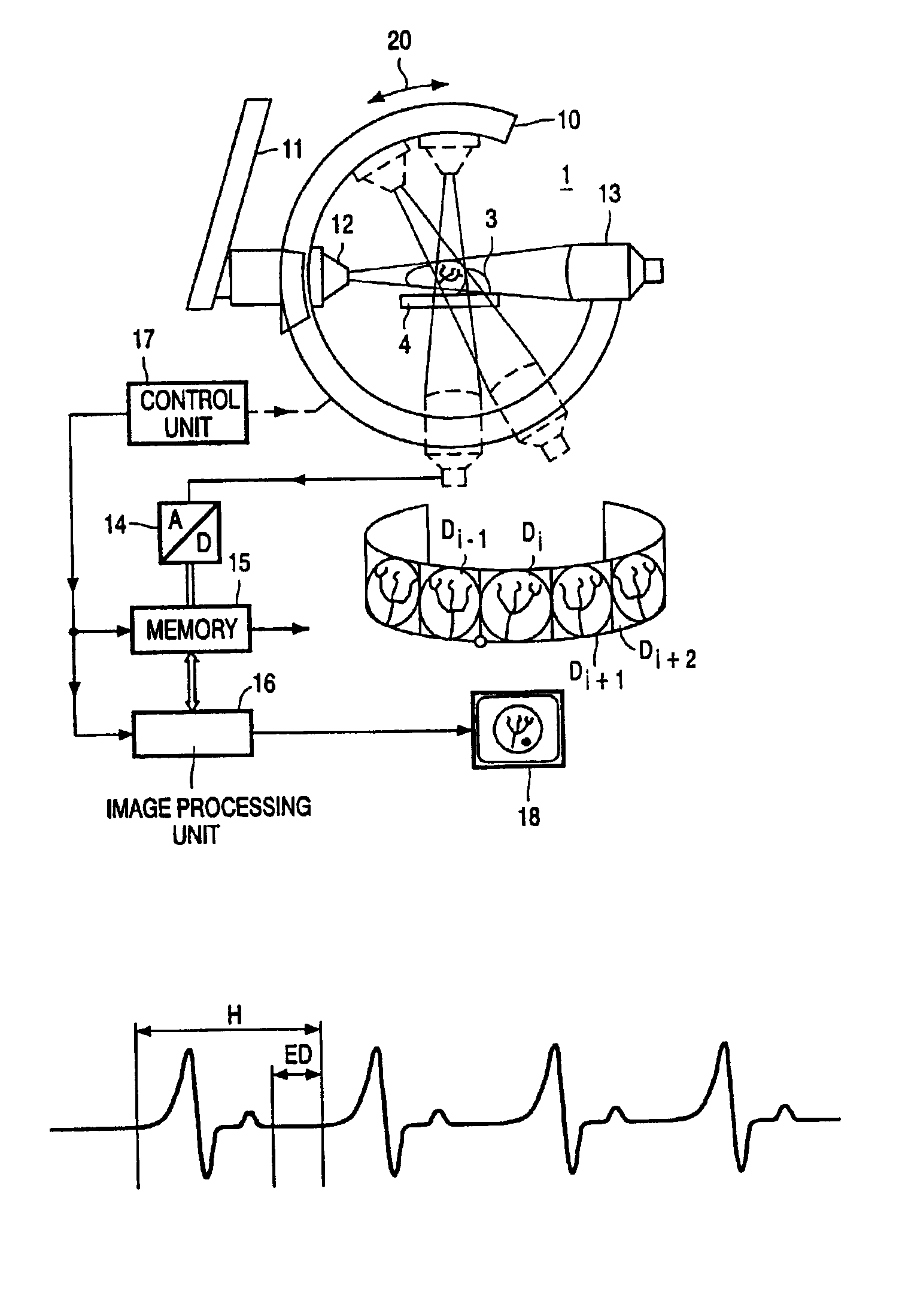
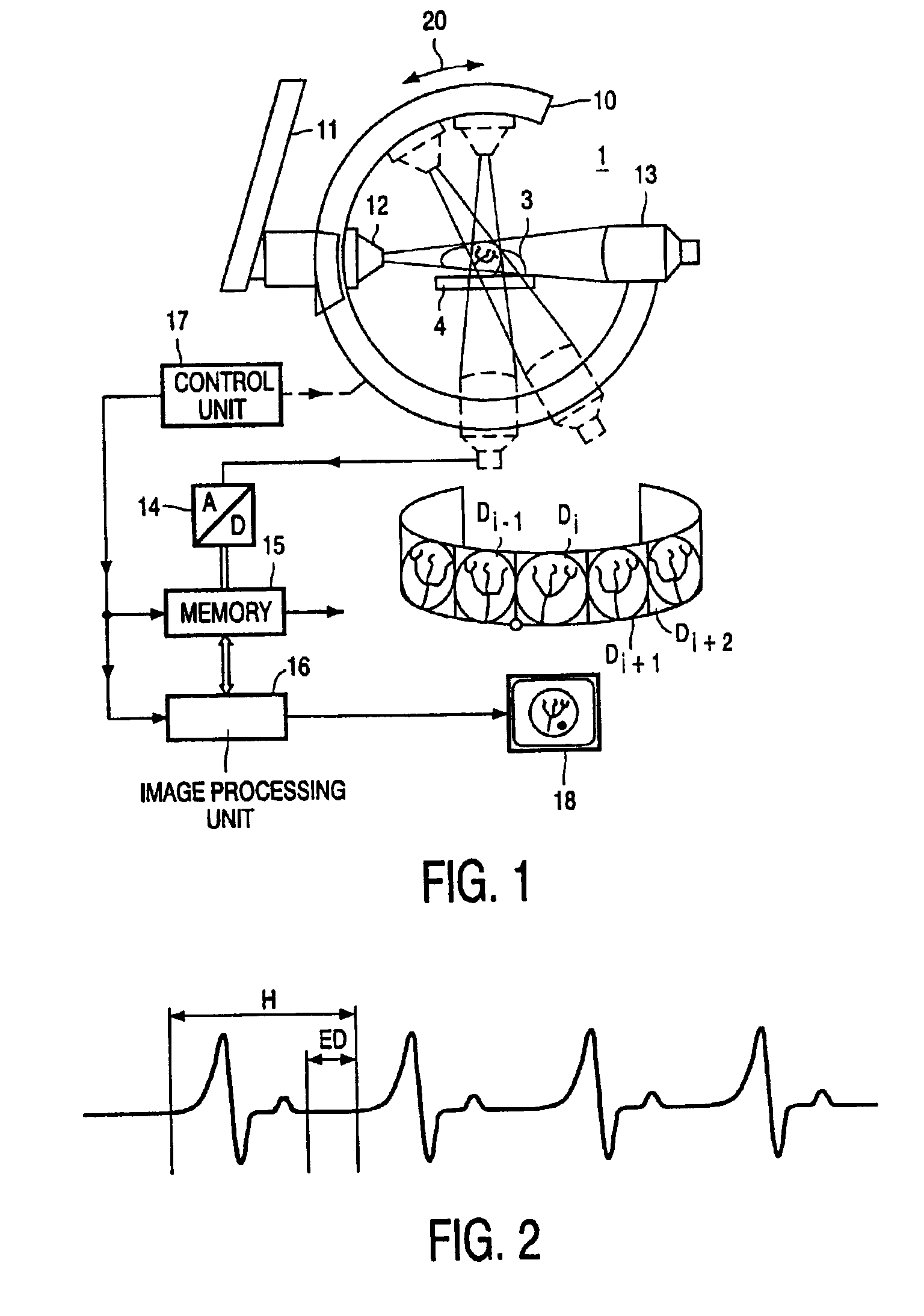
[0012]The imaging apparatus of FIG. 1 serves to form two-dimensional X-ray images of an object to be examined, particularly a periodically moving object such as the heart and the coronary vascular system of the heart of a patient, from which two-dimensional images via reconstruction a 3-dimensional volume of the object can be obtained.

[0013]The imaging apparatus 1 includes a circular C-arm 10 which is mounted on a (only partly shown) stand 11. The C-arm can be rotated over an angle of for example 180° around its center in the direction of the double arrow 20 by means of a motor drive (not shown in the figure). The C-arm 10 accommodates an X-ray source 12 and an X-ray image pick-up device 13, which are aligned relative to each other in such a manner that an X-ray image can be formed of a volume to be examined around said center. A plurality of X-ray images can thus be formed. This plurality of X-ray images shows the volume to be examined from different angular positions (some of whic...


PUM


Abstract

An X-ray imaging method comprises forming 2-dimensional X-ray images of an object to be examined, for example the coronary vascular system of a patient, and reconstruction of a 3-dimensional volume thereof. With a relatively long run length of a scan rotation over substantially 180° of at least 15 sec. and preferably about 20 sec. A sufficient number of images is obtained to perform a more accurate volume reconstruction. This reconstruction method may be combined with existing modelling techniques.

Description

FIELD OF THE INVENTION[0001]The present invention relates to an X-ray imaging method comprising forming a set of 2-dimensional X-ray images of an object to be examined, for example the coronary vascular system of a patient, by means of a scan rotation of an X-ray source around said object over a run length, said X-ray images being acquired at predetermined characteristic time moments in cardiac cycle of the object and a reconstruction of a 3-dimensional volume thereof. The invention further relates to a 3D-rotational X-ray apparatus for applying this method, the apparatus comprising a circular C-arm with a drive, the C-arm accommodating an X-ray source and an X-ray image pick-up device and being rotatable over an angle of substantially 180° around its center, triggering means for triggering the X-ray images at predetermined characteristic time moments in the cardiac cycle of the object and means for processing the images obtained to reconstruct a 3-dimensional volume of the object.B...

Claims


Application Information

Patent Timeline
Patent Type & Authority Patents(United States)
IPC IPC(8): A61B6/00
CPCA61B6/481A61B6/504A61B6/541
Inventor RASCHE, VOLKERWEISS, STEFFENOP DE BEEK, JOHANNES CATHARINA ANTONIUS
Owner KONINKLIJKE PHILIPS ELECTRONICS NV
Why Eureka
Try Eureka