
Method for differentiated investigation of diverse structures in preferably biological preparations

a biological/medical preparation and structure technology, applied in the field of different structure examination, can solve the problems of difficult examination after radiation of biological/medical preparation, unusually difficult reproduction of examination, and inability to reproduce examination

Inactive Publication Date: 2005-08-23
LEICA MICROSYSTEMS CMS GMBH
View PDF15 Cites 21 Cited by
  • Summary
  • Abstract
  • Description
  • Claims
  • Application Information

AI Technical Summary

Benefits of technology

[0004]The objective of the present invention is therefore to configure a method of the type in question for differentiated examination of various structures in preferably biological preparations, in particular via confocal laser scanning microscopy, such that the reproduction of the marking or of the result of the examination is ensured, even after prolonged radiation. The problems occurring in fluorescence microscopy or in connection with fluorescent die binding are to be thereby prevented.

Problems solved by technology

However, the fluorescence microscopy previously employed is in practice exceptionally problematic, particularly since the fluorescent dies used therein fade over time, and specifically have a fading characteristic that prevents the reproduction of examinations.
This not only makes a reproduction of the examination impossible, but, what is more, it also makes any examination subsequent to radiation of the biological / medical preparation more difficult or—in terms of a reliable evaluation—makes such an examination nearly impossible.

Method used


Image

  • Method for differentiated investigation of diverse structures in preferably biological preparations

Examples


Embodiment Construction

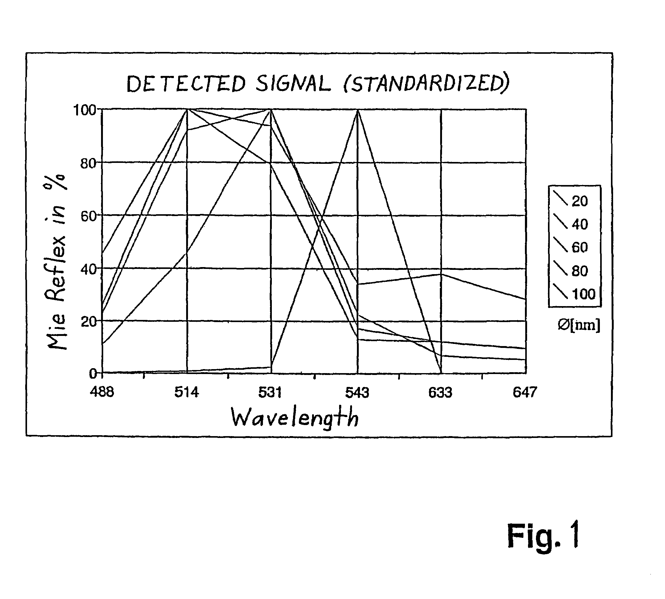
[0010]According to the invention, it has been recognized that the problem occurring within the context of fluorescence microscopy is mainly attributable to the fading characteristic of the usable fluorescent dye. According to the invention, there is a deviation from the marking method typically used in the biomedical field; specifically, the structures involved in the preparation are not marked with any kind of dyes, but with particles having a specific diameter and specific material characteristics. While the fluorescent dye attachment depends on the fluorescence behavior of the fluorescent dyes assigned to the structures, the optical characteristics of the particles at some point play no role. What's more, this depends on the diameter and the material characteristics of the particles.

[0011]The particles are thus—insofar as required—assigned to the structures or areas of the preparations in question, it being possible to provide the particles with binding means that with certain st...


PUM

PropertyMeasurementUnit
diametersaaaaaaaaaa
diametersaaaaaaaaaa
diametersaaaaaaaaaa

Abstract

The invention relates to a method for examining different structures in preferably biological preparations in a differential manner, especially by means of confocal laser scanning microscopy. The method is characterized in that particles having a specific diameter and specific characteristics are assigned to the structures and in that said structures are detected by detecting the particles which have specifically bonded in or to the preparations. The detection process is carried out in an advantageous manner by marking the structures with metal particles with diameters of 10 nm to 1,500 nm and detecting Mie scattering or a plasmon signal.

Description

CROSS REFERENCE TO RELATED APPLICATIONS[0001]The present application is the U.S. national phase under 35 U.S.C. 371 of International Application No. PCT / DE99 / 03946 filed Dec. 10, 1999 claiming priority of German Patent Application No. 198 58 431.8 filed Dec. 17, 1998 and German Patent Application No. 199 50 909.3 filed Oct. 22, 1999.FIELD OF THE INVENTION[0002]The invention relates to a method for the differentiated examination of various structures in preferably biological preparations, in particular via confocal laser microscopy.BACKGROUND OF THE INVENTION[0003]Fundamentally, this involves a detection / marking method, in particular a method as is applied within the context conventional fluorescence microscopy within the biomedical field. However, the fluorescence microscopy previously employed is in practice exceptionally problematic, particularly since the fluorescent dies used therein fade over time, and specifically have a fading characteristic that prevents the reproduction of ...

Claims


Application Information

Patent Timeline
Patent Type & Authority Patents(United States)
IPC IPC(8): G02B21/00G01N21/55G01N33/483G01N21/27G01N21/39G01N21/49G02B21/06G02B21/36
CPCG01N21/554G01N21/6458G01N21/648G02B21/0068G02B21/0072G02B21/0076
Inventor ENGELHARDT, JOHANN
Owner LEICA MICROSYSTEMS CMS GMBH
Learn More