
Apparatus and method for non-invasively measuring cardiac output

a non-invasive, cardiac output technology, applied in the field of non-invasive means of determining cardiac output in patients, can solve the problems of high morbidity and mortality consequences, inability of sedated or unconscious patients to actively participate in inhaling and exhaling into a bag, and inability to accurately measure cardiac output. , to achieve the effect of modest recovery time and reasonable monitoring system cos

Inactive Publication Date: 2005-06-21
RIC INVESTMENTS LLC
View PDF16 Cites 32 Cited by
  • Summary
  • Abstract
  • Description
  • Claims
  • Application Information

AI Technical Summary

Benefits of technology

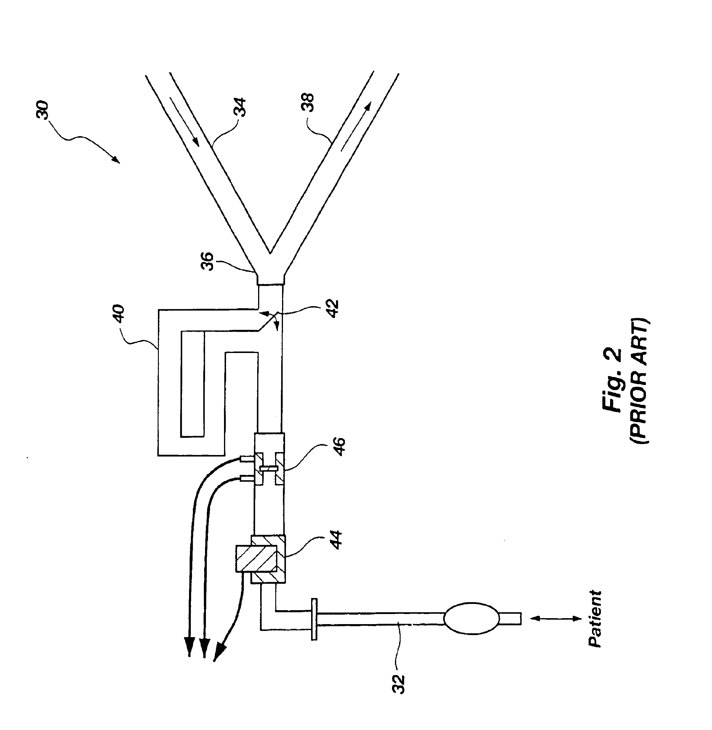
[0011]In accordance with the present invention, apparatus and methods for measuring cardiac output using a modified Fick Equation are provided where the amount of deadspace which is provided in the apparatus can be adjusted to increase or decrease the volume of exhalate to be re-breathed by the patient, thereby decreasing ventilation without changing airway pressure. The apparatus and methods of the present invention also provide an adjustability factor which enables the apparatus to be adjusted to suit any size or capacity of patient. The apparatus of the present invention also employs significantly less expensive elements of construction, thereby rendering the device disposable.
[0013]In previous partial re-breathing methods, a deadspace, usually comprising an additional 50-250 ml capacity of air passage, was provided in the ventilation circuit to decrease the effective alveolar ventilation. In the present invention, a ventilation apparatus is provided with an adjustable deadspace to provide the necessary change in ventilation for determining accurate changes in CO2 production and end-tidal CO2 commensurate with the requirements of differently sized patients. In one embodiment of the ventilation apparatus, selectively adjustable deadspace is provided through which the patient exhales and inhales. Thus, the adjustable deadspace of the apparatus permits easy adjustment of the deadspace to accommodate any size or capacity of patient, from a small to a large adult. As a result, the patient is provided with a volume of re-breathable gas commensurate with the patient's size which decreases effective ventilation without changing the airway pressure. Because airway and intra-thoracic pressure are not affected by the re-breathing method of the present invention, cardiac output is not significantly affected by re-breathing. In an alternative method, the deadspace may be effectively lessened by selectively leaking exhaled gas from the ventilation system to atmosphere or to a closed receptacle means during inspiration.
[0016]In an alternative embodiment, a tracheal gas insufflation apparatus is used to provide the change in ventilation necessary to calculate pulmonary CO2 changes using the differential Fick Equation. Tracheal gas insufflation (“TGI”) apparatus is commonly used to flush the deadspace of the alveolar spaces of the lungs and to replace the deadspace with fresh gas infused through insufflation means. That is, fresh gas is introduced to the central airway to improve alveolar ventilation and / or to minimize ventilatory pressure requirements. TGI apparatus is interconnected to a ventilator system and includes a means of introducing fresh gas into the breathing tube as it enters the patient's lungs. The TGI apparatus may be used in the methods of the present invention to determine baseline measurements of VCO2 and etCO2 during TGI. When the TGI system is turned off, a deadspace is formed by the patient's trachea and the endotracheal tube of the TGI apparatus which allows measurement of a change in CO2 to be evaluated in accordance with the invention. Further, the catheter of the TGI apparatus may be variably positioned within the trachea of the patient to further adjust the deadspace volume.
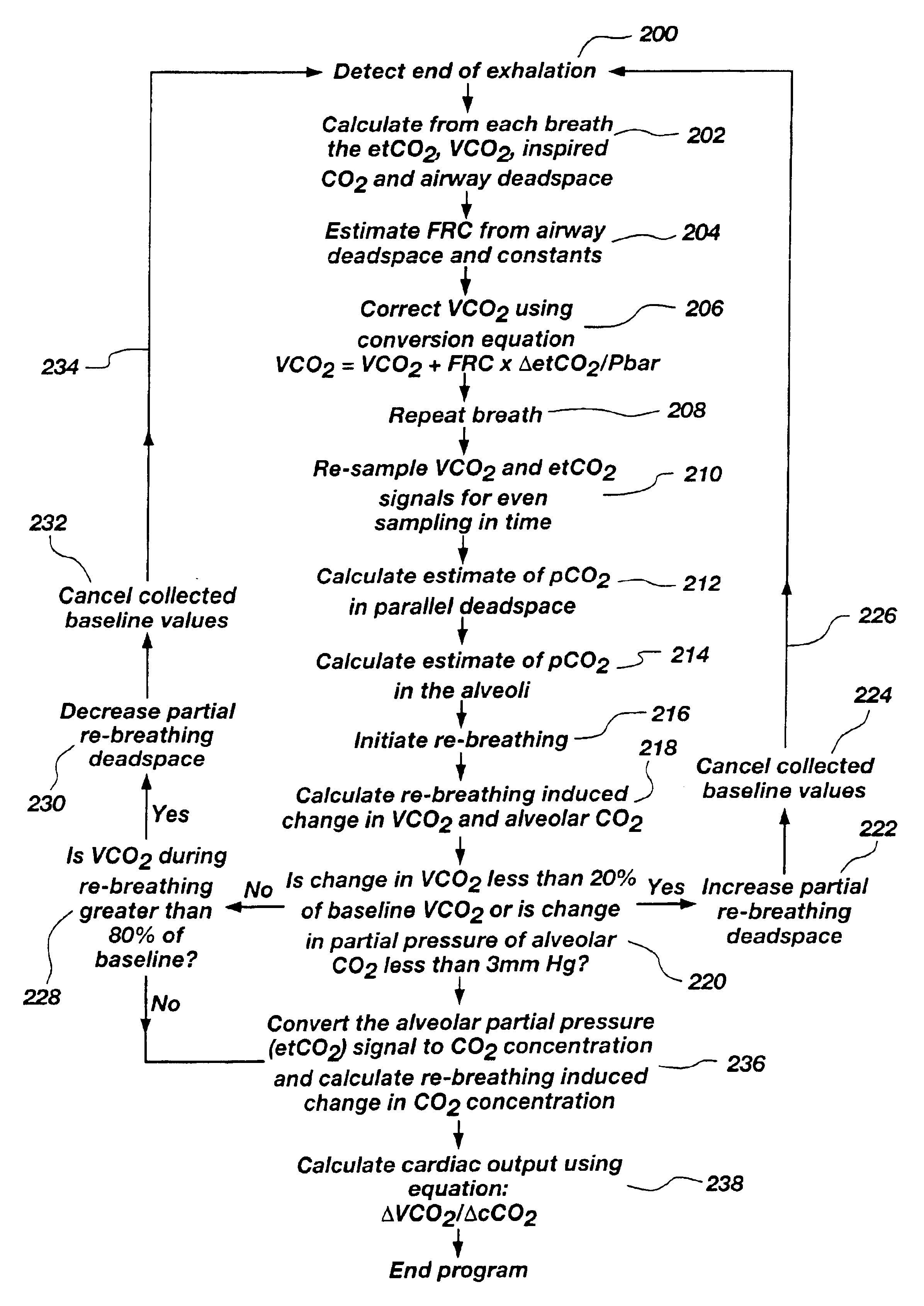
[0018]Cardiac output is determined, in the present invention, by estimating alveolar CO2 concentration rather than basing output on end-tidal CO2 concentration, as is practiced in the prior art. Partial pressure values that are obtained from CO2 measurements are converted to a value for gas content in the blood using the dissociation equation known in the art. Thus, a more accurate cardiac output can be determined. In addition, the accuracy of cardiac output is increased by correcting VCO2 values to account for flow of CO2 into the functional residual capacity of the lungs, defined as the volume of gas left in the lungs at the end of an expired breath. The determination of values based on experiential data is processed by the software program to determine cardiac output.
[0019]The ventilation apparatus of the present invention employs inexpensive yet accurate monitoring systems as compared to the systems currently used in the art. The methods of the invention allow automatic adjustability of the apparatus for accommodating patients of different sizes and provide consistent monitoring with modest recovery time. Further, the present apparatus and methods can be used equally with non-responsive, intubated patients as well as non-intubated, responsive patients.

Problems solved by technology

Although such techniques can produce a reasonably accurate result, the invasive nature of the procedure has high potential for morbidity and mortality consequences.
That method of re-breathing has not proven to be a satisfactory means of measuring cardiac output because the patient is required to breathe directly into and from a closed volume in order to produce the necessary effect.
However, it is usually impossible for sedated or unconscious patients to actively participate in inhaling and exhaling into a bag.
However, known partial re-breathing methods have significant disadvantages as well.
Specifically, known methods 1) are less accurate with non-intubated or spontaneously breathing patients, 2) only allow intermittent measurements (usually about every four minutes), 3) result in an observed slight, but generally clinically insignificant, increase in arterial CO2 levels, and 4) do not permit measurement of shunted blood flow (that is, blood which does not participate in gas exchange).
Further, known apparatus used for partial re-breathing techniques are of standard construction and do not compensate for differences in patient size or capacities.
In addition, many devices employ expensive elements, such as three-way valves, which render the devices too expensive to be used as disposable units.

Method used


Image

  • Apparatus and method for non-invasively measuring cardiac output
  • Apparatus and method for non-invasively measuring cardiac output
  • Apparatus and method for non-invasively measuring cardiac output

Examples


Embodiment Construction

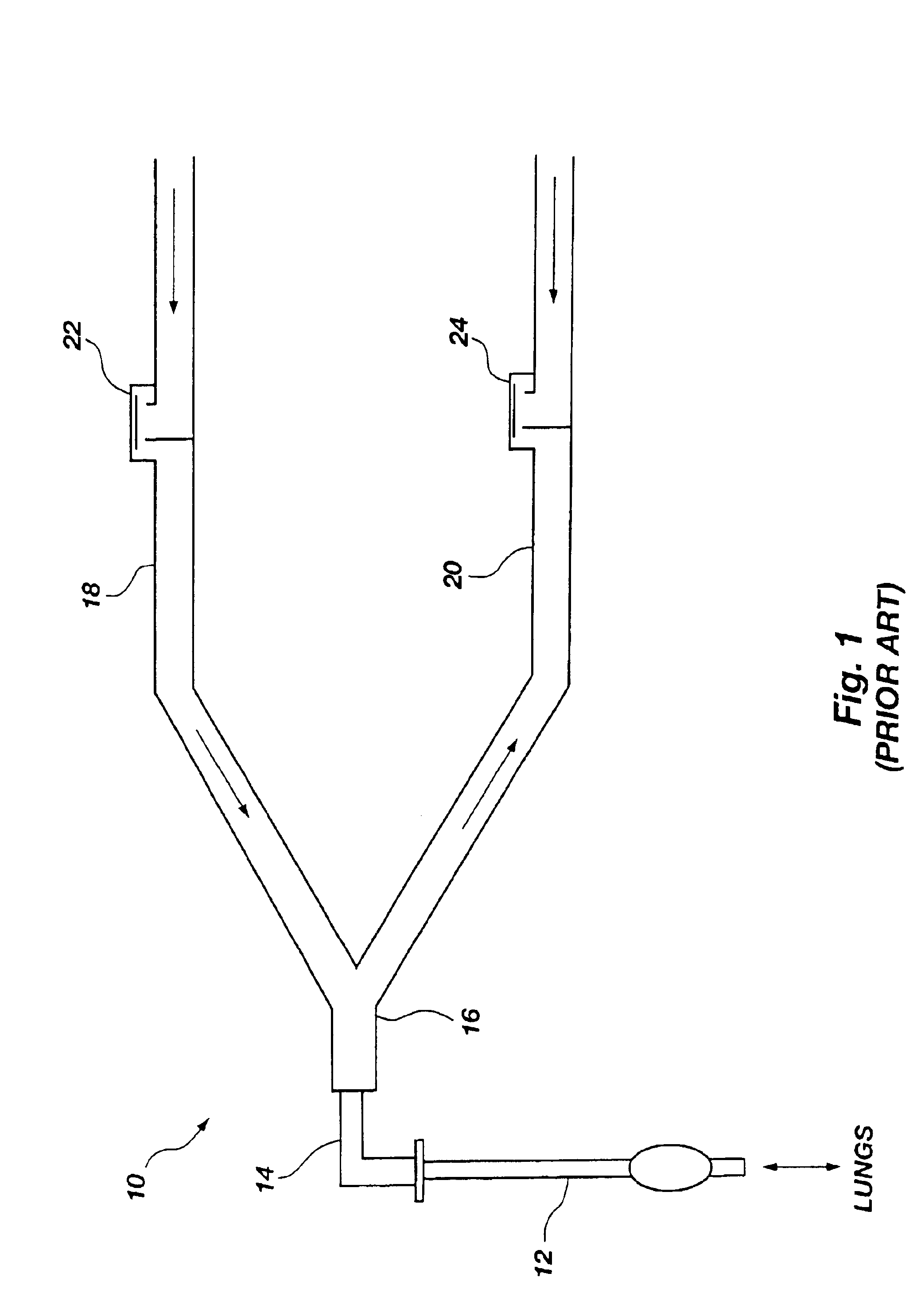
[0033]For comparative purposes, FIG. 1 schematically illustrates a conventional ventilation system which is typically used with patients who require assisted breathing during an illness, a surgical procedure or recovery from a surgical procedure. The conventional ventilator system 10 includes a tubular portion 12 which is inserted into the trachea by intubation procedures. The distal end 14 of the tubular portion 12 is fitted with a Y-piece 16 which interconnects an inspiratory hose 18 and an expiratory hose 20. Both the inspiratory hose 18 and expiratory hose 20 are connected to a ventilator machine (not shown) which delivers air to the inspiratory hose 18. A one-way valve 22 is positioned on the inspiratory hose 18 to prevent exhaled gas from entering the inspiratory hose 18 beyond the valve 22. A similar one-way valve 24 on the expiratory hose 20 limits movement of inspiratory gas into the expiratory hose 20. Exhaled air flows passively into the expiratory hose 20.

[0034]In known ...


PUM


Abstract

Apparatus and methods for non-invasively determining cardiac output using partial re-breathing techniques are disclosed in which the apparatus is constructed with an instantaneously adjustable deadspace for accommodating differences in breathing capacities of various patients. The apparatus is constructed of inexpensive elements, including a single two-way valve which renders the apparatus very simple to use and inexpensive so that the unit may be readily disposable. The method of the invention provides a novel means of estimating cardiac output based on alveolar CO2 values rather than end-tidal CO2 values as previously practiced. A program for calculating cardiac output is also disclosed.

Description

CROSS-REFERENCE TO RELATED APPLICATIONS[0001]This application is a divisional of application Ser. No. 09 / 767,363, filed Jan. 23, 2001, now U.S. Pat. No. 6,648,831, issued Nov. 18, 2003, which is a continuation of application Ser. No. 08 / 770,138, filed Dec. 19, 1996, now U.S. Pat. No. 6,306,098, issued Dec. 23, 2001.BACKGROUND OF THE INVENTION[0002]1. Field of the Invention[0003]This invention relates to non-invasive means of determining cardiac output in patients, and specifically relates to partial re-breathing systems and methods for determining cardiac output in patients.[0004]2. Background of Related Art[0005]It is important in many medical procedures to determine or monitor the cardiac output of a patient. Techniques are known and used in the art which employ the use of catheters inserted at certain arterial points (e.g., femoral artery, jugular vein, etc.) to monitor blood temperature and pressure in order to determine cardiac output of the patient. Although such techniques ca...

Claims


Application Information

Patent Timeline
IPC IPC(8): A61M16/00A61B5/029A61B5/026A61B5/08A61B5/083A61B5/087A61B5/097A61B10/00G01N33/00
CPCA61B5/029A61B5/0836A61M16/0045G01N33/004A61M2016/0036A61M2230/432A61M16/0833Y02A50/20
Inventor ORR, JOSEPH A.KOFOED, SCOTT A.WESTENSKOW, DWAYNEJAFFE, MICHAEL B.
Owner RIC INVESTMENTS LLC
Learn More