In situ heat induced antigen recovery and staining apparatus and method

a heat-induced antigen and staining technology, applied in the field of samples, can solve the problems of high cumbersome and inefficient separation apparatus, no currently available automated or semi-automated staining instruments specifically teach

Inactive Publication Date: 2005-02-15
ANGROS LEE
View PDF30 Cites 100 Cited by
  • Summary
  • Abstract
  • Description
  • Claims
  • Application Information

AI Technical Summary

Benefits of technology

FIG. 3A is a cross-sectional view of the apparatus of FIG. 1 (shown with a reaction compartment having a raised slide support surface) taken through line 3A—3A.

Problems solved by technology

Due to excessive evaporation from the bath, the patent teaches that the solution should not drop below the biological sample on the slide because drying out of the sample is deleterious.
The disclosure of U.S. Pat. No. 5,244,787 is limited to the use of a microwave oven as the source of heating.
This pre-treatment process of heating and removing the slides from the heating device for staining in a separated apparatus is highly cumbersome and inefficient.
As noted herein, no currently available automated or semi-automated staining instruments specifically teach the ability to heat an aqueous or non-aqueous liquid for the unmasking of antigens.
The instruments that do automated or semi-automated staining limit their scope to that task alone, and don't address the task of HIER or antigen retrieval pre-treatments.
If heated to near boiling conditions the liquid would evaporate through all four open sides, immediately causing the biological sample to dry.
300 microliters) is again minimal, and if heated to boiling, would cause the aqueous layer to dry out leaving only the oil layer present thus damaging the biological sample unless more aqueous reagent was applied during the treatment process.
There is no teaching of high temperature heating of a liquid for a substantial amount of time.
Further, even if one would increase the temperature of the slide, the loosely rotating top of the chamber would allow so much evaporative loss that the solution would never reach boiling or near boiling temperatures, nor would it maintain the boiling conditions for 10 minutes or longer.
As a result, none of these systems could hold sufficient liquid on top of a slide (e.g., 4 ml) and are enclosed in a chamber which is properly vented to minimize the energy loss from evaporation to cause sufficient heating to boil the liquid on the slide for the length of time generally required to cause antigen unmasking (e.g., 10-30 minutes).
Of the automated stainers available today, there is not one instrument that has the ability to overcome the inherent problems of heating an aqueous or non-aqueous solution at a sufficient volume without the undesirable effect of evaporative heat loss and subsequent volume decrease of the solution.

Method used


Image

  • In situ heat induced antigen recovery and staining apparatus and method
  • In situ heat induced antigen recovery and staining apparatus and method
  • In situ heat induced antigen recovery and staining apparatus and method

Examples


Embodiment Construction

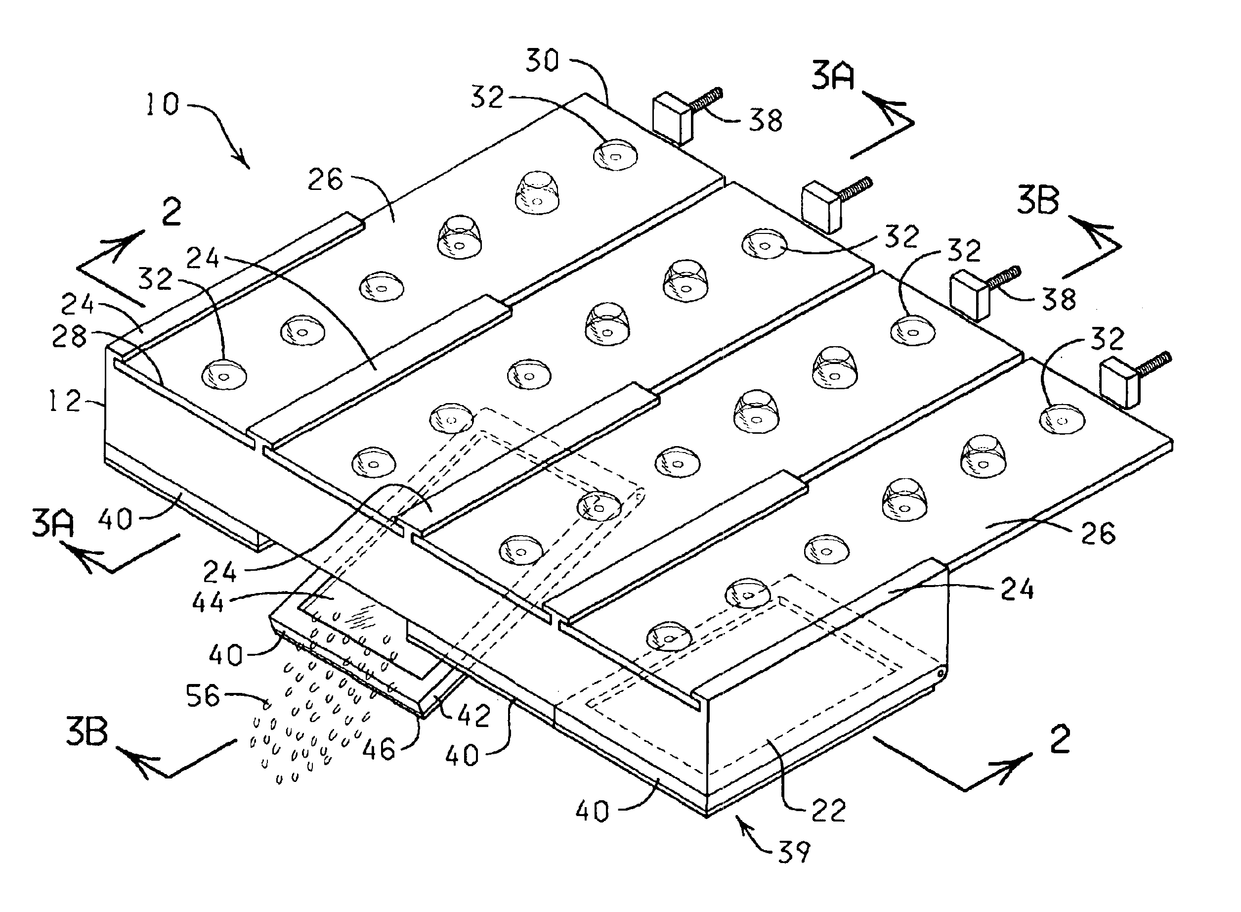
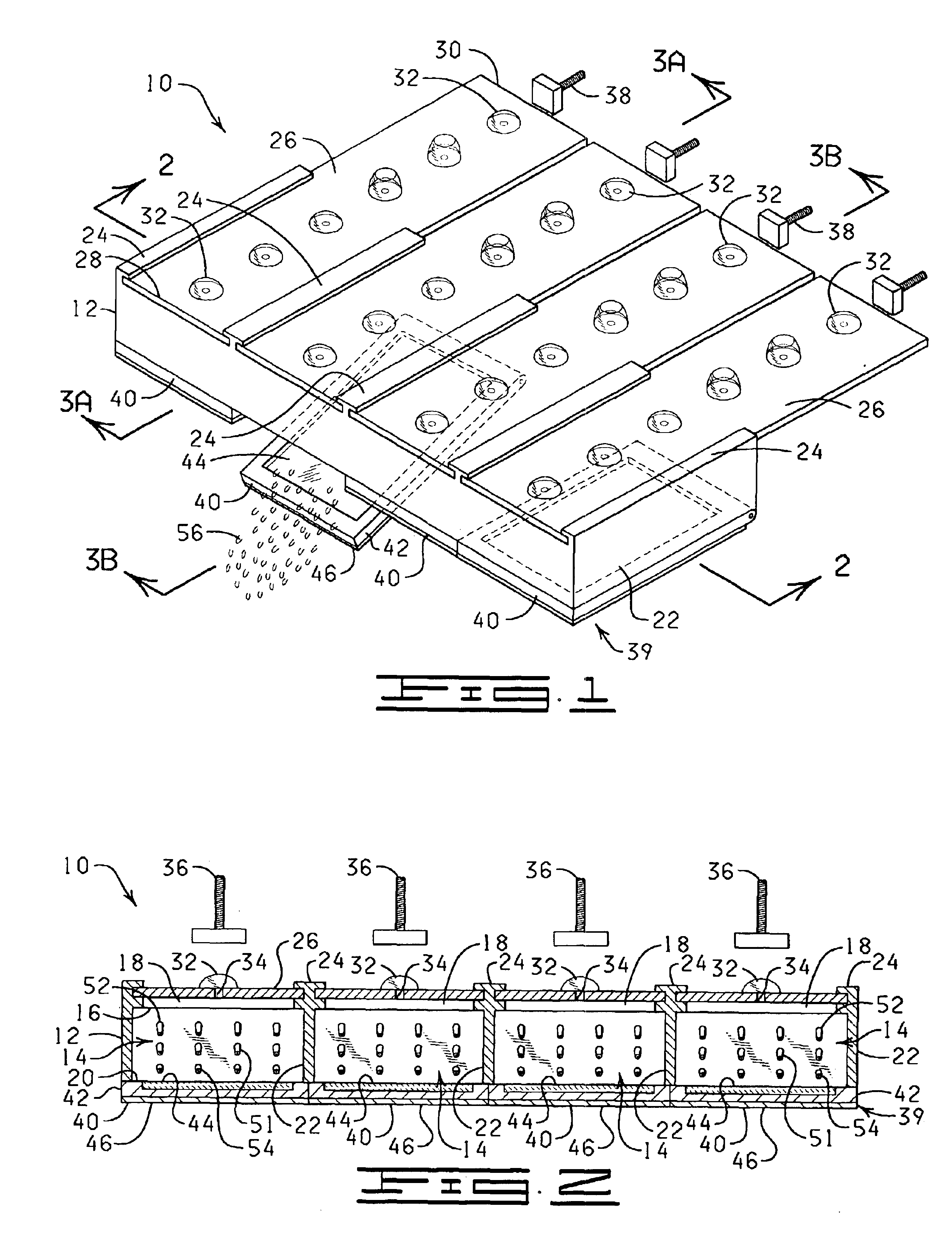
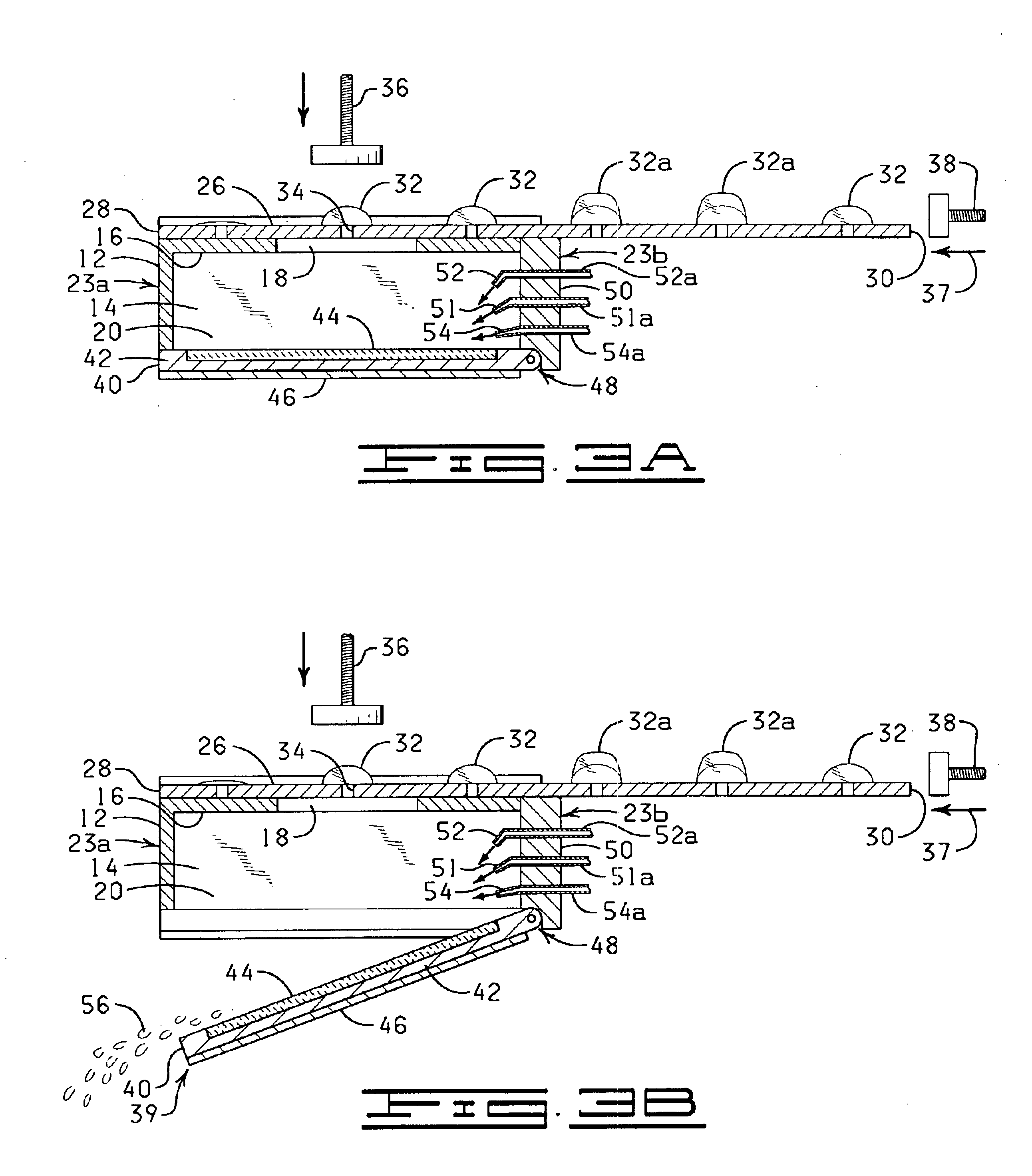
The present invention is directed to an automated method and apparatus for treating biological samples on microscope slides for unmasking (“retrieving” or “recovering”) epitopes or antigens of the biological samples and then staining or otherwise treating the biological samples. The automated apparatus comprises an array of individual reaction compartments, each of which is used to treat a single microscope slide (also referred to herein as a “slide”), wherein each reaction compartment preferably can function and can be controlled independently of the other reaction compartments in the array. Each reaction compartment in the array comprises a support element comprising a surface upon which a microscope slide can be supported and positioned adjacent or inserted into the compartment for treatment with a reagent. The support element further comprises, in a preferred embodiment, a conduction type heating element for heating the microscope slide to a predetermined treatment temperature w...


PUM


Abstract

An automated in situ heat induced antigen recovery and staining method and apparatus for treating a plurality of microscope slides. The process of heat induced antigen recovery and the process of staining the biological sample on the microscope slide are conducted in the same apparatus, wherein the microscope slides do not need to by physically removed from one apparatus to another. Each treatment step occurs within the same reaction compartment. The reaction conditions of each reaction compartment for treating a slide can preferably be controlled independently, including the individualized application of reagents to each slide and the individualized treatment of each slide. The reagents are preferably held in a reagent dispensing strip similar to a “blister pack”.

Description

STATEMENT REGARDING FEDERALLY SPONSORED RESEARCH OR DEVELOPMENTNot ApplicableBACKGROUNDThe present invention is related to the field of treating samples on microscope slides and more specifically to the field of heat induced antigen recovery and staining.Antigen recovery, also known as antigen unmasking, antigen epitope unmasking, antigen retrieval or heat induced epitope recovery (HIER) is a process in which biological samples (e.g., cells, tissues, blood, fluids) are treated under heat with a series of aqueous or non-aqueous reagents and buffers (e.g., citrate, EDTA, and urea) for the purpose of exposing the presence of specific types of antigens or biochemical features in the biological samples. HIER is regarded as a pre-treatment procedure to be performed prior to the beginning of a specific staining protocol to identify cellular components.Biological samples must be preserved after removal from the body. This preservation process, known as fixation, kills and localizes the biol...

Claims


Application Information

Patent Timeline
IPC IPC(8): G01N1/30G01N1/31G01N33/52
CPCG01N33/52G01N33/521G01N1/30G01N33/5306G01N1/44G01N33/53G01N1/312B01L9/52Y10T436/114165Y10T436/2575Y10T436/111666Y10T436/110833Y10T436/112499Y10T436/114998Y10T436/25Y10T436/11Y10T436/119163G01N35/00029G01N35/00584G01N2035/00138G01N2035/00346
Inventor ANGROS, LEE
Owner ANGROS LEE
Why Eureka
Try Eureka