Free floating cryostat sections for use in light and electron microscopy and method

a cryostat and free floating technology, applied in the field of specimen viewing, can solve the problems of inability to view the exact same tissue sample with both devices, no medium has been developed, and the comparison of two separate samples is inherent limitation

Inactive Publication Date: 2003-04-29
GOVERNMENT OF THE U S AS REPRESENTED BY THE SEC OF THE ARMY U S MEDICAL RES & MATERIEL COMMAND THE
View PDF2 Cites 3 Cited by
  • Summary
  • Abstract
  • Description
  • Claims
  • Application Information

AI Technical Summary

Benefits of technology

An object of the invention is to provide a type of specimen for viewing that can bridge the gap between light and electron microscope use.
Another object of the invention is to provide a method that provides ease of progressive processing of the same tissue specimen from light microscopy to electron microscopy.
Another object of the invention is to provide a method that is useful when results of immunolabeling experiments need to be elucidated quickly at histological and ultrastructural levels as required for diagnostic and accelerated investigative strategies.

Problems solved by technology

Unfortunately, there is no known way to view the exact same tissue sample with both devices.
No medium has been developed that allows the scientist to move a single tissue specimen from one device to the other.
However, the comparison of two separate samples has inherent limitations.
In regard to paraffin processing, thick sections subsequently processed for electron microscopy are structurally compromised as a result of the necessary exposure to highly volatile reagents required for adequate paraffin processing (zylenes, toluenes).
These procedures (cryofixation and cryoultramicrotomy) are terribly expensive (cryoultratomes) and extremely labor intensive requiring committed technical dedication by a well-trained technical staff.
The types of unfixed tissues that can be vibratomed successfully are terribly restricted with skin being the least desirable.
As stated, there are particular types of tissues that are especially difficult to work with for purposes of microscope viewing.
However, inherent limitations of conventional frozen sections, including compromised morphology and requirement for glass slide-mounting, usually limit study solely to the light microscopic level.
This process is both expensive, time consuming and requires comparison of two separate and distinct tissue specimens.
A single specimen cannot be subsequently used for viewing with the electron microscope.
This concern becomes a special problem when antigenic epitopes of proteins are vulnerable to damage by chemical fixation rendering antigenic binding sites insensitive to antibody labeling.
Since proteins of the skin basement membrane zone are susceptible to chemical fixation-induced damage, immunoidentification of basement membrane proteins is restricted largely to unfixed cryostat sections mounted onto microscope slides.
This useful, but arduous technique causes loss of valuable material when tissue sections do not separate from the slide cleanly and often yields poor ultrastructural detail (Bretschneider et al., 1981).

Method used


Image

  • Free floating cryostat sections for use in light and electron microscopy and method
  • Free floating cryostat sections for use in light and electron microscopy and method
  • Free floating cryostat sections for use in light and electron microscopy and method

Examples


example 1

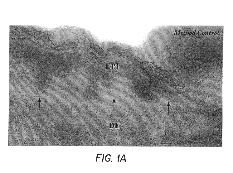
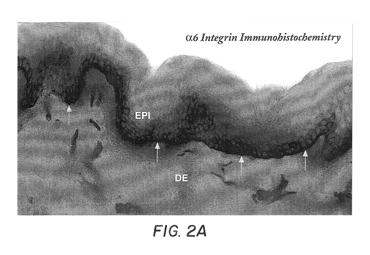
Fresh skin samples frozen in the chlorodifluoromethoane refrigerant Freon 22 were sectioned at 60.mu. and individually placed in wells filled with cryoprotectant solution as formulated by deOlmos and colleagues. DeOlmos, J. S., H. Hardy and L. Heimer: (1978) J. Comp. Neurol. 181: 213-244. Immunohistochemistry was performed using the avidin-biotin-peroxidase complex (ABC) method described by Hsu and coworkers. Hsu, S-M, L. Raine and H. Fanger (1981) J Histo Cyto. 29(4): 577-580. The steps of immunohistochemistry are as follows:

Sections were washed thoroughly in three changes of PBS to remove excess cryoprotectant solution, and sequentially incubated in 5% normal serum, primary antibody, biotinylated-secondary antibody, ABC solution and DAB. Primary antibodies used were .alpha.6 integrin (1:100), laminin (1:100), collagen type IV (1:100) and collagen type VII (1:100). All secondary antibodies were applied at dilutions of 1:200. Sections processed without primary antibody were used as ...


PUM

PropertyMeasurementUnit
thickaaaaaaaaaa
volumeaaaaaaaaaa
thicknessaaaaaaaaaa

Abstract

A method is described of viewing a single tissue sample with a light microscope and an electron microscope. A tissue sample is cryopreserved, cryo sectioned and then floated in a cryoprotectant solution on a surface of a glass slide or in a well and is viewed by light microscopy. The step of floating the tissue sample does not destroy said tissue sample for subsequent electron microscope viewing. The same tissue sample is then prepared by conventional ultrastructural processing for viewing by electron microscopy. The data from the light microscope and electron microscope images of selected sites of the same tissue sample can be compared and analyzed.

Description

This invention relates to the field of histology and immuno-histology using immunoelectron microscopy. More specifically, this invention relates to the field of free-floating cryostat sections for use in light and electron microscopy to bridge the gap between these two viewing mediums.In many instances, a scientist studying a histological sample desires to view a tissue specimen with both a light microscope and also with an electron microscope to obtain information that is unique to each discipline. Unfortunately, there is no known way to view the exact same tissue sample with both devices. No medium has been developed that allows the scientist to move a single tissue specimen from one device to the other.In the past, two separate tissue samples from the same subject were required for viewing with the separate devices. A comparison had to be made to bridge the gap between the results of the light microscope and the electron microscope. However, the comparison of two separate samples...

Claims


Application Information

Patent Timeline
Patent Type & Authority Patents(United States)
IPC IPC(8): G01N1/30
CPCG01N1/30
Inventor PETRALI, JOHN P.KAN, ROBERT KWAIHAMILTON, TRACEY A.OGLESBY-MEGEE, SUSAN B.
Owner GOVERNMENT OF THE U S AS REPRESENTED BY THE SEC OF THE ARMY U S MEDICAL RES & MATERIEL COMMAND THE
Why Eureka
Try Eureka