
Methods for volumetric tissue ablation

a volumetric tissue and ablation technology, applied in the field of volumetric tissue ablation, can solve the problems of difficult to heat the tumors to a lethal temperature, limited number of patients, and uncertain long-term survival benefits, so as to avoid significant heating and other surgical effects, facilitate the penetration of tissue, and reduce the number of patients.

Inactive Publication Date: 2002-10-22
BOARD OF RGT UNIV OF NEBRASKA
View PDF84 Cites 102 Cited by
  • Summary
  • Abstract
  • Description
  • Claims
  • Application Information

AI Technical Summary

Problems solved by technology

Unfortunately, only a limited number of patients are candidates for surgical resection.
While percutaneous probes are being developed, they are currently capable only of treatment of smaller lesions.
While this therapy improves the response to systemic chemotherapy, it has uncertain benefit for long-term survival.
One limitation of hyperthermia is that it is difficult to heat the tumors to a lethally high temperature.
Moreover, tumor cells tend to become thermoresistant if they survive early treatments.
Such small diameters are insufficient for the vast majority of lesions encountered clinically thus requiring multiple fiber placement and prolonged procedure times.
It can therefore be seen that the treatment of primary and metastatic liver tumors and other solid tumors elsewhere in the body, remains problematic.
The percutaneous methods have the virtue of being less invasive, so they can be appropriately used for a larger spectrum of patients, but current percutaneous methods all suffer from a limited ability to ablate a large volume of tissue in a single procedure with a single probe passage.

Method used


Image

  • Methods for volumetric tissue ablation
  • Methods for volumetric tissue ablation
  • Methods for volumetric tissue ablation

Examples


Embodiment Construction

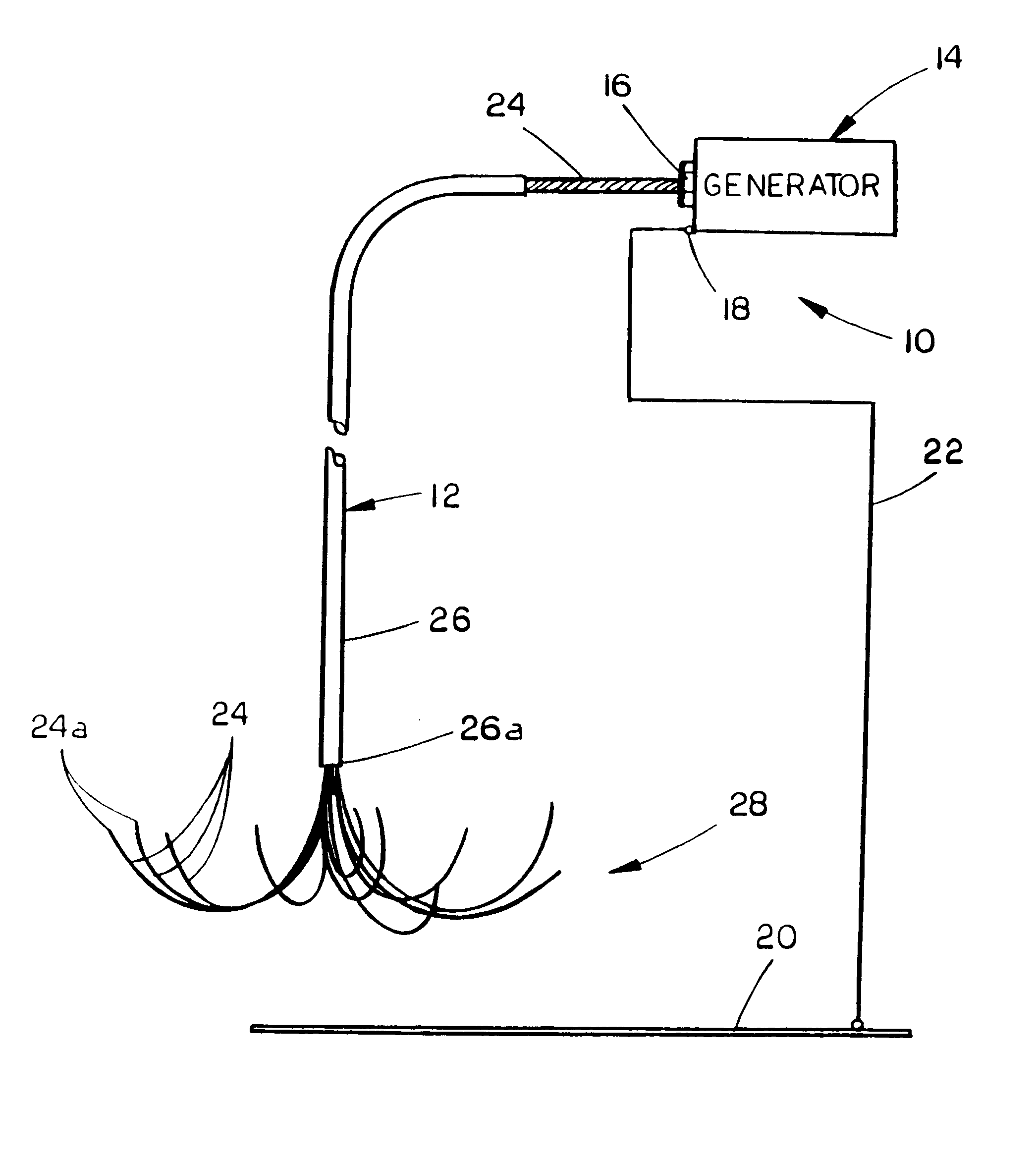
Referring now to the drawings, in which similar or corresponding parts are identified with the same reference numeral, and more particularly to FIG. 1, the volumetric tissue ablation apparatus of the present invention is designated generally at 10 and includes a probe 12 electrically connected to a generator 14.

In experiments with a prototype of the present invention, the inventor utilized a Bovie.RTM. X-10 electrosurgical unit for generator 14, to generate radio frequency current at specific energies, using the probe 12 as the active electrode and placing the tissue sample on a dispersive or ground plate. Thus, generator 14 includes at least an active terminal 16 and a return terminal 18, with a dispersive or ground plate 20 electrically connected by conductor 22 to terminal 18.

Probe 12 is comprised of a plurality of electrically conductive wires 24 which are bundled at a proximal end and connected to terminal 16 to conduct RF current therefrom. Wires 24 are threaded through an ele...


PUM


Abstract

A volumetric tissue ablation apparatus includes a probe having a plurality of wires journaled through a catheter with a proximal end connected to the active terminal of a generator and a distal end projecting from a distal end of the catheter. The probe wire distal ends are arranged in an array with the distal ends located generally radially and uniformly spaced-apart from the catheter distal end. A conductor connected to the return terminal of the generator is located relative to the probe wire array to form a closed electrical circuit through tissue to be ablated. Preferably, the probe wire array includes 10 wires, each formed in an arch from the catheter distal end. The conductor can be either a conventional ground plate upon which the tissue is supported, or a conductor wire extending through the probe and electrically insulated from the probe wires.

Description

The present invention relates generally to radio frequency electrodes for tissue ablation, and more particularly to an improved RF electrode having a spreading array of wires to ablate large volumes of tissue.The liver is a common repository for metastasis from many cancers, including those of the stomach, bowel, pancreas, kidney, and lung. In colorectal cancer the liver is the initial site of spread in more than one-third of patients, and is involved in more than two-thirds at the time of death. While patients with untreated colorectal metastasis to the liver have no five year survival, patients undergoing surgical resection have approximately a 25-30% five year survival. Unfortunately, only a limited number of patients are candidates for surgical resection.Cryosurgery is also used for the treatment of hepatic metastasis. Cryosurgery, which relies on a freeze-thaw process to nonselectively kill cells, has been found equally effective as surgical resection but is more tissue sparing...

Claims


Application Information

Patent Timeline
Patent Type & Authority Patents(United States)
IPC IPC(8): A61B18/18A61B18/14A61N5/02A61N5/04A61B18/12
CPCA61B18/1477A61B18/1485A61B18/1492A61B18/18A61N5/045A61B18/1402A61B2018/00214A61B2018/1425A61B2018/143A61B2018/1432A61B2018/1475A61B18/12
Inventor LEVEEN, ROBERT F.FOX, RANDY
Owner BOARD OF RGT UNIV OF NEBRASKA
Learn More