
Polysaccharide sponges for cell culture and transplantation

a cell culture and polysaccharide technology, applied in the field of polysaccharide sponges for cell culture and transplantation, can solve the problems of inability to prepare functional organ equivalents made up of multilayer cell aggregates, and inability to carry a very thin layer of cells

Inactive Publication Date: 2002-07-30
BEN GURION UNIVERSITY OF THE NEGEV
View PDF10 Cites 86 Cited by
  • Summary
  • Abstract
  • Description
  • Claims
  • Application Information

AI Technical Summary

Benefits of technology

It is another aim of the invention to provide polysaccharide matrices which are biodegradable but which degrade only slowly in vivo and thereby permit the cells carried thereby to become established and to form their own tissue matrix at the site of transplant to the point where they no longer require the polysaccharide matrix; or when the matrix is used alone as an implant, it is to be stable for sufficient time for the surrounding tissue to invade it and proliferate thereon to the point where the invasive cells have become established and have formed their own tissue matrix. thereby replacing the originally deficient tissue; or when the matrix is used alone as a first stage of an implant, it is to be stable for sufficient time to allow for vascularization from the surrounding tissue into the implant to occur by invasion thereof by blood vessels, and to allow for the second stage in which cells of choice can be injected into the vascularized matrix and subsequently proliferate thereon to the point where these cells have become established, have formed their own tissue matrix, and thereby have replaced, at least functionally, the originally deficient or damaged tissue.
Yet another aim of the present invention is to provide such polysaccharide sponges of a highly porous nature that may be readily invaded by blood vessels and / or cells and subsequently which may adequately support the growth, proliferation and biological function of such implanted or transplanted cells, both in vitro and afterwards in vivo when used in implants or transplants, such polysaccharide sponges being of a morphology such that their internal volume is optimally utilized, and further providing such sponges which do not require any form of external coating or the like for the purposes of implantation or transplantation, and hence the sponges will not be simply vehicles for the encapsulation of cells but rather would serve as a matrix or scaffold for the cells which they carry and permit free transport of oxygen and nutrients into the cells and in vivo provide for vascularization of the cells for the purposes of nutrient supply and subsequent tissue regeneration from these transplanted cells. Hence, the polysaccharide sponges of the present invention are designed not to serve as merely encapsulation devices for cells but rather as effective matrices, supports or scaffolds for optimal use in implantations and transplantations for tissue repair, as noted above.

Problems solved by technology

However, in most of the porous matrices described to date, the ones that have been successfully prepared and used in implants or transplants have been limited to those which carry a very thin layer of cells, being principally those which serve as skin substitutes or replacements (see, for example, Yannas, 1990).
However, when such matrices are to be used with cells such as, for example, hepatocytes, which grow in aggregates of cells and with a thickness greater than the thickness which these earlier matrices are designed to support, a serious problem arises as regards the adequate diffusion of oxygen and nutrients to the inner cells within the matrix, with the result that these inner or lower layers of cells usually die.
Thus, these earlier matrices may be useful for preparing skin equivalents, but are much less useful for preparing functional organ equivalents made up of multilayer cell aggregates, both in vitro and with subsequent transplantation use in vivo.
The collagen-based matrices have several disadvantages, including: they degrade at relatively rapid rate; many disappearing as early as 4 weeks postimplantation (see Olde Damink et al., 1995; Ben-Yishay et al., 1995).
Although the rate of degradation of the collagen matrix may be reduced by cross-linking with glutaraldehyde, the resulting cross-linked matrices, however, exhibited immunogenicity, calcification, and fibrous scarring when implanted for long periods (see Timple et al., 1980).
Furthermore, collagen matrices are also not suitable for prolonged in vitro cultivation of cells, due to a significant contraction of the collagen scaffold, which occurs after approximately one week of incubation, rendering this collagen scaffold less amenable to surgical handling when intended for use as a transplantation matrix (Ben-Yishay, 1995).
Other synthetic biodegradable foams based on poly(D, L-Lactic-co-glycolic acid) have been developed as scaffolds for tissue engineering, as noted above, but because these polymers are hydrophobic, when a cell suspension or culture media is placed on these foams or injected into their interior, the majority of their pores remain empty, resulting in the underutilization of the volume of these foams.
In addition, studies have also shown that the degradation of these biodegradable foams results in the significant accumulation of acid products which significantly decreases the internal pH within the foam to less than pH 3.0 (see Park Lu and Crotts, 1995), which acidity is very harmful to the growing cells.
This coating, however, greatly reduces the microcapsule permeability towards nutrients, which leads to the death of the encapsulated cell.

Method used


Image

  • Polysaccharide sponges for cell culture and transplantation
  • Polysaccharide sponges for cell culture and transplantation

Examples


example 2

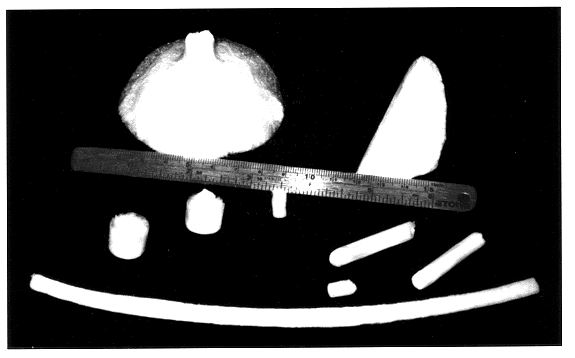
Morphology of Alginate Sponges

The morphology of the alginate sponges was investigated by scanning electron microscopy (SEM, JEOL JSM-35CF). Samples of the various alginate sponges were mounted on aluminum stubs and coated with an ultrathin (100 A) layer of gold in a Polaron E 5100 coating apparatus. The parameters of the sponge microstructure were investigated by geometrical measurements on the SEM-micrographs. The pore length, width and wall thickness (i.e., the average distance between neighboring pores), were measured by a stereo microscope (Bausch & Lomb) equipped with an optical micrometer. The effective size of the pores was calculated, using the equation:

d=l.multidot.h

where 1, h--are the average length and width of the pores, respectively. Wall thickness measurements were performed as this parameter characterizes the distance between the pores, and hence the microstructure of the sponges.

Initially, the shape of the sponges was determined by the dish in which the processing of...

example 3

Mechanical Properties and the Pore Compressibility of the Alginate Sponges

The mechanical properties of alginate sponges were determined at 22.degree. C. (room temperature) by compressing the sample sponges at a constant deformation rate of 2 mm / min, using a standard test apparatus. This apparatus is illustrated schematically in FIG. 5 and comprises a load cell, a deformation cell, and indentor, and a table. The sample sponge is placed between the indentor and the table and is subjected to a load which is measured by the measuring apparatus to which the table is connected at its other end ("translation"). With such an apparatus, the load and deformation were monitored with high accuracy down to loads of 1 g and at deformations of less than 0.05 mm. In this apparatus, the diameter of the indentor (7 mm) was usually smaller than that of the sample (sample sponges being usually about 15 mm), or greater when subjected to compression, and hence the influence of sample diameter variations ...

example 4

Degradation of Alginate Sponges, in Vitro:

In order to determine the degradation of the alginate sponges of the present invention over a period of time, the sponges were incubated in a complete culture medium and samples were withdrawn at different time intervals for the purposes of assaying the concentration of soluble alginate by way of a calorimetric assay. This assay was based on the meta-chromatic change induced by alginate binding to the dye 1,9-dimethyl methylene blue (DMMB) (see Halle et al., 1993).

In this assay, the alginate sponges were cultured in a standard culture medium being the standard complete medium usually used for culturing cells such as fibroblasts and hepatocytes, at 37.degree. C. The cultured sponges were of two kinds, the one kind having been seeded with cells and the other being without any cells. Results of this assay show that alginate sponges, with or without seeded cells, cultured in culture medium at 37.degree. C. maintained their physical stability for...


PUM

PropertyMeasurementUnit
pore sizeaaaaaaaaaa
thicknessaaaaaaaaaa
viscosityaaaaaaaaaa

Abstract

A polysaccharide sponge characterized by having: (i) an average pore size in the range between about 10 mum to about 300 mum; (ii) an average distance between the pores being the wall thickness of the pores in the range between about 5 mum to about 270 mum; and (iii) an E-modulus of elasticity being a measure of the rigidity of the sponge in the range of about 50 kPa to about 500 kPa.

Description

The present invention concerns new bioresorbable polysaccharide sponges, a method for their preparation and uses thereof for the cultivation of mammalian cells in vitro, as well as the use thereof as matrices, supports or scaffolds for implantation into a patient to replace damaged or removed tissue, the polysaccharide sponge implant serving as a substrate, matrix or scaffold for surrounding host tissue to invade it, proliferate thereon and eventually form an active part of the tissue or organ in which the implant was made, or the implant serving as an initial substrate for vascularization from the surrounding host tissue, and once vascularized, cells of choice grown it vitro or obtained from the host may be injected into the vascularized implant to enable a rapid acclimatization and proliferation of the cells which will subsequently form an active replacement for the organ or tissue that was damaged or removed. The polysaccharide sponge can also serve as a substrate, matrix or scaf...

Claims


Application Information

Patent Timeline
Patent Type & Authority Patents(United States)
IPC IPC(8): A61F2/10A61K9/12A61K9/00A61L27/60A61L27/38A61L27/00A61L27/20C08J9/00C08J9/26A61F2/00A61F2/02C12N1/12C08J9/28C12N5/04C12N5/07C12N5/071
CPCA61F2/105A61K9/0024A61K9/122A61L27/20A61L27/3804A61L27/60C08J9/26C08L5/02A61F2210/0004A61F2250/0067C08J2305/02A61L27/56
Inventor SHAPIRO, LILIAGLICKLIS, RACHELCOHEN, SMADAR
Owner BEN GURION UNIVERSITY OF THE NEGEV
Learn More