
Two-way mirror display for dental treatment system

a technology of dental treatment system and mirror display, which is applied in the direction of patient positioning for diagnostics, image enhancement, instruments, etc., can solve the problems of degraded image quality, blurred or otherwise, difficult completion of x-ray image acquisition procedures, etc., and achieves less positioning errors, faster treatment planning, and improved image quality.

Active Publication Date: 2022-05-05
PALODEX GROUP
View PDF1 Cites 1 Cited by
  • Summary
  • Abstract
  • Description
  • Claims
  • Application Information

AI Technical Summary

Benefits of technology

The patent describes a system that helps dental professionals position patients properly for x-ray image acquisition, resulting in better quality images and reduced positioning errors. This system can also help inexperienced operators and increase patient satisfaction by allowing them to position themselves. Additionally, it can provide patient-specific workflows to accommodate different patient needs. Overall, this system improves the efficiency and accuracy of dental x-ray image acquisition.

Problems solved by technology

X-ray image acquisition procedures can be difficult to complete.
Patient movement may result in degraded image quality.
Shaking, rocking, talking, or other patient movement may result in blurred or otherwise low-quality images.
In some cases, a patient may become fearful, agitated, or tired.
In some cases, the patient may feel isolated and feel that he or she is receiving inadequate feedback from a dentist or dental technician regarding the image of acquisition or the position of the patient.

Method used


Image

  • Two-way mirror display for dental treatment system
  • Two-way mirror display for dental treatment system
  • Two-way mirror display for dental treatment system

Examples


example 2

[0059] the dental x-ray image acquisition system of example 1, wherein the two-way mirror is also positioned between the camera and the patient.

example 3

[0060] the dental x-ray image acquisition system any of examples 1-2, wherein the camera is a component of the display.

[0061]Example 4: the dental x-ray image acquisition system of example 1, wherein the camera is a three-dimensional camera.

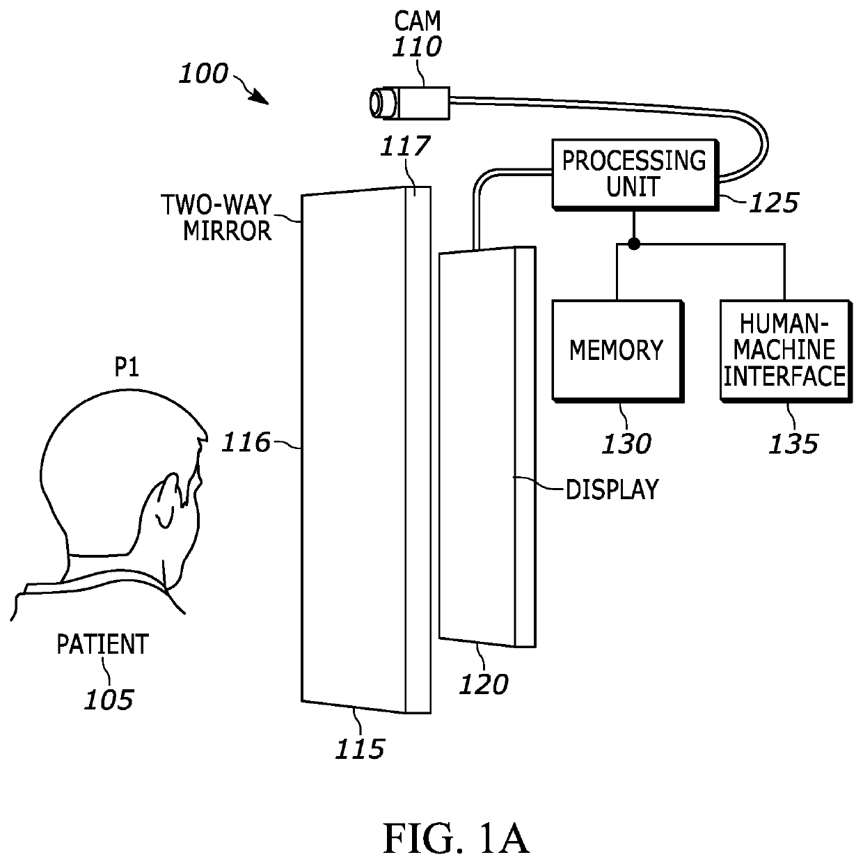
[0062]Example 5: a dental x-ray image acquisition system, the system comprising at least one camera configured to capture an image of a patient, a display, a two-way mirror positioned in between a patient location and the display, and an electronic processor coupled to the camera and the display, the electronic processor configured to select, based upon a user input, an operating mode for the display and, based upon the selected operating mode, display at least one image on the display.

[0063]Example 6: the system of example 5, wherein the operating mode is an operating mode selected from a group of operating modes consisting of a mirror mode, an augmented mode, a light mode, and a monitor mode.

[0064]Example 7: the system of example 6, wherein the...

example 13

[0070] the system of example 12, wherein the movement guide illustrates alignment of the head of the patient with respect to two or more anatomical planes.

[0071]Example 14: the system of any of examples 12-13, wherein a graphical element of the movement guide is changed when the head of the patient is aligned with the at least one anatomical plane.

[0072]Example 15: a method for positioning a patient for x-ray image acquisition, the method comprising receiving, with an electronic processor, image data from at least one camera; identifying, with the electronic processor, at least one facial feature of the patient in the image data; determining, with the electronic processor, if a face of the patient is aligned with at least one anatomical plane based upon the at least one facial feature; and displaying, with the electronic processor, at least one movement guide on a display based upon the determined alignment of the face of the patient.

[0073]Example 16: the method of example 15, where...


PUM


Abstract

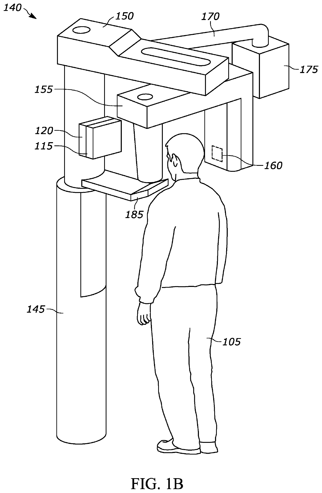
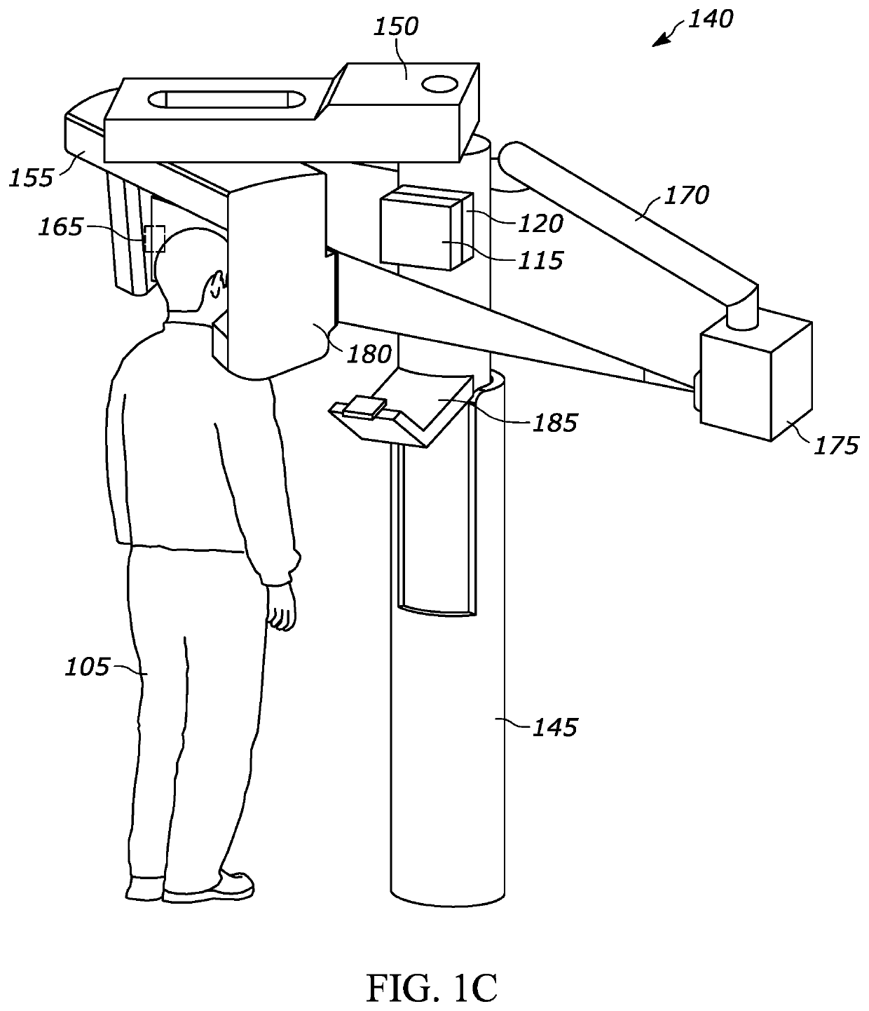
Systems and methods utilizing a two-way mirror display for patient self-positioning for dental x-ray image acquisition. The system includes a camera configured to capture an image of a patient, a display, a two-way mirror positioned between a patient location and the display, and an electronic processor. The electronic processor is configured to select, based upon a user input, an operating mode for the display; and based upon the selected operating mode, displaying at least one image on the display. The method includes receiving image data from a camera, identifying at least one facial feature of the patient in the image data, determining if a face of the patient is aligned with at least one anatomical plane based upon the at least one facial feature, and displaying at least one movement guide on a display based upon the determined alignment of the face of the patient.

Description

BACKGROUND OF THE INVENTION[0001]Many dental procedures involve a patient's teeth being imaged by a camera or other image acquisition device, for example, an x-ray or optical scanner or camera. The imaging results in the generation of images. Depending on the type of image acquisition device used and the procedure performed, patients may be standing, sitting, or lying down on a treatment table during x-ray image acquisition.[0002]X-ray image acquisition procedures can be difficult to complete. In many cases, precise and still positioning of the patient is needed to capture high-quality and accurate images of an anatomical region of interest. Patient movement can result in capturing images of areas other than a desired anatomical region of interest. Patient movement may result in degraded image quality. Shaking, rocking, talking, or other patient movement may result in blurred or otherwise low-quality images. In some cases, a patient may become fearful, agitated, or tired. A patient ...

Claims


Application Information

Patent Timeline
Patent Type & Authority Applications(United States)
IPC IPC(8): G06T7/00G06T19/00G06T7/33A61B6/14A61B6/00A61B6/04A61B6/51
CPCG06T7/0012G06T19/006G06T7/33A61B6/14A61B6/461A61B6/04G06T2210/41G06T2207/10028G06T2207/30201G06T2207/10116G06T2207/30036G06T2200/24A61B6/467A61B6/032A61B6/462A61B6/08A61B6/51A61B2090/365G06T7/70A61B6/488
Inventor POLLARI, MIKAALHONNORO, TUOMASINKILÄINEN, VILLERANTALA, MAARIAHARHANEN, LAURISAVOLAINEN, JORMAMETSO, ARI
Owner PALODEX GROUP
Learn More