Method for preparing biological tissue for surgical implantation

Pending Publication Date: 2022-03-10
PF PROD FEATURES GMBH
View PDF0 Cites 2 Cited by
  • Summary
  • Abstract
  • Description
  • Claims
  • Application Information

AI Technical Summary

Benefits of technology

The present invention provides a new method for preparing biological tissue for sterile dry storage, allowing for surgical implantations without the need for a liquid preservation solution. This method also reduces calcification, prevents biofilm adherence over pericardium, and improves the mechanical properties of bioprosthesis comprising the biological tissue.

Problems solved by technology

However, biological tissues obtained from the abattoir, in particular porcine and bovine cadavers, begin to degrade immediately.
However, there are certain disadvantageous associated with the above mentioned tissue preparation methods.
For example, upon implantation a biological tissue, especially aldehyde fixed tissue, is susceptible to the formation of degenerative calcific deposits.
Calcification, in particular pathologic calcification, of soft biological tissues due to deposition of calcium phosphate mineral salts in an implanted tissue is undesirable and the deposition of the calcific deposits can have severe consequences on device performance.
Calcification of implants can lead to stiffening, structural instability and ultimately to device failure.
However, a large amount of bioprosthetic heart valves fail due to pathologic calcification of the valve causing tissue stiffening and tearing.

Method used


Image

  • Method for preparing biological tissue for surgical implantation
  • Method for preparing biological tissue for surgical implantation
  • Method for preparing biological tissue for surgical implantation

Examples


example 1-1

[0094]40 membranes with 1×1 cm of a biological tissue selected from bovine pericardium “sample 180702-1” (P+F Brasil, EDQM certified) has been at first removed from 0.625% glutaraldehyde solution (P+F GmbH / Biocollagen) and after that soaked in cold 0.9% saline solution (JP Pharma) for 3 minutes. The soaked tissue has then been immersed in Hydrogen Peroxide 0.5% per volume (Sigma-Aldrich) at 18° C. for 60 minutes. As a next step, the tissue was contacted with cold (10° C.) PBS pH 7.4 and 0.5% by weight EDTA for 3 minutes (Sigma-Aldrich). After that the tissue was immersed in 99% per volume ethanol (Sigma-Aldrich) at 22° C. temperature for 60 seconds with intense stirring, and then immersed in a mixture of glycerol / ethanol (50 / 50) and 0.5% by weight EDTA (Sigma-Aldrich) at 22° C. temperature for 60 minutes with slow stirring. Further, the tissue was immersed in glycerol 99% (Sigma-Aldrich) at 22° C. temperature for 120 minutes with slow stirring. Next, the tissue was soaked with absol...

example 1-2

[0095]40 membranes with 1×1 cm of a biological tissue selected from bovine pericardium “sample 180702-2” has been prepared according to procedure described in Example 1-1.

example 1-4

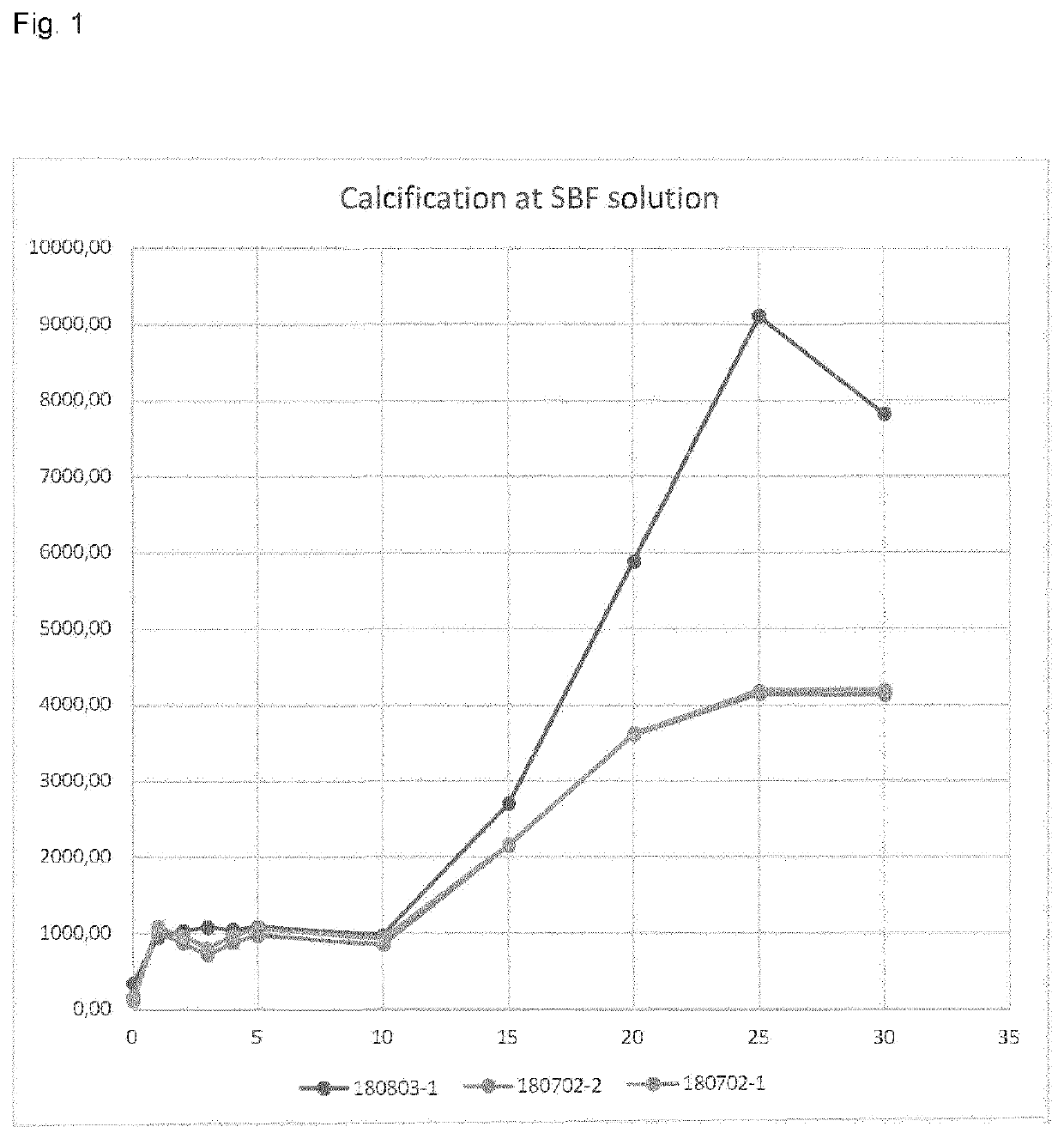
[0097]Bovine pericardium membranes from Examples 1-1 to 1-3 (number of samples: 180702-2 and 180702-1 are according to the present invention, and 180803-1 is a comparative example) were soaked in saline solution for 2 minutes and then immersed in a Simulated Body Fluid (SBF), which is a solution with ionic concentration similar to that of human blood plasma such as demonstrated in Table 3, maintained under the same physiological conditions of pH and temperature (pH 7.4 and temperature of 36.5° C.). The immersion time varied between 1 and 30 days.

[0098]The samples were digested in a closed flask conductive heating system called CHDS. The calibration curve was prepared from the Specsol® 1000 mg. L−1 standard solution to contain 0.0-20 mg. L−1 Ca.

[0099]The determination of Ca presented in Table 1 and FIG. 1 was performed on a ContrAA 300 High Resolution Continuous Source Atomic Absorption Spectrometer (HR-CS FAAS) (Analytic Jena, Jena, Germany), equipped with a short arc lamp of Xe as ...


PUM


Abstract

The present invention relates to a method for treating biological tissue and a biological tissue obtained by the treatment method, and specifically to a method for treating biological tissue so as to suppress the calcification, risk of biofilm adherent over pericardium and strength reduction of the tissue due to treatment. The invention is also directed to bioprosthesis and transcatheter heart valves containing the biological tissue.

Description

CROSS REFERENCE TO RELATED APPLICATIONS[0001]This application is a national stage application under 35 U.S.C. 371 of PCT Application No. PCT / EP2020 / 053001 having an international filing date of 6 Feb. 2020, which PCT application claimed the benefit of European Patent Application No. 19155986.3 filed 7 Feb. 2019, the entire disclosure of each of which are hereby incorporated herein by reference.FIELD OF THE INVENTION[0002]The present invention is directed at a method for treating biological tissue and a biological tissue obtained by the treatment method, and specifically at a method for treating biological tissue so as to suppress the calcification, risk of biofilm adherent over pericardium and strength reduction of the tissue due to treatment. The invention is also directed at bioprosthesis and transcatheter heart valves containing the biological tissue.BACKGROUND OF THE INVENTION[0003]Biological tissues are widely used to make prosthetic replacements for heart valves and blood vess...

Claims


Application Information

Patent Timeline
IPC IPC(8): A61L27/36A61L2/20A61L2/18
CPCA61L27/3625A61L27/3687A61L2/206A61L2400/02A61L2430/20A61L2430/40A61L2/186A61L27/50
Inventor KISS, KATHARINAAGRELI, GUILHERME
Owner PF PROD FEATURES GMBH
Why Eureka
Try Eureka