
Devices and methods for minimally invasive surgical procedures

a surgical procedure and minimally invasive technology, applied in the field of minimally invasive surgical procedures, can solve the problems of increasing the risk of surgical site infections, attempting to overcome cumbersome challenges, and the utilization of additional abdominal wall incisions in the mainstream laparoscopic and robotic techniques, and achieve the effect of increasing the openings

Pending Publication Date: 2021-12-23
NICE SURGICAL SOLUTIONS PTE LTD
View PDF0 Cites 1 Cited by
  • Summary
  • Abstract
  • Description
  • Claims
  • Application Information

AI Technical Summary

Benefits of technology

This patent describes a method for extracting a specimen through the rectum using a cylindrical device that can expand to fit the rectum. The device has a drawstring and a sheath to help with insertion. The technical effect is a safer and more effective way to extract specimens from the rectum.

Problems solved by technology

In cases in which a diseased segment of the alimentary track needs to be removed, one major drawback of the current mainstream laparoscopic and robotic techniques is the utilization of an additional abdominal wall incision.
The overwhelming majority of current practices do not utilize natural-orifice assisted methods to remove the specimen, nor do they attempt to overcome the cumbersome challenges of achieving a safe and successful intracorporeal anastomosis (ICA).
There are many disadvantages to bringing a portion of the bowel outside the body cavity, including the possibility of trauma or traction injury to the bowel wall or associated blood vessels, inability for the end of the bowel to reach the abdominal wall in certain anatomical situations, and potential for contamination of the incision with bowel contents, thereby increasing the risk of surgical site infections.
The placement and utilization of an additional trans-abdominal incision also carries numerous disadvantages and significantly adds to the invasiveness of the procedure.
For example, such an incision may result in interruption of the flow of the procedure, as the introduction of an incision results in loss of pneumoperitoneum and therefore the minimally invasive platform is paused.
There is also potential for contamination of the abdominal wall incision, traction injury of the bowel, inadequate resection of the specimen, additional procedure time, increased post-operative pain, increased need for opioid use, and increased morbidity rates.

Method used


Image

  • Devices and methods for minimally invasive surgical procedures
  • Devices and methods for minimally invasive surgical procedures
  • Devices and methods for minimally invasive surgical procedures

Examples


example 1

rifice Intracorporeal Anastomosis with Transrectal Extraction

[0130]The following methods of bowel division and natural orifice intracorporeal anastomosis with transrectal extraction are performed without the use of an abdominal wall incision (only port incisions are used), in a stepwise approach designed for a robotic platform. The following steps are performed once the diseased portion of the bowel has been mobilized and the mesentery of this portion has been divided.

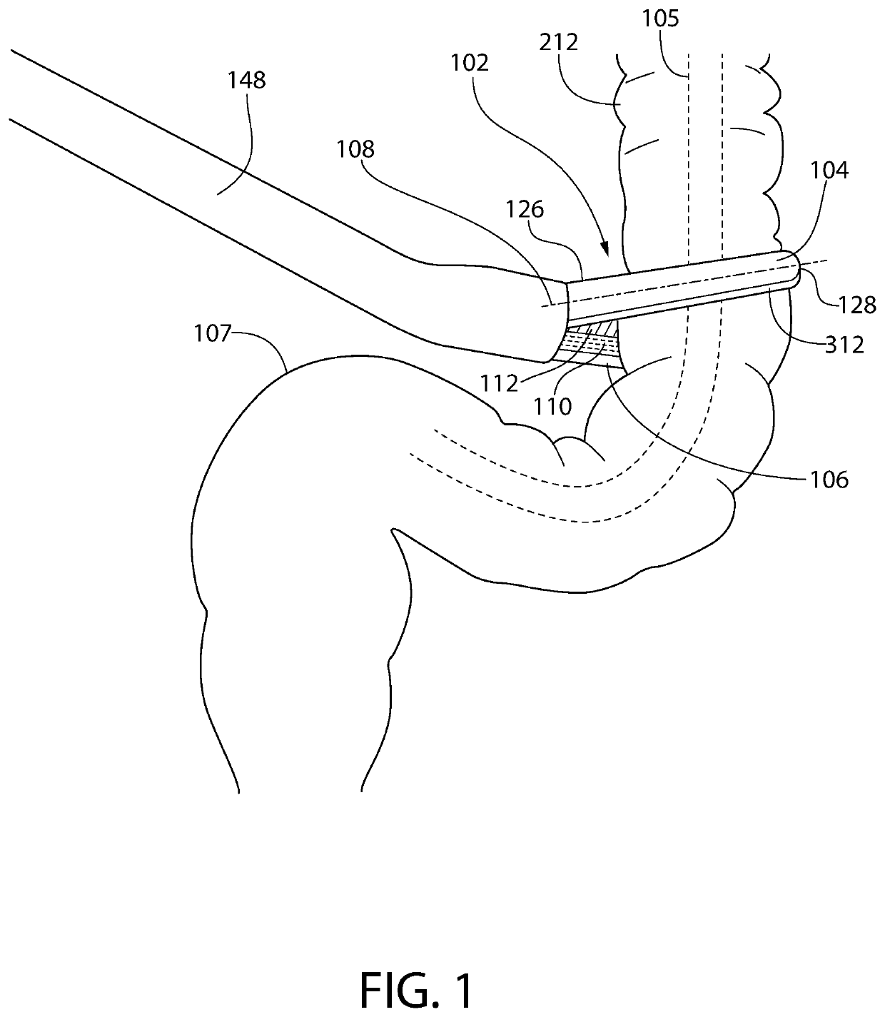
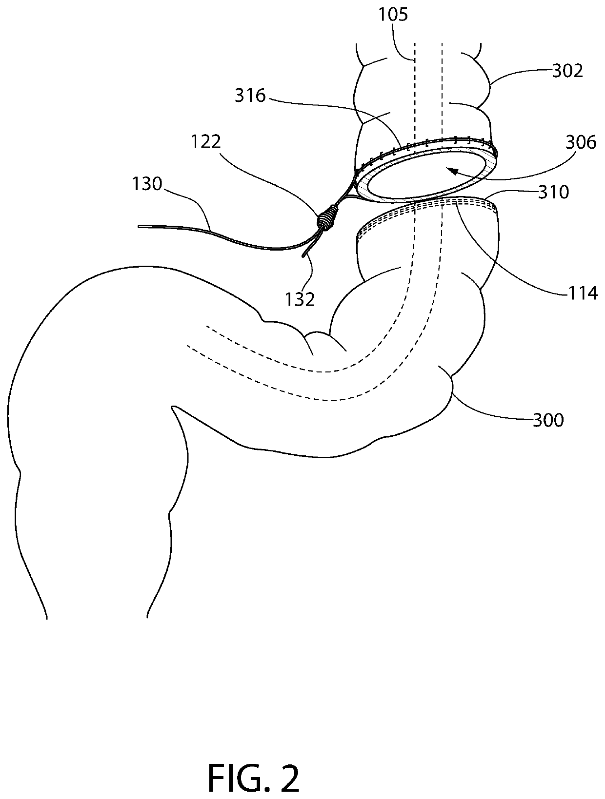
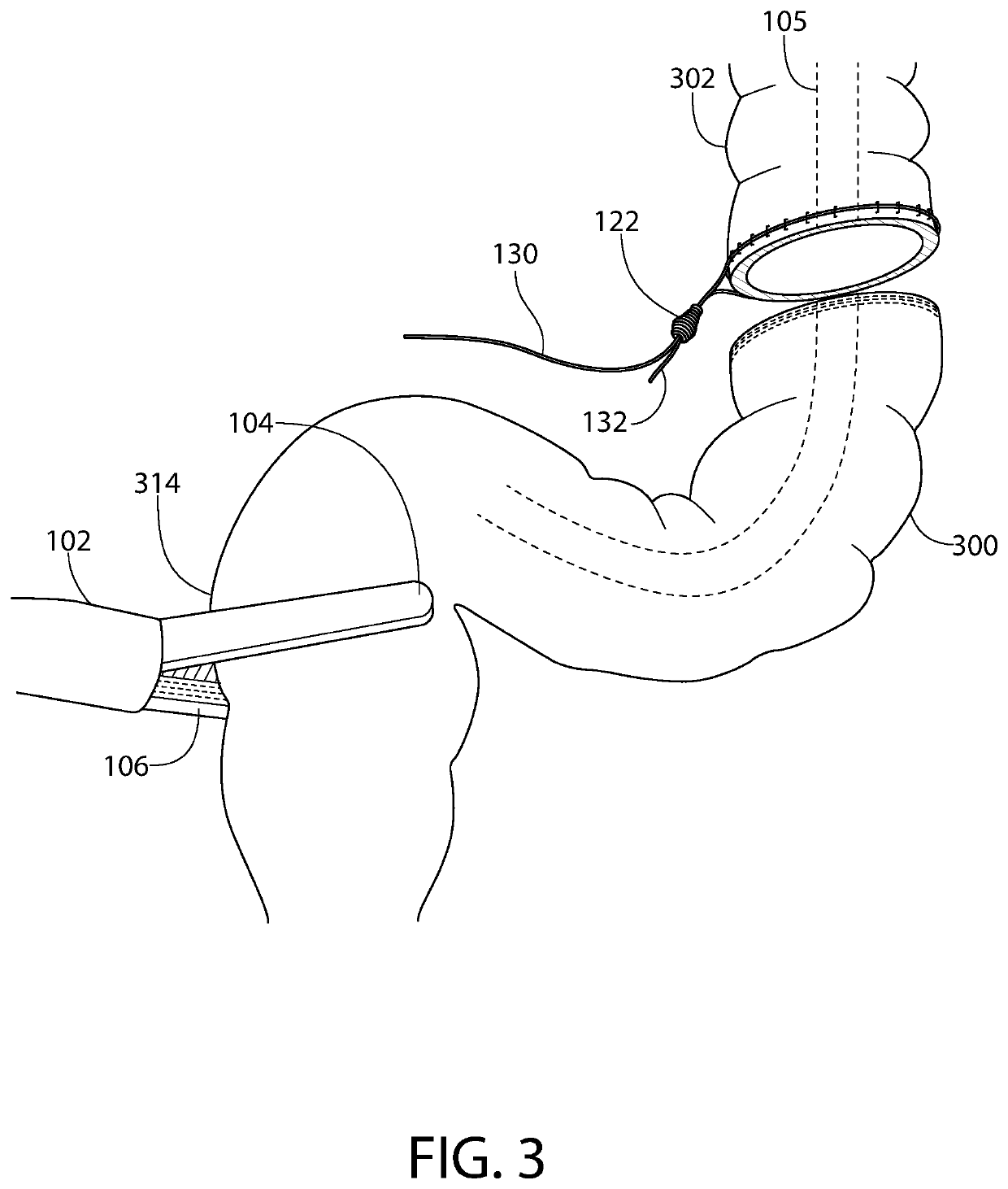
[0131]Step 1: The surgical device 102 (having a suturing mechanism 172 (FIG. 22), cutting mechanism 176 (FIGS. 22 and 26) and stapling mechanism 174 (FIG. 26) as described herein) is placed across the bowel wall at the proposed proximal margin of resection 312 (FIG. 1) and deploys a closed linear staple line across the side of the specimen 300 and attaches a pursestring suture with a slip-knot 122 to the proximal bowel portion 302 (FIG. 2). As an alternative, the bowel is divided by any known method and the suture clip...


PUM


Abstract

A suture clip applier for applying a suture around a tubular tissue adjacent to an open lumen. The suture clip applier includes an elongated body having a tissue clinching mechanism at one end of the body. The tissue clinching mechanism comprises two arms that are movable between open and clinched positions. The suture clip applier includes a plurality of deformable suture clips mounted along a longitudinal length of the elongated body, wherein each suture clip is configured to be detachably positioned within an interior of the two arms and to be deformed between open and clinched positions in response to the two arms being moved between the open and clinched positions. The suture clip applier further includes a length of suture comprising a pre-formed adjustable fastening and a loop extending from the pre-formed adjustable fastening, the loop extending between the two arms.

Description

CROSS-REFERENCE TO RELATED APPLICATIONS[0001]This application claims the benefit of U.S. Provisional Patent Application No. 62 / 747,809 filed Oct. 19, 2018 entitled “Devices and Methods for Minimally Invasive Surgical Procedures”, which is incorporated by reference herein in its entirety.FIELD OF THE INVENTION[0002]The present application relates to minimally invasive surgical methods, and devices for performing those methods. In particular, embodiments of the present invention relate to devices and methods for performing resection of a diseased segment of a colon and / or rectum, and extraction of the diseased segment through the rectum.BACKGROUND OF THE INVENTION[0003]Surgery in which a diseased portion of the intestine requires resection is common and is performed for a multitude of diseases including colorectal cancer, polyps, diverticulitis, Crohn's Disease, ulcerative colitis, rectal prolapse, endometriosis and others. The purpose of such a procedure is to remove the diseased por...

Claims


Application Information

Patent Timeline
IPC IPC(8): A61B17/04A61B17/11A61B17/115A61B17/29A61B17/10
CPCA61B17/0487A61B17/1114A61B17/1155A61B2017/07285A61B17/105A61B2017/0488A61B17/29A61B17/122A61B17/07207A61B2017/1142A61B2017/0464A61B2017/0409A61B17/12013A61B17/0401A61B2017/07271A61B2017/07228A61B17/1285A61B2017/1132
Inventor HAAS, ERIC
Owner NICE SURGICAL SOLUTIONS PTE LTD
Learn More