
Specific murine and humanized monoclonal antibodies detecting pathology associated secondary structure changes in proteins and peptides

a monoclonal antibody and protein technology, applied in the field of antibodies and binding fragments thereof, can solve the problems of cell dysfunction, cell death, and fibrillary structure that becomes, and achieve the effects of reducing pathology, effective detection, monitoring and treating ndd, and reducing pathology

Pending Publication Date: 2021-10-21
NEW YORK UNIV
View PDF0 Cites 0 Cited by
  • Summary
  • Abstract
  • Description
  • Claims
  • Application Information

AI Technical Summary

Benefits of technology

These antibodies effectively recognize and bind to toxic oligomeric forms of amyloidogenic proteins, potentially reducing amyloid burden and ameliorating symptoms of amyloid diseases with improved safety and efficacy compared to existing treatments.

Problems solved by technology

This conformational change leads to the generation of a toxic oligomeric form and ultimately to a fibrillary structure that becomes insoluble and precipitates in the milieu where the change was initiated.
In some cases the amyloid protein / peptide accumulates intracellularly, resulting in cell dysfunction and ultimately cell death.
Currently there is no effective therapy for any of these disorders.
Although conventional anti-Aβ specific immunotherapy held great promise as a means of reducing amyloid deposition, it, unfortunately, has been accompanied by major obstacles.
Specific problems associated with immunotherapy that were identified in a clinical trial for AD include the potential of toxicity from encephalitis (related to excessive cell mediated immunity), the immunological targeting of both the normal and abnormal Aβ peptide, the failure to address tau related pathology, and the apparent poor efficacy.

Method used


Image

  • Specific murine and humanized monoclonal antibodies detecting pathology associated secondary structure changes in proteins and peptides
  • Specific murine and humanized monoclonal antibodies detecting pathology associated secondary structure changes in proteins and peptides
  • Specific murine and humanized monoclonal antibodies detecting pathology associated secondary structure changes in proteins and peptides

Examples


example 1

n of Monoclonal Antibodies Against Conformational β-Sheet Secondary Structures Common to Oligomeric Forms of Different Pathological Peptides and Proteins of Various Neurodegenerative Diseases

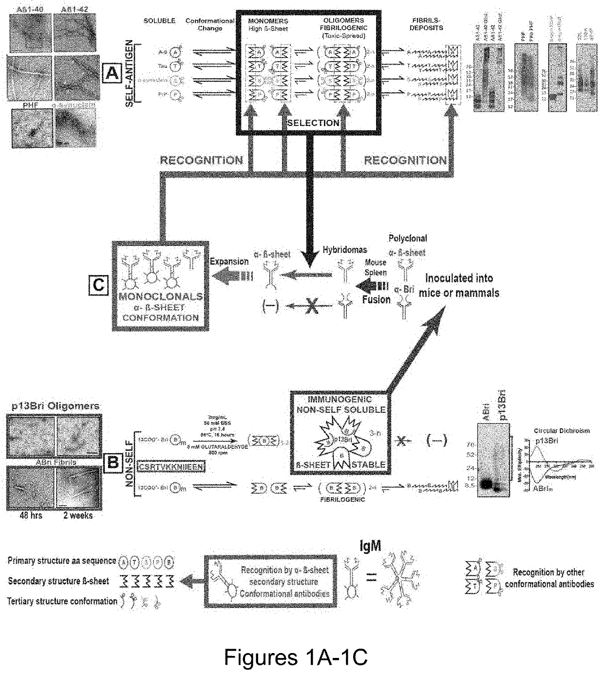
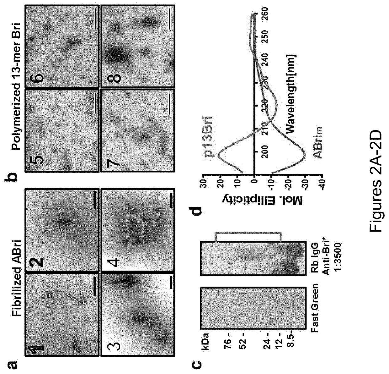
[0215]Synthesis and Polymerization of 13-mer Bri Peptide (pBri). The procedure was performed as previously published (Goñi et al, “Immunomodulation Targeting Abnormal Conformation Reduces Pathology in a Mouse Model of Alzheimer's Disease,” PLoS One 5(10): e13391 (2010), which is hereby incorporated by reference in its entirety). Briefly, the 13 residue peptide that corresponds to the carboxyl terminus of ABri (Cys-Ser-Arg-Thr-Val-Lys-Lys-Asn-Ile-Ile-Glu-Glu-Asn; SEQ ID NO: 111) was synthetized on an ABI 430A peptide synthesizer (AME Bioscience, Chicago, Ill.) at the Keck peptide synthesis facility at Yale University, CT. Mass spectroscopy of the lyophilized end-product was used to verify the expected molecular weight.

[0216]To have a stable oligomeric conformation and make the 13 mer Bri peptide ...

example 2

ization of Anti-β-Sheet Conformational mAb aβComAb WG-3D7

[0253]The purification of the anti-β-sheet conformational monoclonal antibody aβComAb WG-3D7 is depicted in FIGS. 11A-11B. FIG. 11A shows western blots of the conformational monoclonal antibody WG-3D7 purified with a llama anti-μ column. The far left panel shows Fast Green staining for protein loading control, the middle panel shows anti-mouse IgM μ specific reactivity, and the right panel shows anti-mouse Kappa reactivity. Lanes 1 and 2 of each panel contain un-reduced and DTT reduced proteins, respectively (IgMk p=pentamer, Hμ r=reduced heavy chain, and Kr=Kappa light chain reduced). FIG. 11B contains graphs of ELISA data showing the reactivity of the purified conformational monoclonal antibody WG-3D7 against (in order from left to right on the graphs) Aβ40, oligomerized Aβ42, and PHF (left graph), and positive and negative controls showing that the antigen specificity of the WG-3D7 antibody (solid bars) is not an artifact o...

example 3

ization of Anti-β-Sheet Conformational mAb aβComAb FT-11F2

[0259]The purification of the anti-β-sheet conformational monoclonal antibody FT-11F2 with SAS and quantification of the antibody binding levels to pathological conformers is depicted in FIGS. 17A-17B. FIG. 17A shows Western blots of the conformational monoclonal antibody FT-11F2, which was initially purified using 50% SAS. The left panel shows Fast Green staining for protein loading, the middle panel shows anti-mouse IgM μ specific reactivity, and the right panel shows anti-mouse Kappa specific reactivity. Lane 1 contains un-reduced sample (IgMk p=pentamer) and lane 2 contains 0.1 M dithiothreitol (DTT) reduced sample (Hμ, r=reduced heavy chain and Kr=Kappa light chain reduced). FIG. 17B are graphs of ELISA data showing the reactivity of the purified conformational monoclonal antibody FT-11F2 against (in order from left to right on the graph) Aβ40, oligomerized Aβ42, PHF, and PrP (left graph). The right graph of FIG. 17B sho...


PUM


Abstract

The present invention relates to antibodies and binding fragments thereof that bind the β-sheet secondary structure of a pathological monomeric or oligomeric non-fibrillar proteins without binding to the non-toxic, non-pathological forms of these proteins or peptides. These antibodies and binding fragments thereof are suitable for the diagnosis, prevention, and treatment of protein conformational disorders including all amyloid diseases.

Description

[0001]This application claims the priority benefit of U.S. Provisional Patent Application Ser. No. 62 / 365,465, filed on Jul. 22, 2016.[0002]This invention was made with government support under grant number NS073502 awarded by the National Institutes of Health. The government has certain rights in this invention.FIELD OF THE INVENTION[0003]The present invention relates to antibodies and binding fragments thereof that bind β-sheet secondary structures on pathological proteins, such as the β-sheet secondary structures found in toxic oligomeric forms of amyloidogenic proteins or in infectious, toxic or necrotic protein complexes. These antibodies and binding fragments thereof are suitable for the diagnosis, prevention, and treatment of protein conformational disorders including all amyloid disease and some foreign infective complexes.BACKGROUND OF THE INVENTION[0004]Amyloidosis broadly encompasses a variety of diseases that are characterized by the extracellular or intracellular deposi...

Claims


Application Information

Patent Timeline
Patent Type & Authority Applications(United States)
IPC IPC(8): C07K16/44A61P25/28C07K7/06C07K16/18C07K14/00
CPCC07K16/44A61P25/28C07K7/06C07K16/18A61K2039/505C07K2317/33C07K2317/34C07K2317/30C07K14/00
Inventor WISNIEWSKI, THOMAS M.GONI, FERNANDO
Owner NEW YORK UNIV
Learn More