Method for measuring a biomarker in a biological sample of an ipaf patient
a biomarker and patient technology, applied in the field of method for measuring a biomarker in a biological sample of an ipaf patient, can solve the problems of reducing vital capacity and gas exchange capacity, affecting the discrimination between ctd-ild and iips, and affecting the quality of life of patients
- Summary
- Abstract
- Description
- Claims
- Application Information
AI Technical Summary
Benefits of technology
Problems solved by technology
Method used
Image
Examples
example 1
[0123](1) Biological Sample
[0124]Sera of 102 patients with interstitial pneumonia were used as biological samples. Of the 102 patients, 16 patients were diagnosed with CTD-ILD (CTD-ILD group), 35 patients were diagnosed with IPAF (IPAF group), and 51 patients were diagnosed with IPF (IPF group).
[0125](2) Measurement of Biomarkers
[0126](2.1) Measurement of CXCL9
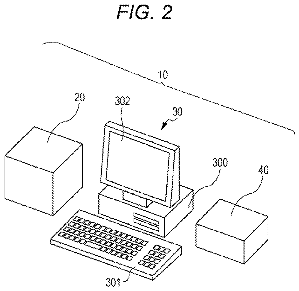
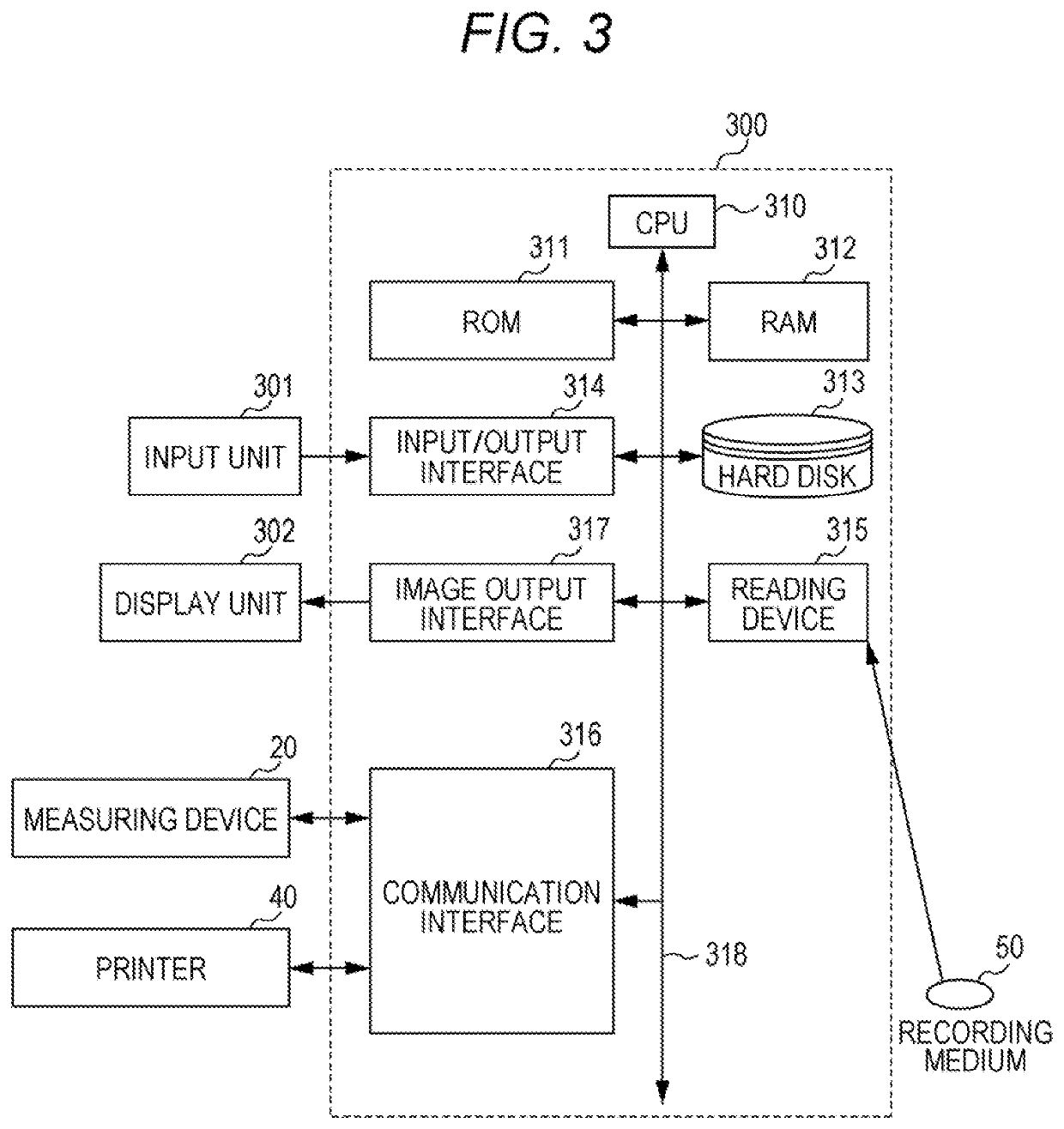
[0127]The serum CXCL9 concentration of each patient was measured using a fully automated immunoassay system HISCL-5000 or HISCL-2000i (Sysmex Corporation) using following R1 to R5 reagents.
[0128]R1 Reagent
[0129]An anti-MIG monoclonal antibody (Randox Laboratories Ltd.) was digested with pepsin or IdeS protease by a conventional method to obtain a Fab fragment. The Fab fragment was labeled with biotin by a conventional method and dissolved in a buffer containing 1% bovine serum albumin (BSA) and 0.5% casein to obtain an R1 reagent.
[0130]R2 Reagent
[0131]Magnetic particles on which streptavidin was immobilized on its surface (her...
example 2
[0141](1) Discrimination Between CTD-ILD and IPAF
[0142]For each concentration of CXCL9 and CXCL10 in the serum, an optimal cut-off value for distinguishing between the CTD-ILD group (16 patients) and the IPAF group (35 patients) in Example 1 was determined by ROC analysis. The sensitivity, specificity and area under the curve (AUC) for determination using the set cut-off value were calculated. In this determination, when the concentration of CXCL9 or CXCL10 was greater than or equal to the cut-off value, the patient was determined to have CTD-ILD. When the concentration of CXCL9 or CXCL10 was lower than the cut-off value, the patient was determined not to have CTD-ILD. The obtained ROC curves are shown in FIGS. 7 and 8. Table 1 shows the cut-off value (pg / mL), sensitivity (%), specificity (%) and AUC for determination of each biomarker.
TABLE 1SensitivitySpecificityBiomarkerCut-off value(%)(%)AUCCXCL932.587490.72CXCL10287.160800.75
[0143]From Table 1, in the determination based on eac...
example 3
[0148](1) Biological Sample
[0149]The sera of the interstitial pneumonia patients in Example 1 were used as biological samples.
[0150](2) Measurement of Biomarkers
[0151](2.1) Measurement of CXCL9 and CXCL10
[0152]The measured values acquired in Example 1 were used as the serum CXCL9 and CXCL10 concentration values of each patient.
[0153](2.2) Measurement of KL-6
[0154]The serum KL-6 concentration of each patient was measured using HISCL-5000 or HISCL-2000i (Sysmex Corporation) using a HISCL KL-6 reagent (Sekisui Medical Co., Ltd.). The measurement procedure of KL-6 was similar to the measurement of CXCL9 in Example 1. The HISCL KL-6 reagent included an R1 reagent containing biotin-labeled mouse anti-KL-6 monoclonal antibody, an R2 reagent containing STA-bound magnetic particles, an R3 reagent containing ALP-labeled mouse anti-KL-6 monoclonal antibody, a HISCL R4 reagent as a measurement buffer solution, and a HISCL R5 reagent containing CDP-Star (registered trademark), a HISCL washing so...
PUM
| Property | Measurement | Unit |
|---|---|---|
| pH | aaaaa | aaaaa |
| pH | aaaaa | aaaaa |
| concentration | aaaaa | aaaaa |
Abstract
Description
Claims
Application Information
 - R&D
- Intellectual Property
- Life Sciences
- Materials
- Tech Scout
- Unparalleled Data Quality
- Higher Quality Content
- 60% Fewer Hallucinations
Browse by: Latest US Patents, China's latest patents, Technical Efficacy Thesaurus, Application Domain, Technology Topic, Popular Technical Reports.
© 2025 PatSnap. All rights reserved.Legal|Privacy policy|Modern Slavery Act Transparency Statement|Sitemap|About US| Contact US: help@patsnap.com


