
Mapping ciliary activity using phase resolved spectrally encoded interferometric microscopy

Active Publication Date: 2020-08-06
RGT UNIV OF CALIFORNIA
View PDF0 Cites 6 Cited by
  • Summary
  • Abstract
  • Description
  • Claims
  • Application Information

AI Technical Summary

Benefits of technology

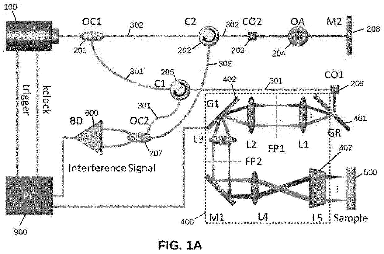
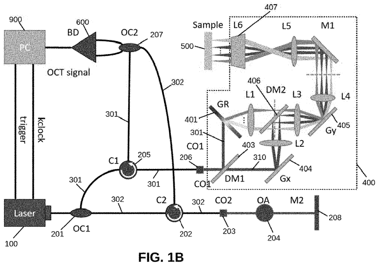
The present invention is a system that uses a special technique called swept source spectrally encoded interferometric microscopy (SS-SEIM) to create high-speed images of cilieating frequency and pattern. This system is capable of mapping the ciliary activity in real-time, providing valuable information about the activity of cilia in tissues. In this study, a swept source laser is used to achieve 91 frames per second, over an image area up to 1 mm2. This is the first system to do so, making it possible to examine the frequency and pattern of ciliary activity in vivo. Overall, this technology can help to improve understanding of cellular behavior and has the potential to impact areas like research and medicine.

Problems solved by technology

Although prior techniques using OCT are able to visualize in-vivo cilia activity within one cross-sectional image, they have low potential to study the surface dynamic of ciliated tissue due to their limited lateral FOV.
A prior study reported a spectrometer based spectrally encoded system to visualize vibration, but the spatial resolution of the system (˜7 μm) is not enough to resolve ciliary movement.
Additionally, recent studies have discovered that the spectrometer-based Doppler is inferior to swept source based in terms of the maximum measurable Doppler velocity.

Method used


Image

  • Mapping ciliary activity using phase resolved spectrally encoded interferometric microscopy
  • Mapping ciliary activity using phase resolved spectrally encoded interferometric microscopy
  • Mapping ciliary activity using phase resolved spectrally encoded interferometric microscopy

Examples


example

[0060]The following is a non-limiting example of the present invention. It is to be understood that said example is not intended to limit the present invention in any way. Equivalents or substitutes are within the scope of the present invention.

[0061]The present invention features the first PRD-SEIM system to do real time en face imaging of ciliated tissue. This invention has been developed and validated by two trachea samples and one oviduct sample ex-vivo, where the microscopic surface dynamic of these tissues were visualized at different temperatures and drug applications.

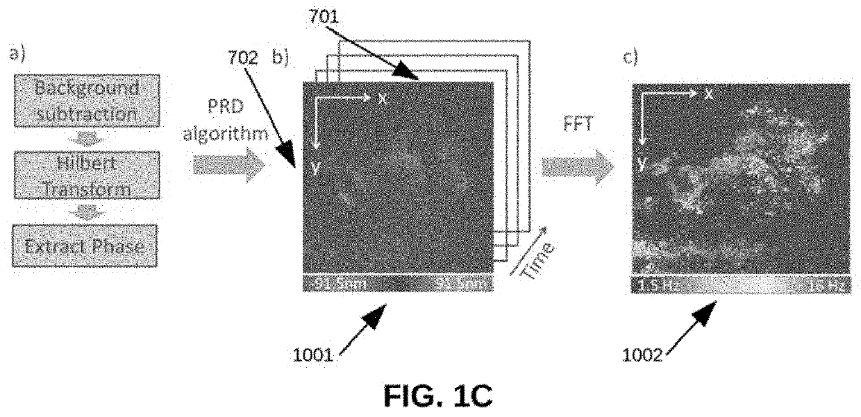
[0062]The invention features a PRD-SEIM method of measuring and quantifying the spatial ciliary beating frequency of ex-vivo rabbit trachea. This technology offers high resolution, high speed and high sensitivity en face image of tissue structure and displacement. To be specific, the PRD algorithm allows for nanometer displacement sensitivity, ensures accurate measurement of the microscopic ciliary movement. Fur...


PUM


Abstract

Ciliary motion in the upper airway is the primary mechanism by which the body transports foreign particulate out of the respiratory system. The ciliary beating frequency (CBF) is often disrupted with the onset of disease. Current imaging of ciliary motion relies on microscopy and high speed cameras, which cannot be easily adapted to in-vivo imaging. M-mode optical coherence tomography (OCT) imaging is capable of visualization of ciliary activity, but the field of view is limited. The present invention features the development of a spectrally encoded interferometric microscopy (SEIM) system using a phase-resolved Doppler (PRD) algorithm to measure and map the ciliary beating frequency within an on face region. This novel high speed, high resolution system allows for visualization of both temporal and spatial ciliary motion patterns.

Description

CROSS-REFERENCES TO RELATED APPLICATIONS[0001]This application is a non-provisional and claims benefit of U.S. Provisional Application No. 62 / 800,284 filed Feb. 1, 2019, the specification(s) of which is / are incorporated herein in their entirety by reference.STATEMENT REGARDING FEDERALLY SPONSORED RESEARCH OR DEVELOPMENT[0002]This invention was made with government support under Grants No. R01HL-125084 and No. R01HL-127271 awarded by the National Institutes of Health and Grant No. FA9550-17-1-0193 awarded by the Air Force Office of Scientific Research. The government has certain rights in the invention.BACKGROUND OF THE INVENTION[0003]The human airway serves not only as a passage for oxygen intake, but also as the first line of defense to protect the lungs from harmful foreign particulate. The main defense mechanism is the mechanical clearance of mucus through ciliary activity and regulation of airway surfaces. Specifically, ciliary beat frequency and amplitude are considered the mai...

Claims


Application Information

Patent Timeline
IPC IPC(8): A61B5/11G01B9/02A61B1/00A61B1/267A61B1/06A61B1/005A61B5/00G02B21/00G02B21/36
CPCG02B21/0056A61B1/00149A61B1/00172G02B21/0028G02B21/365A61B1/267A61B1/005A61B5/1128A61B1/00177A61B1/063G01B9/02004G01B9/02041G02B21/0048A61B5/0066A61B1/00096A61B5/08A61B5/7257A61B5/7253A61B5/7214A61B1/002A61B1/07A61B1/0638A61B1/00006A61B1/00009G01P3/36G01B9/02091G01B9/02045G01B9/02027
Inventor CHEN, ZHONGPINGHE, YOUMINJING, JOSEPHQU, YUEQIAOHEIDARI, ANDREW EMONCHEN, JASON
Owner RGT UNIV OF CALIFORNIA
Learn More