
Medical image processing device, medical image processing method, and medical image processing program

Active Publication Date: 2020-06-18
FUJIFILM CORP
View PDF0 Cites 5 Cited by
  • Summary
  • Abstract
  • Description
  • Claims
  • Application Information

AI Technical Summary

Benefits of technology

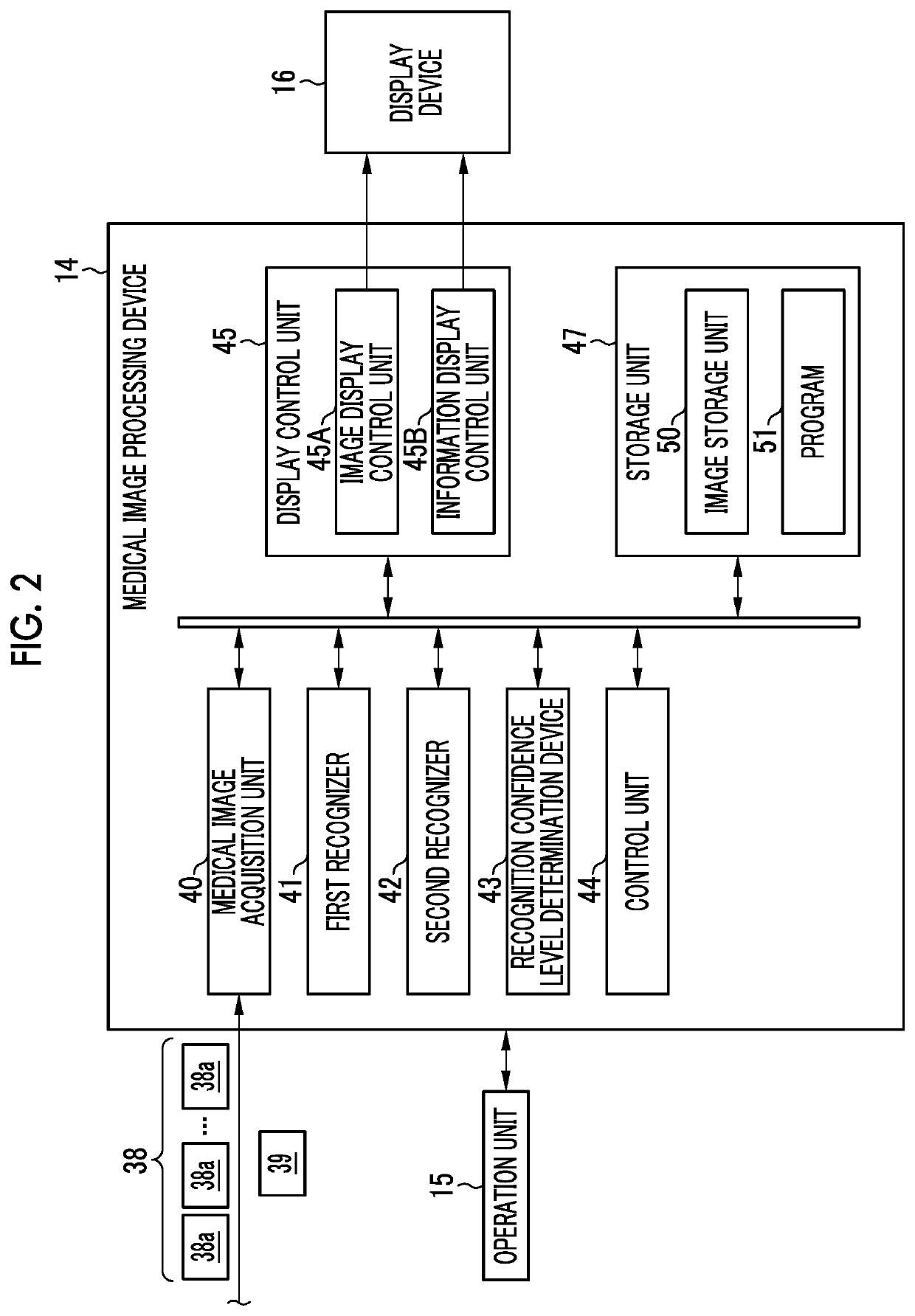
The invention is a medical image processing device, method and program that can speed up image recognition and improve recognition accuracy. It uses two recognizers, one with a lower load of the recognition process and one with higher recognition accuracy. In case of low confidence level for the recognition result, the second recognizer is used to improve the recognition accuracy. The second recognizer has a greater number of layers including filters, which makes the processing time longer but increases the confidence level of the recognition result. The control unit can also decide a category priority of the plurality of categories on the basis of the category recognition result of the second recognizer and change the display order of the plurality of categories accordingly. Overall, this invention enhances the efficiency and accuracy of medical image recognition.

Problems solved by technology

However, there is a problem in that the recognition accuracy of the image becomes lowered in the recognizer which performs the image recognition process at high speed.
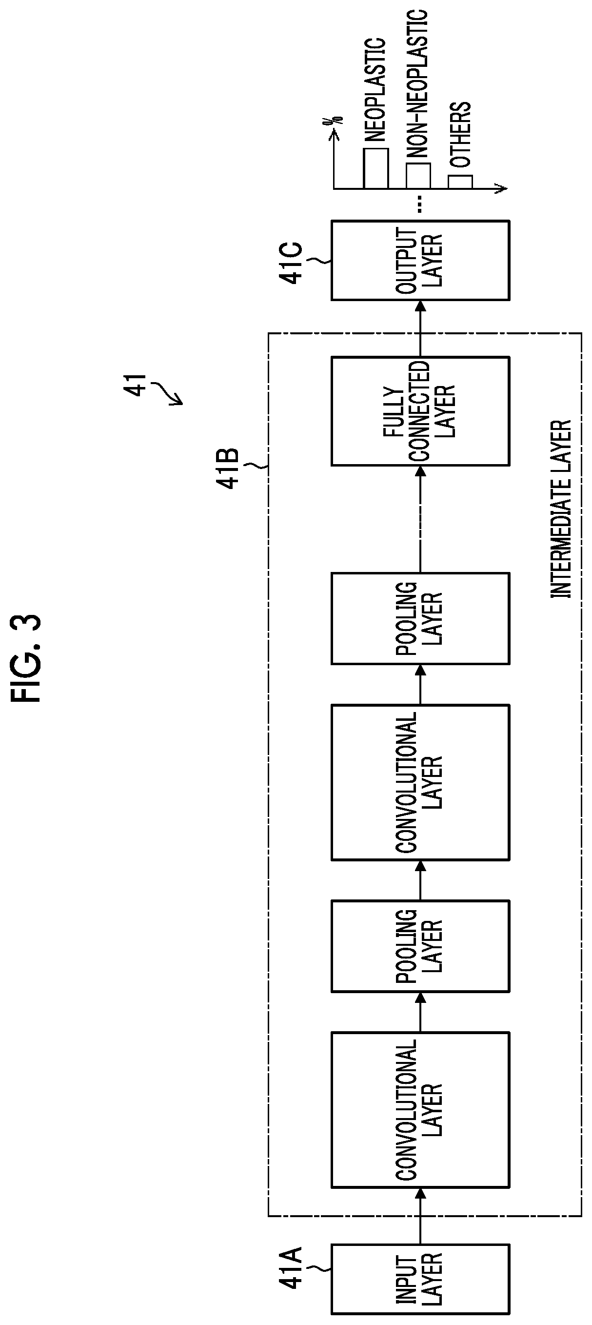
For example, in case of the CNN, the number of layers of the multiple layer structure is required to be reduced for speeding up, but there is a problem in that the recognition accuracy of the image becomes lowered.
Meanwhile, in a case where the number of layers of the layer structure is increased to improve the recognition accuracy of the image, there is a problem in that the amount of calculation is increased and the operation load on the recognizer is increased so that the recognition process is delayed.
Accordingly, in the information processing device disclosed in WO2010 / 050333, in a case where the recognizer which performs the image recognition process at high speed (recognizer with low recognition accuracy of the image) is applied, there is a problem in that the confidence level for the recognition result becomes lowered and the warning information is frequently displayed.

Method used


Image

  • Medical image processing device, medical image processing method, and medical image processing program
  • Medical image processing device, medical image processing method, and medical image processing program
  • Medical image processing device, medical image processing method, and medical image processing program

Examples


Embodiment Construction

[0046]Hereinafter, preferred embodiments of a medical image processing device, a medical image processing method, and a medical image processing program according to the invention will be described with reference to accompanying drawings.

[0047][Entire Configuration of Endoscope System]

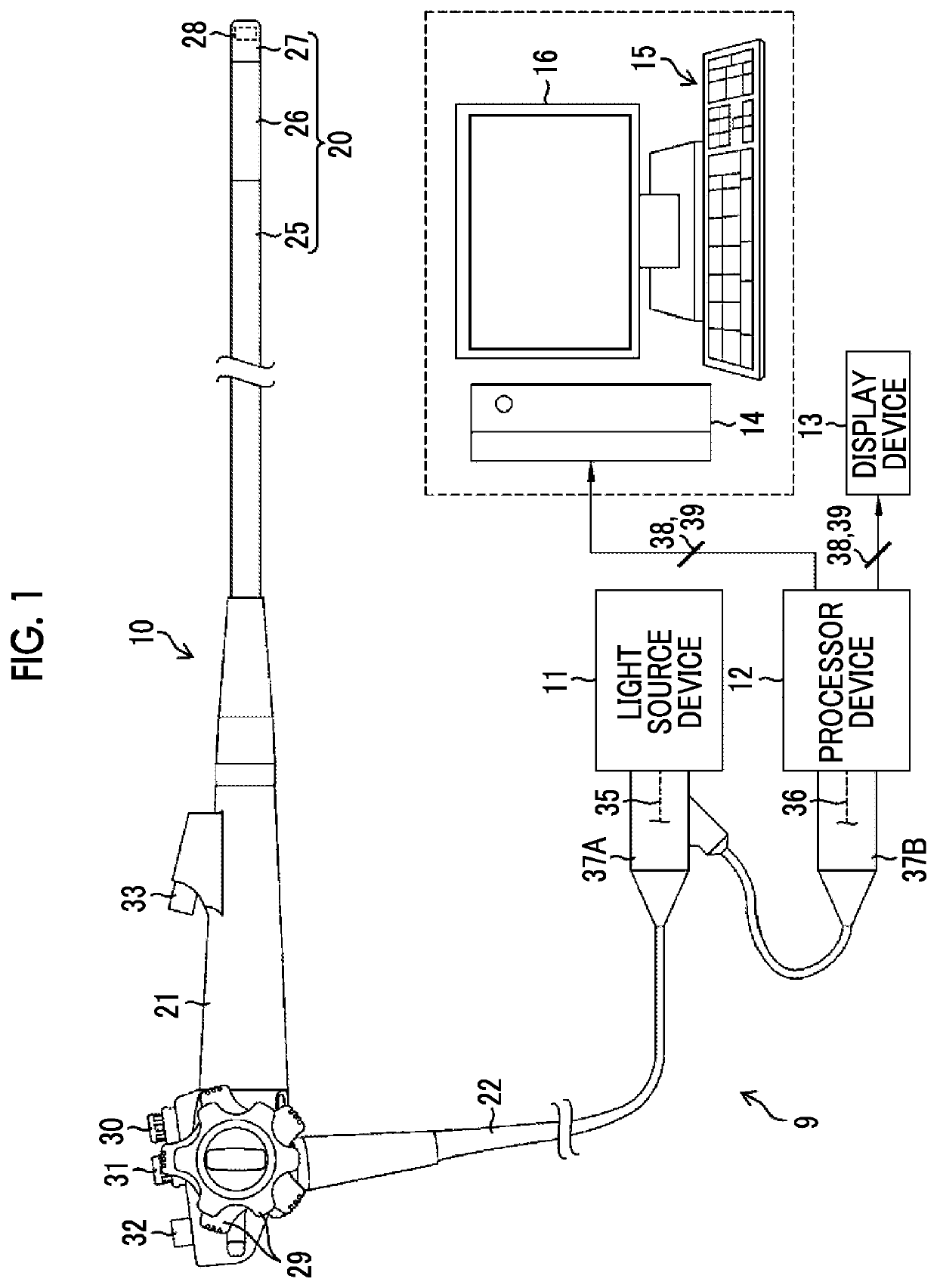
[0048]FIG. 1 is a schematic diagram showing the entire configuration of an endoscope system 9 including a medical image processing device according to an embodiment of the invention. As shown in FIG. 1, the endoscope system 9 includes an endoscope 10 which is an electronic endoscope, a light source device 11, a processor device 12, a display device 13, a medical image processing device 14, an operation unit 15, and a display unit 16.

[0049]The endoscope 10 corresponds to a medical device of an embodiment of the invention, and is a flexible endoscope, for example. The endoscope 10 includes an insertion part 20 that is to be inserted into an object to be examined and has a distal end and a proximal end, a...


PUM


Abstract

A medical image processing device having a processor configured to: acquire a medical image including an image of a subject; perform a first recognition of the medical image using a first recognizer; determine a confidence level for a recognition result of a first recognition by the first recognition; and perform a second recognition of the medical image using a second recognizer according to the confidence level for the recognition result of the first recognition, the second recognition having higher recognition accuracy than the first recognition.

Description

CROSS-REFERENCE TO RELATED APPLICATIONS[0001]This application is a Continuation of PCT International Application No. PCT / JP2018 / 026945 filed on Jul. 18, 2018, which claims priority under 35 U.S.C § 119(a) to Japanese Patent Application No. 2017-177491 filed on Sep. 15, 2017. Each of the above application(s) is hereby expressly incorporated by reference, in its entirety, into the present application.BACKGROUND OF THE INVENTION1. Field of the Invention[0002]The invention relates to a medical image processing device, a medical image processing method, and a non-transitory computer readable medium for storing a medical image processing program, and more particularly relates to a technique for performing a recognition process of a medical image by using a recognizer.2. Description of the Related Art[0003]In the medical field, inspection using an endoscope system is performed. In recent years, a system of performing recognition of a lesion included in an image by image analysis has been k...

Claims


Application Information

Patent Timeline
IPC IPC(8): G06K9/62G06T7/00G06T7/70G06V10/25G06V10/764G06V10/776G06V10/80
CPCG06K2209/05G06T7/0012G06T2207/30096G06T7/70G06K9/6262G06T2207/20084A61B1/045G06T2207/10068G06T2207/10024G06V10/454G06V10/25G06V2201/03G06V10/82G06V10/80G06V10/776G06V10/764G06F18/25G06F18/217
Inventor OOSAKE, MASAAKI
Owner FUJIFILM CORP
Learn More