
System for automatic tumor detection and classification

a tumor detection and automatic technology, applied in the field of machine learning, can solve the problems of time-consuming and intensive pathology, a large amount of time for the slides representing one tissue, and the process of diagnosis for a particular slide containing potentially cancerous tissue is time-consuming and intensive for pathologists performing diagnosis,

Active Publication Date: 2020-03-05
APPLIED MATERIALS INC
View PDF0 Cites 4 Cited by
  • Summary
  • Abstract
  • Description
  • Claims
  • Application Information

AI Technical Summary

Benefits of technology

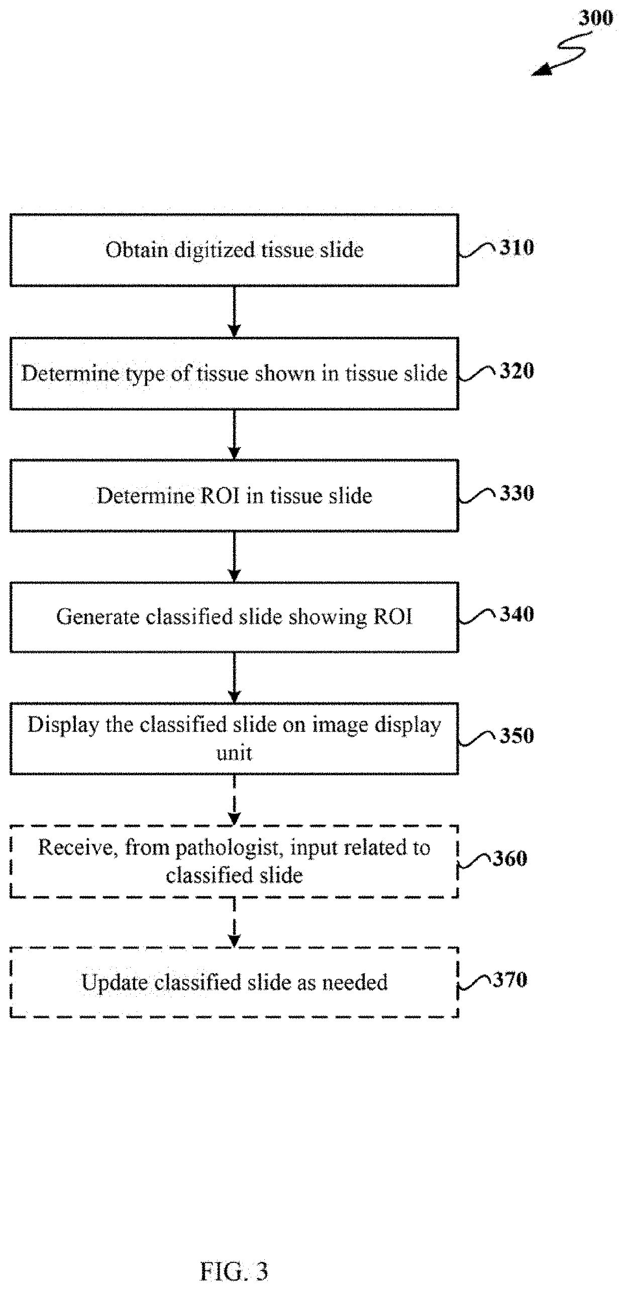
The patent text describes a method for automatically detecting and classifying tumor regions in a tissue slide. The method involves obtaining a digitized tissue slide, determining the type of tissue based on the output of a tissue classification module, identifying a region of interest (ROI) within the tissue as tumor based on a tumor classification model, and estimating the diameter of the ROI. The method then generates a classified slide that shows theROI with a corresponding tumor class based on the diameter, and transmits it to a pathologist assistance system. The technical effects of this method include improved efficiency and accuracy in detecting and classifying tumor regions, which can aid in the diagnosis and treatment of cancer.

Problems solved by technology

Currently, the process of diagnosis for a particular slide containing potentially cancerous tissue is time consuming and intensive for pathologists performing the diagnosis.
As a result, in order to determine if a given tissue does or does not include a tumor can take a good deal of time for the slides representing one tissue.
Further, if the pathologist detects a tumor, further time is necessary to analyze the tumor.
In such cases, it may take days to get the slides to the pathologist before analysis even begins.
However, existing machine learning systems are incapable of effectively identifying and classifying tumors depicted in tissue slides.
Further, existing systems are incapable of properly annotating or marking tissue slides to assist the pathologist in diagnosis.
As a result, none or only very little pathologist time may be effectively saved by existing machine learning systems.

Method used


Image

  • System for automatic tumor detection and classification
  • System for automatic tumor detection and classification
  • System for automatic tumor detection and classification

Examples


Embodiment Construction

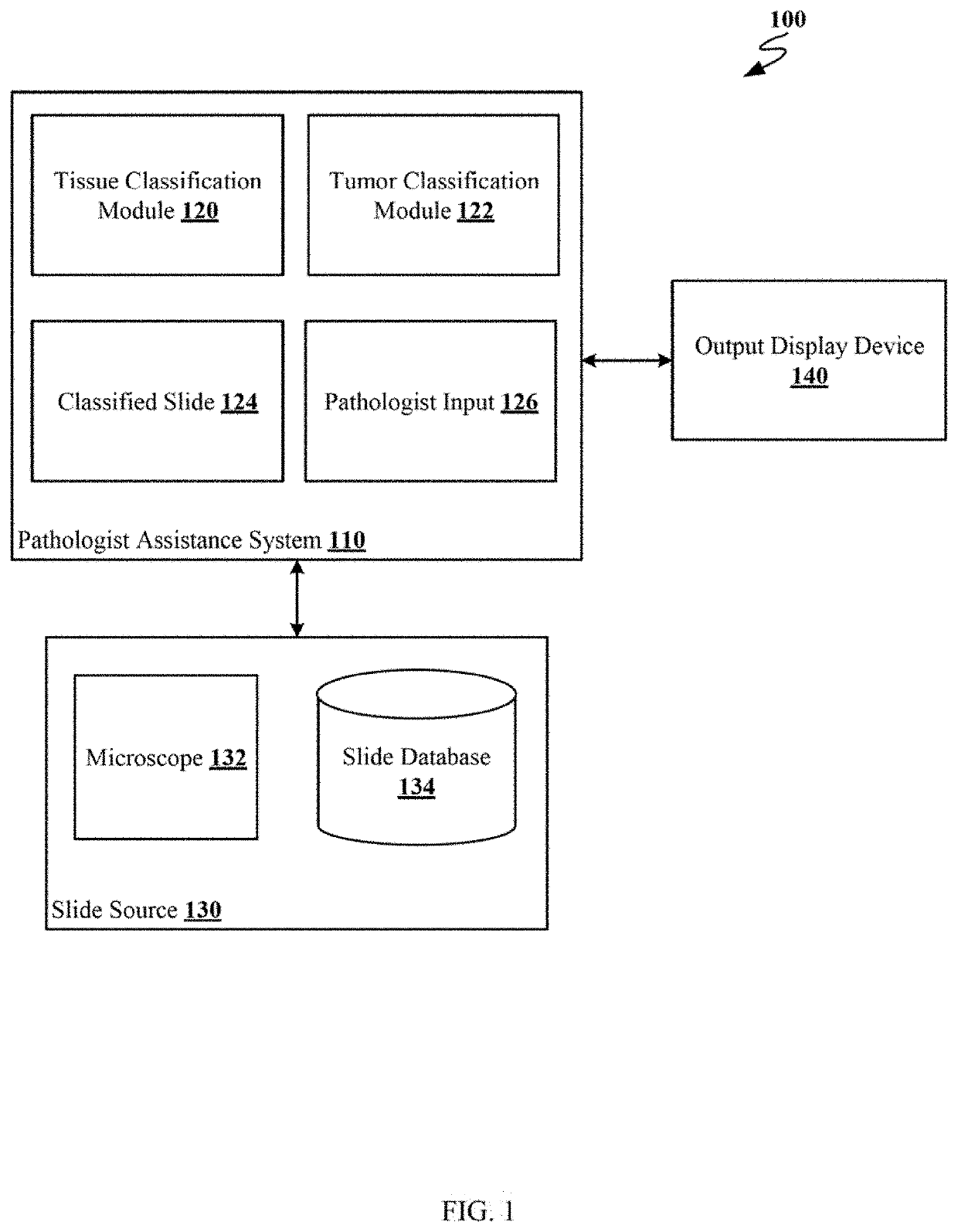
[0014]FIG. 1 depicts an example computing environment 100 for the automatic detection and classification of tumors. As shown, computing environment 100 includes pathologist assistance system 110, slide source 130 and output display device 140. Although shown as separate entities, in other examples the functions of pathologist assistance system 110, slide source 130 and output display device 140 may be performed by a single computing device or by a distributed computing system. In other embodiments, pathologist assistance system 110, slide source 130 and output display device 140 may be connected via a network such as, for example, a local area network (LAN), a wide area network (WAN) or the Internet.

[0015]Pathologist assistance system 110 is a computing device comprising at least a processor and a memory, capable of executing software resident in the memory by the processor. Pathologist assistance system 110 includes tissue classification module 120, tumor classification module 122,...


PUM


Abstract

Certain aspects of the present disclosure provide techniques for automatically detecting and classifying tumor regions in a tissue slide. The method generally includes obtaining a digitized tissue slide from a tissue slide database and determining, based on output from a tissue classification module, a type of tissue of shown in the digitized tissue slide. The method further includes determining, based on output from a tumor classification model for the type of tissue, a region of interest (ROI) of the digitized tissue slide and generating a classified slide showing the ROI of the digitized tissue slide and an estimated diameter of the ROI. The method further includes displaying on an image display unit, the classified slide and user interface (UI) elements enabling a pathologist to enter input related to the classified slide.

Description

CROSS-REFERENCE TO RELATED APPLICATIONS[0001]This application claims benefit of Indian Provisional Patent Application No. 201841032562, filed Aug. 30, 2018, the entirety of which is herein incorporated by reference.BACKGROUND[0002]Aspects of the present disclosure relate to machine learning and in particular to the classification of images using machine learning pattern recognition.[0003]Currently, the process of diagnosis for a particular slide containing potentially cancerous tissue is time consuming and intensive for pathologists performing the diagnosis. In the current process, a pathologist has to manually analyze many images of tissue slides. Each individual slide is typically scanned in across multiple magnification and zoom levels. As a result, in order to determine if a given tissue does or does not include a tumor can take a good deal of time for the slides representing one tissue. Further, if the pathologist detects a tumor, further time is necessary to analyze the tumor....

Claims


Application Information

Patent Timeline
IPC IPC(8): G06K9/00G06N20/00G06T7/00
CPCG06K9/00147G06N3/02G06T7/11G06T2207/20081G06N20/00G06T7/0012G06T2207/30024G06N3/08G06T2207/20084G06V20/698G06V10/25G06V2201/03G06V10/82G06V10/987G06V10/7784G06V10/764G06N3/045G16H50/20A61B5/7275
Inventor PRABHUDESAI, PARIJAT PRAKASHMOHANUR RAGHUNATHAN, GANESH KUMARJHA, SUMIT KUMARSISTA, ADITYACHANDAN, NARASIMHA MURTHY
Owner APPLIED MATERIALS INC
Learn More