Non-invasive frequency domain optical spectroscopy for neural decoding

Inactive Publication Date: 2019-11-07
HI LLC
View PDF0 Cites 13 Cited by
  • Summary
  • Abstract
  • Description
  • Claims
  • Application Information

AI Technical Summary

Benefits of technology

The present invention provides an optical non-invasive measurement system that can measure the intensity of light scattered by an anatomical structure, such as a brain, during a single measurement period. The system uses an optical source to intensity modulate sample light at multiple frequencies, and a controller to instruct the optical source to sequentially intensity modulate the sample light over a frequency range. The system also includes an optical detection assembly to detect the scattered light, an optical detector to output an electrical signal, and an amplifier to amplify the signal. The detected signal light is then converted to digital data using an analog-to-digital converter. The system can analyze the detected signal light in the frequency or time domain to determine the occurrence and spatial depth of a physiological event in the anatomical structure. The technical effects of the invention include improved accuracy and non-invasiveness for measuring brain activity and other physiological events, as well as the ability to measure the intensity of scattered light in real-time.

Problems solved by technology

However, because optical imaging techniques rely on light, which scatters many times inside brain, skull, dura, pia, and skin tissues, the light paths occurring in these techniques comprise random or “diffusive” walks, and therefore, only limited spatial resolution can be obtained by a conventional optical detector, often on the order of centimeters, with penetration depths being limited to a few millimeters.
The reason for this limited spatial resolution is that the paths of photons striking the detector in such schemes are highly variable and difficult, and even impossible, to predict without detailed microscopic knowledge of the scattering characteristics of the brain volume of interest, which is typically unavailable in practice (i.e., in the setting of non-invasive measurements through skull for brain imaging and brain interfacing).
In summary, light scattering has presented challenges for optical imaging techniques in achieving high spatial resolution deep inside tissue.
Moreover, the diffusive nature of light propagation also creates challenges for measurements of fast changes in optical scattering inside tissue, since essentially all paths between source and detector are highly scattered to begin with.
However, nearly all diffusive optical imaging techniques to date offer relatively poor temporal resolution (100 ms-1 sec per sample), as they are primarily designed to detect hemodynamics that vary on a similarly slow time scale.
However, because this approach only samples the brain tissue at one modulation frequency, the detection sensitivity of fast-optical signals in the brain tissue is not maximized.
Furthermore, this approach does not acquire spatial depth information of the fast-optical signals.
Although the FDPM technique described in O'Sullivan intensity modulates the light source at multiple frequencies, O'Sullivan discloses no means for measuring fast-optical signals within brain tissue using the FDPM technique, and furthermore, does not disclose any means for using the frequency information to obtain spatial depth information of any biologically inherent signals.
However, these techniques utilize holographic methods, mixing the detected light against a reference beam, thereby requiring a relatively complicated and expensive arrangement of components.
Further, while the iNIRS or SS-OCT approaches are very sophisticated, they require the detection and measurement of speckles, presenting challenges in a highly attenuating medium, such as the human body, due to the very low number of photons that reach each detector.
Thus, a very large number of detectors (or pixels) are required to individually detect the speckles, thereby further increasing the complexity and expense of the system.

Method used


Image

  • Non-invasive frequency domain optical spectroscopy for neural decoding
  • Non-invasive frequency domain optical spectroscopy for neural decoding
  • Non-invasive frequency domain optical spectroscopy for neural decoding

Examples


Embodiment Construction

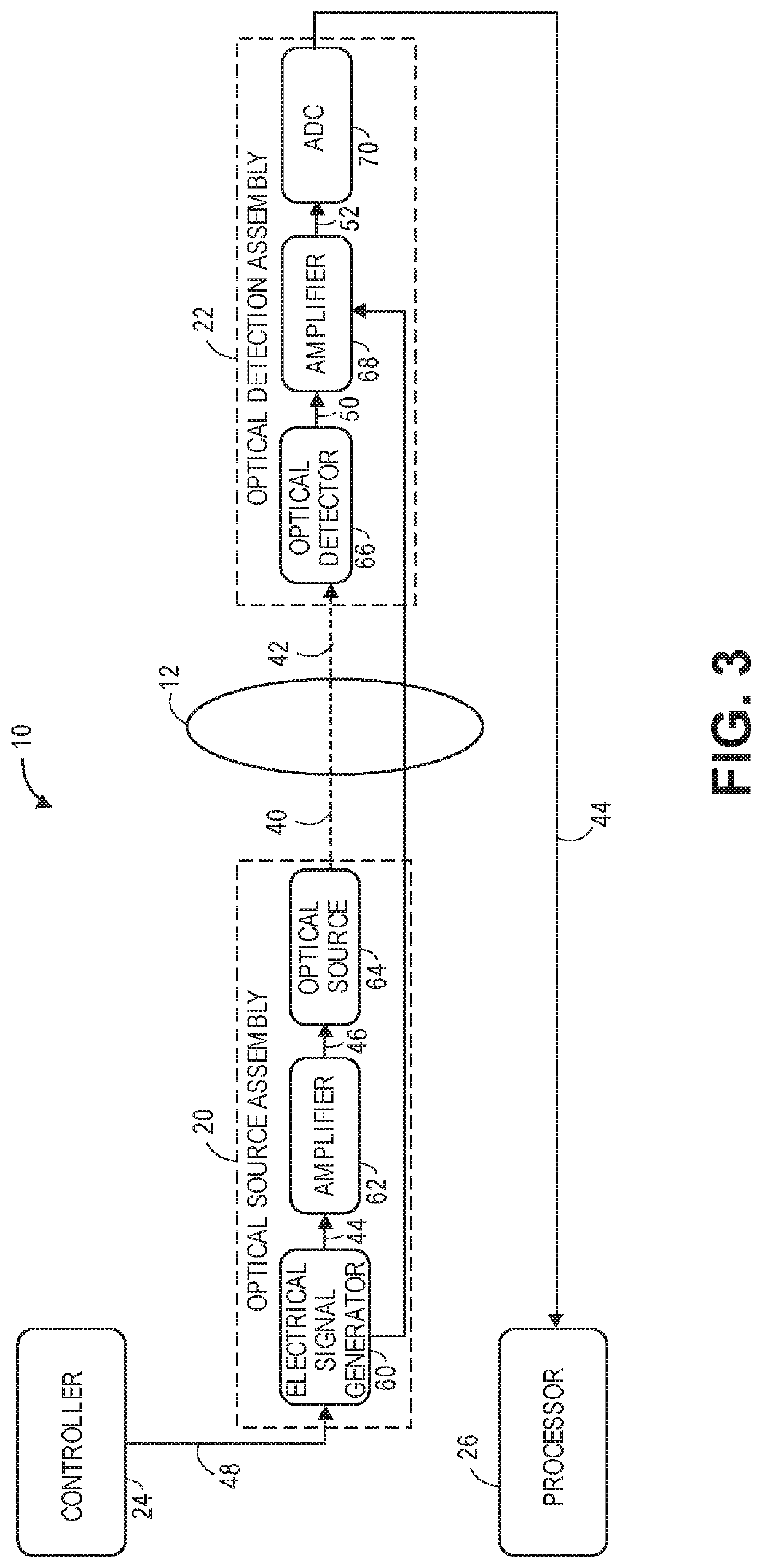
[0068]Referring first to FIG. 1, one embodiment of an optical non-invasive measurement system 10 constructed in accordance with the present inventions will now be described. The optical measurement system 10 is designed to non-invasively detect and localize a physiological event in an anatomical structure 12. In the illustrated embodiments, the anatomical structure 12 is a brain. Although for exemplary purposes, the optical measurement system 10 is described herein as being used to detect and localize a physiological event in brain tissue, variations of the optical measurement system 10 can be used to detect and localize a physiological event in other anatomical parts of a human body, animal body and / or biological tissue.

[0069]Although the optical non-invasive measurement system 10 is initially described as creating one optical path 14 through the brain 12, in a practical implementation, variations of the optical non-invasive measurement system 10 described herein will create multip...


PUM


Abstract

An optical measurement system comprises an optical source assembly configured for intensity modulating sample light at multiple frequencies within a frequency range, and delivering the intensity modulated sample light along an optical path of an anatomical structure during a single measurement period, such that the intensity modulated sample light is scattered by the anatomical structure, resulting in signal light that exits the anatomical structure. The optical measurement system further comprises an optical detection assembly configured for detecting the signal light over the frequency range within the measurement period. The optical measurement system further comprises a processor configured for analyzing the detected signal light, and, based on this analysis, determining an occurrence and spatial depth of a physiological event in the anatomical structure.

Description

RELATED APPLICATION DATA[0001]Pursuant to 35 U.S.C. § 119(e), this application claims the benefit of U.S. Provisional Patent Application 62 / 666,926, filed May 4, 2018, and U.S. Provisional Patent Application 62 / 692,074, filed Jun. 29, 2018, which are expressly incorporated herein by reference.FIELD OF THE INVENTION[0002]The present inventions relate to methods and systems for non-invasive measurements in the human body, and in particular, methods and systems related to detecting physiological events in the human body, animal body, and / or biological tissue.BACKGROUND OF THE INVENTION[0003]Measuring neural activity in the brain is useful for medical diagnostics, neuromodulation therapies, neuroengineering, or brain-computer interfacing. Conventional methods for measuring neural activity in the brain include diffusive optical imaging techniques, which employ moderate amounts of near-infrared or visible light radiation, thus being comparatively safe and gentle for a biological subject i...

Claims


Application Information

Patent Timeline
IPC IPC(8): A61B5/00
CPCA61B5/0042A61B5/0082A61B5/4064A61B5/0066A61B5/6814A61B2562/04
Inventor ALFORD, JAMUHORSTMEYER, ROARKEMARBLESTONE, ADAM
Owner HI LLC
Why Eureka
Try Eureka