Three Dimensional Bioprinted Tumor Models for Drug Testing

a three-dimensional bioprinting and tumor technology, applied in the field of three-dimensional bioprinting tumor models for drug testing, can solve the problems of loss of heterogeneity that is required to accurately mimic human disease, short life span, inbreeding, etc., and achieve the effect of accurate screening

Pending Publication Date: 2019-10-10
ORGANOVO +1
View PDF5 Cites 6 Cited by
  • Summary
  • Abstract
  • Description
  • Claims
  • Application Information

AI Technical Summary

Benefits of technology

The invention provides a 3D model for studying biological cancer in a human environment. This model can be used to measure the potential of tumor tissue to grow and engage with other tissues. The model can also be used to test different therapies for their effectiveness. The model is made using a stromal microenvironment and bioprinting technology, which allows for a longer analysis period compared to animal-based models. The model provides a better screening tool for testing cancer therapeutics. The model is firm enough to allow for precise incision and deposition of cellular materials. Overall, this invention provides a more accurate and reliable tool for studying cancer and its treatment.

Problems solved by technology

Dissociation of human tumor samples causes fundamental alterations in the cancer cells as they are removed from their normal three-dimensional environment and results in a loss of heterogeneity that is required to accurately mimic the human disease.
A significant drawback to a tumor xenograft mouse models is that it is based on an animal that, in comparison to humans, is significantly smaller, has a much higher metabolic rate, is inbred, and has a short life span.
The presence of the animal stromal cells in the transplanted tumor negatively impacts measurement of biological activities and / or the efficacy of therapeutics being tested, particularly for molecularly targeted therapeutics that disrupt ligand / receptor interactions that rely on species specificity.
Additionally, the use of immunocompromised animals prevents the ability to test immunomodulatory therapies.

Method used


Image

  • Three Dimensional Bioprinted Tumor Models for Drug Testing
  • Three Dimensional Bioprinted Tumor Models for Drug Testing
  • Three Dimensional Bioprinted Tumor Models for Drug Testing

Examples


example 1

ng of Stromal Tissue Block

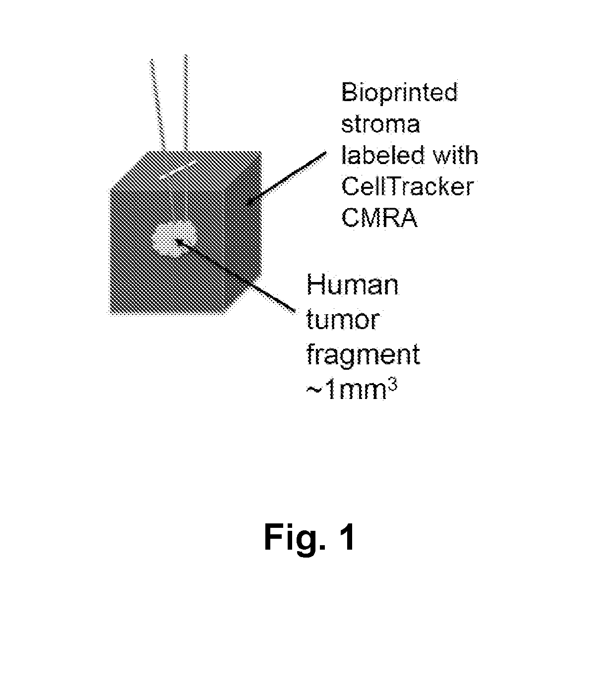
[0365]The stromal compartment comprised human mammary fibroblasts, endothelial cells, and preadipocytes printed as a solid cube measuring approximately 2 mm×2 mm×2 mm. In order to track the position of the bioprinted cells or implanted cells, a red fluorescent dye (CellTracker CMRA Orange (ThermoFisher)) was incorporated into the bioprinted tissue.

example 2

ioning of Stromal Tissue Block

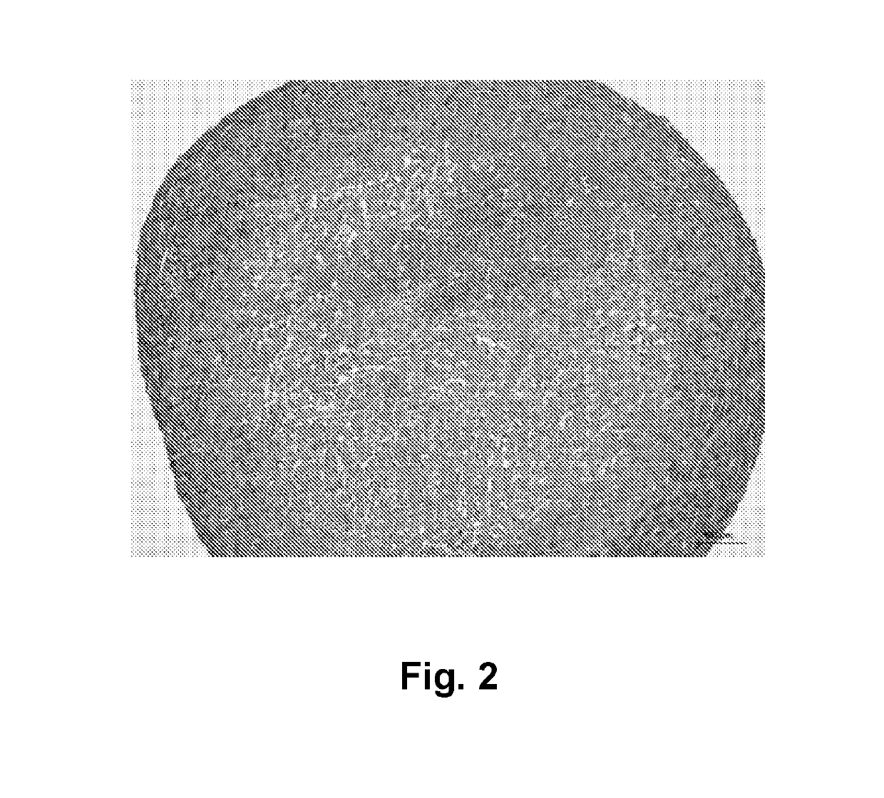
[0366]Bioprinted stromal tissues were cross-linked with 50 mM calcium chloride and cultured in a rolling bioreactor in 50 mL vented cap tubes (CellTreat) at 18 rpm for 3 days. Tissues were treated with 50 mg / mL alginate lyase overnight in the bioreactor tube, and cultured in the bioreactor for 4 additional days in culture media to prepare the stromal tissues. Preconditioned tissues cultured in the rolling bioreactor exhibited a dense capsule of fibroblasts on the outer surface of the tissue that permitted incision with a scalpel. See FIG. 2.

example 3

ion of Primary Human Solid Tumor Fragment into Stromal Tissue Block

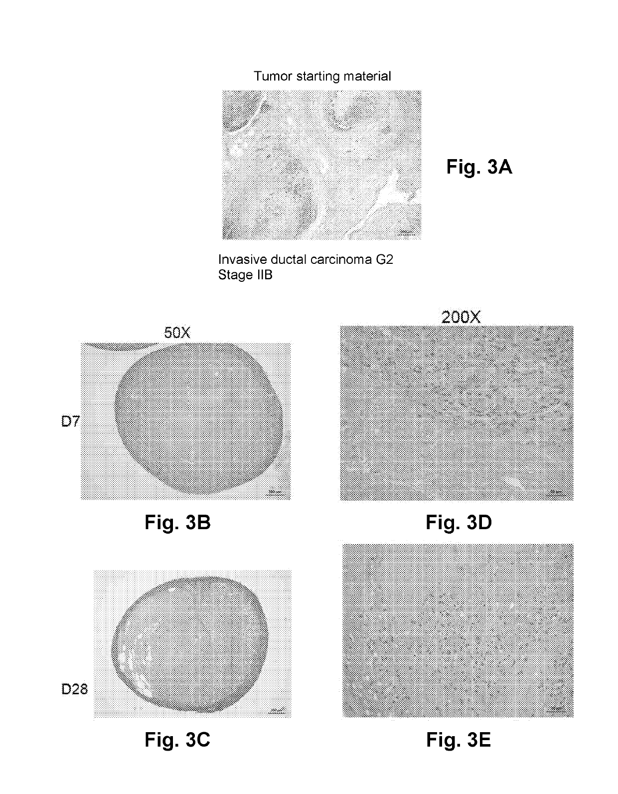
[0367]Small segments of human breast cancer tissue (approximately 0.5 mm×0.5 mm×0.5 mm) were then implanted into the interior of the bioprinted stromal tissue following the creation of a small incision. The wound induced during implantation is self-sealing and does not require a suture or adhesive to retain the tumor tissue in the interior of the structure.


PUM


Abstract

Described are three-dimensional, engineered, biological cancer models, methods of producing the same, and methods of identifying a therapeutic agent for cancer in an individual utilizing the three-dimensional, engineered, biological cancer models.

Description

STATEMENT REGARDING FEDERALLY-SPONSORED RESEARCH AND DEVELOPMENT[0001]This work was performed as part of an SBIR Phase I Contract funded by the NIH / National Cancer Institute (Contract HHSN261201400024C). The U.S. government has certain rights to the invention.BACKGROUND OF THE INVENTION[0002]The interaction between cancer cells and the surrounding stromal cells, comprised of fibroblasts, endothelial cells, adipocytes, and immune cells, plays a critical role in cancer initiation, progression, and metastasis. The stromal cells play a structural support role for the epithelium-derived cancer cells, modulate cell signaling and influence angiogenesis and metastasis to distant target tissues.[0003]Dissociation of human tumor samples causes fundamental alterations in the cancer cells as they are removed from their normal three-dimensional environment and results in a loss of heterogeneity that is required to accurately mimic the human disease. Researchers originally addressed the problem b...

Claims


Application Information

Patent Timeline
IPC IPC(8): C12N5/071C12N5/00C12N5/077C12N5/09A01K67/027G01N33/50A61K49/00B33Y70/00B33Y80/00
CPCC12N5/0697A01K2267/0331C12N5/0693A01K2207/30A01K2207/12A01K2207/15A01K2227/105A61K49/0008C12N2513/00B33Y70/00C12N2503/04B33Y80/00A01K67/0271C12N5/0062C12N2503/02C12N5/0656G01N33/5011A61L27/3604C12N5/0653C12N5/069
Inventor PRESNELL, SHARON C.KING, SHELBY MARIENGUYEN, DEBORAH LYNN GREENEJO, MINJIPELZ, ROSALIE SEARSALLEN-PETERSEN, BRITTANYLANGER, ELLEN
Owner ORGANOVO
Why Eureka
Try Eureka