Simultaneous isolation and preconcentration of exosomes by ion concentration polarization method and apparatus

a technology of exosomes and polarization ion concentration, applied in biochemistry apparatus and processes, laboratory glassware, instruments, etc., can solve the problems of limited frequency with which a region can be sampled, inherently invasive surgical biopsies, and significant risks for patients

Active Publication Date: 2019-09-05
UNIV OF NOTRE DAME DU LAC
View PDF0 Cites 7 Cited by
  • Summary
  • Abstract
  • Description
  • Claims
  • Application Information

AI Technical Summary

Problems solved by technology

Surgical biopsies are inherently invasive and therefore pose significant risks to patients.
This places a limitation on the frequency with which a region can be sampled to check for cancer, and in some cases, the suspected region may even be surgically inaccessible.
Additionally, the tissue sampled by a surgical biopsy tends to be a heterogeneous, rather than a homogeneous, representation of the tissue at large thereby leading to ambiguous conclusions.
Before analyzing exosomes for useful information, they need to be isolated from their resident media, which is difficult given their small size.
Such high speeds require not only large initial capital costs but also large maintenance and operating costs.
In addition to its high expense, ultracentrifugation is a time-consuming and labor-intensive process typically requiring four to six hours of work by a skilled technician or researcher.
In the end, it still does not produce very pure samples and results in yields of only 5-23% [41].
The final exosome sample still suffers from contamination by proteins, and the results tend to be highly variable.
However, it adds significantly to the complexity and time

Method used


Image

  • Simultaneous isolation and preconcentration of exosomes by ion concentration polarization method and apparatus
  • Simultaneous isolation and preconcentration of exosomes by ion concentration polarization method and apparatus
  • Simultaneous isolation and preconcentration of exosomes by ion concentration polarization method and apparatus

Examples


Embodiment Construction

[0035]The following description of example methods and apparatus is not intended to limit the scope of the description to the precise form or forms detailed herein. Instead the following description is intended to be illustrative so that others may follow its teachings.

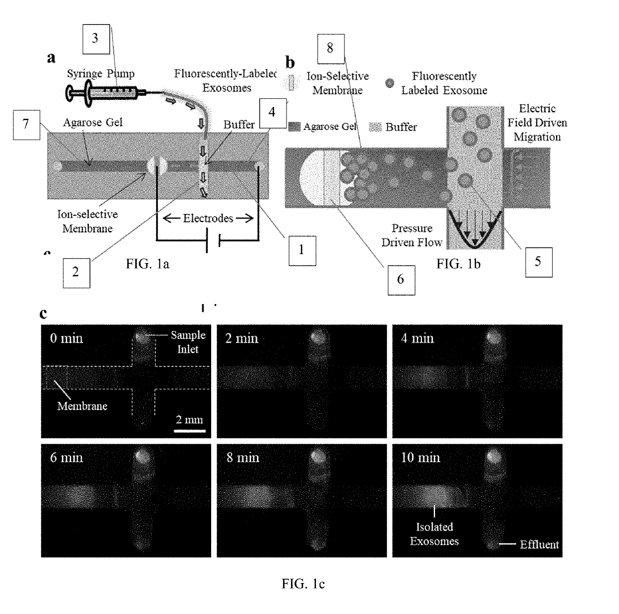
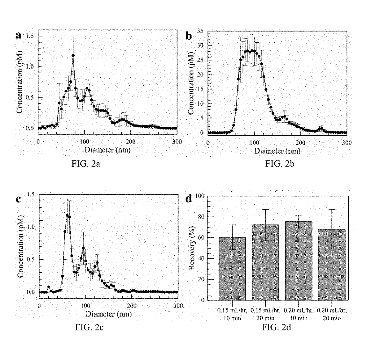
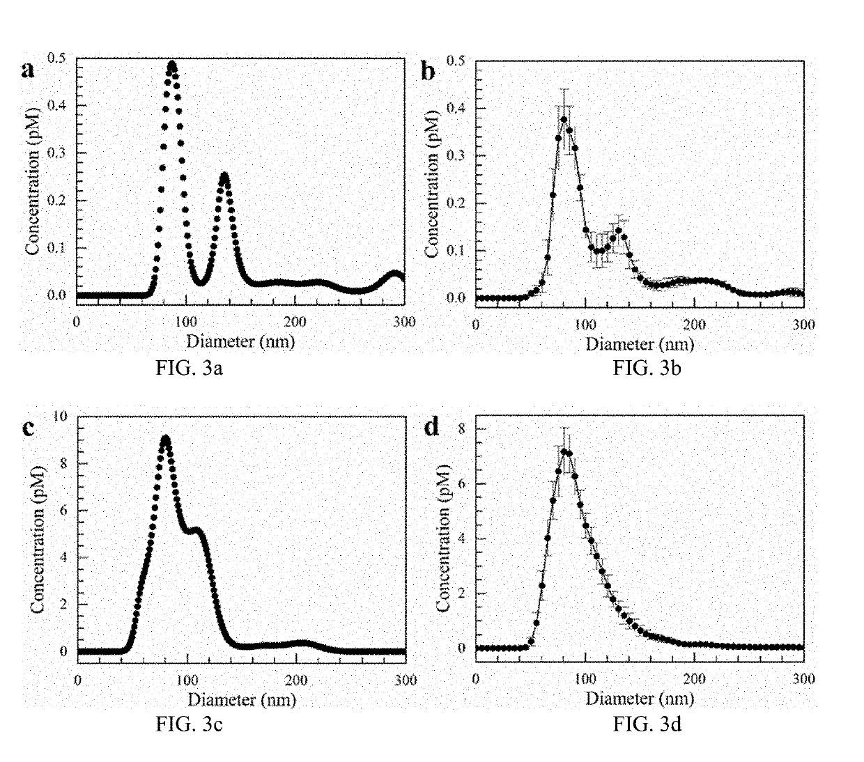
[0036]Exosomes carry microRNA biomarkers, occur in higher abundance in cancerous patients than in healthy ones, and because they are present in most biofluids, including blood and urine, can be obtained non-invasively. Standard laboratory techniques to isolate exosomes are expensive, time-consuming, provide poor purity, and recover on the order of 25% of the available exosomes. We present a new microfluidic technique to simultaneously isolate exosomes and preconcentrate them by electrophoresis using a high transverse local electric field generated by ion-depleting ion-selective membrane. We use pressure-driven flow to deliver an exosome sample to a microfluidic chip such that the transverse electric field forces them ...


PUM

PropertyMeasurementUnit
Fractionaaaaaaaaaa
Fractionaaaaaaaaaa
Fractionaaaaaaaaaa

Abstract

Exosomes carry microRNA biomarkers, occur in higher abundance in cancerous patients than in healthy ones, and because they are present in most biofluids, including blood and urine, can be obtained non-invasively. Standard laboratory techniques to isolate exosomes are expensive, time-consuming, provide poor purity, and recover on the order of 25% of the available exosomes. We present a new microfluidic technique to simultaneously isolate exosomes and preconcentrate them by electrophoresis using a high transverse local electric field generated by ion-depleting ion-selective membrane. We use pressure-driven flow to deliver an exosome sample to a microfluidic chip such that the transverse electric field forces them out of the cross flow and into an agarose gel which filters out unwanted cellular debris while the ion-selective membrane concentrates the exosomes through an enrichment effect. We efficiently isolated exosomes from 1×PBS buffer, cell culture media and blood serum. Using flow rates from 150 μL/hr to 200 μL/hr and field strengths of 100 V/cm, we consistently captured between 60% to 80% of exosomes from buffer, cell culture media, and blood serum as confirmed by both fluorescence spectroscopy and nanoparticle tracking analysis. Our microfluidic chip maintained this recovery rate for more than twenty minutes with a concentration factor of 15 for ten minutes of isolation.

Description

RELATED APPLICATION INFORMATION[0001]This application claims the benefit of and is a continuation of U.S. Provisional Application No. 62 / 637,209, filed Mar. 1, 2018, entitled “Microfluidics for Exosome Concentration” which is incorporated by reference in its entirety.GOVERNMENT FUNDING STATEMENT[0002]This invention was made with government support under Grant No. R21 AI105361, R21 CA206904 and HG009010 awarded by the National Institute for Health. The government has certain rights in the invention.FIELD OF THE DISCLOSURE[0003]The present disclosure relates generally to a connector for microfluidic devices and more particularly to a system and method for isolating exosomes.BACKGROUND OF RELATED ART[0004]In many instances, diagnosis of cancer requires surgical biopsy of a suspected cancerous region. Surgical biopsies are inherently invasive and therefore pose significant risks to patients. This places a limitation on the frequency with which a region can be sampled to check for cancer...

Claims


Application Information

Patent Timeline
IPC IPC(8): G01N1/40B01L3/00C12Q1/6806C12Q1/6825C12Q1/6886
CPCG01N1/4005B01L3/502753B01L3/502761C12Q1/6806C12Q1/6825B01L2400/0436G01N2001/4011G01N2001/4038B01L2400/0481B01L2400/0421C12Q1/6886B01L3/50273B01L2200/0668B01L2300/069B01L2400/0478G01N2001/4088
Inventor RICHARDS, KATHERINE E.MARCZAK, STEVENRAMSHANI, ZEINABHILL, REGINALDGO, DAVID B.CHANG, HSUEH-CHIA
Owner UNIV OF NOTRE DAME DU LAC
Why Eureka
Try Eureka