
Image processing method and apparatus using elastic mapping of vascular plexus structures

a vascular plexus and elastic mapping technology, applied in the field of image processing methods and medical observation devices, can solve the problems of insufficient spatial accuracy of these methods, consuming time and critical tim

Pending Publication Date: 2019-08-15
LEICA INSTR SINGAPORE PTE
View PDF10 Cites 4 Cited by
  • Summary
  • Abstract
  • Description
  • Claims
  • Application Information

AI Technical Summary

Benefits of technology

The invention provides a method and device that allow image-guided surgery to be performed on soft tissue that may easily deform and shift during surgery. This is done by using a structure that matches the pre-operative image data to the in vivo image data, which helps the surgeon align the image data without having to manually adjust it. The device may also include an optical cross-polarizing filter to reduce specular reflections and improve the identification of vascular plexus structures. Overall, this invention improves the precision and effectiveness of image-guided surgery.

Problems solved by technology

The spatial accuracy of these methods is not sufficient, however, if surgery is performed on soft tissue, which can shift and deform relative to the markers.
This consumes time and time is critical for all surgery.

Method used


Image

  • Image processing method and apparatus using elastic mapping of vascular plexus structures

Examples


Embodiment Construction

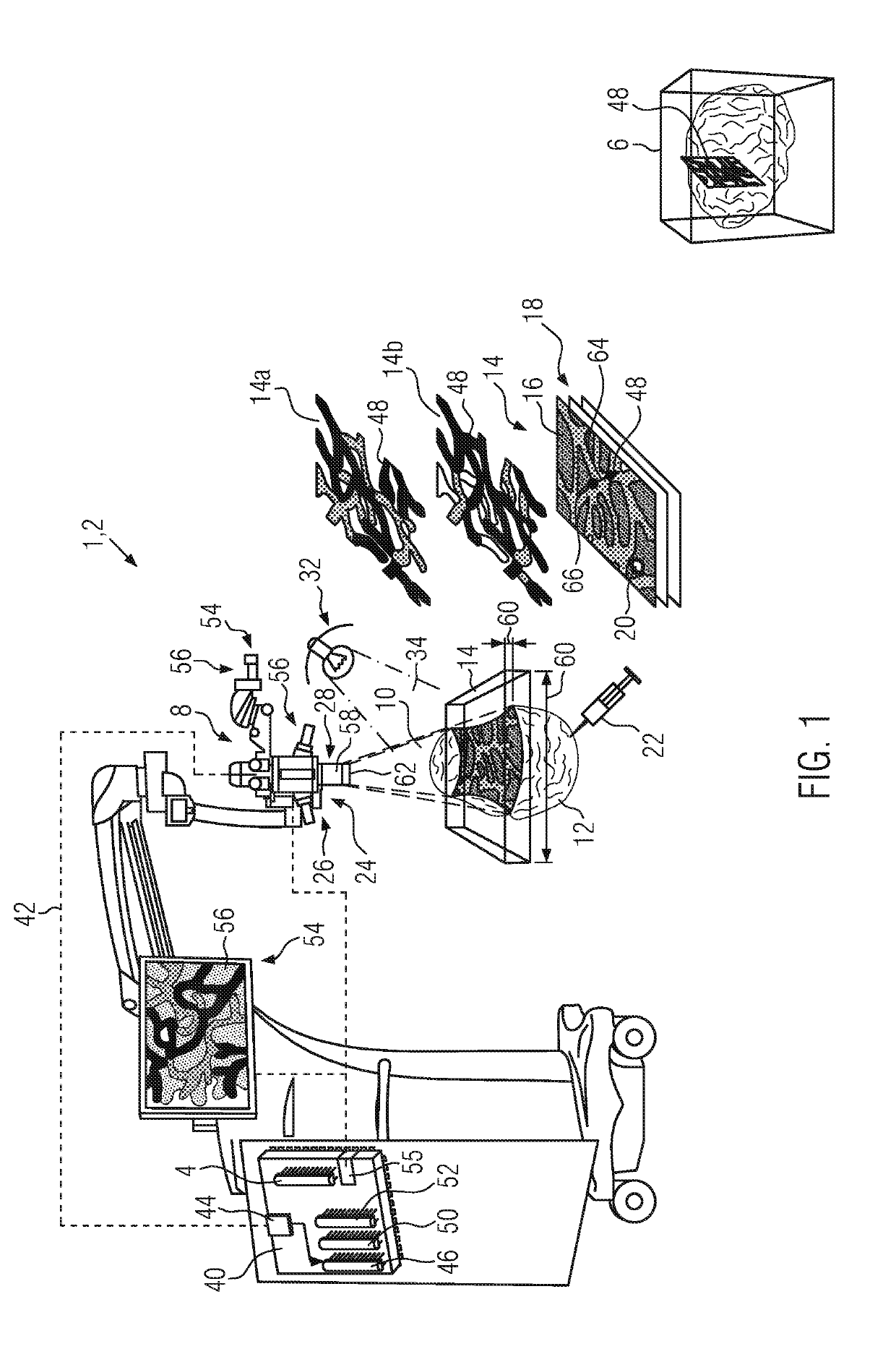
[0031]The configuration and function of an optical observation device 1 for observing live tissue, in particular during surgery, is explained. The medical observation device is shown to be a microscope 2 just for the purposes of explanation. The medical observation device 1 may also be an endoscope (not shown).

[0032]The medical observation device 1 comprises a memory assembly 4, in which pre-operative three-dimensional image data 6 are stored. The memory assembly 4 may comprise standard computer memory.

[0033]The medical observation device 1 further comprises a camera assembly 8, which has a field of view 10. During surgery, soft biological tissue 12, such as brain tissue, muscle tissue, lymph tissue or tissue of an internal organ or of other soft body parts, may be arranged in the field of view 10. During surgery, the camera assembly 8 acquires interoperative image data 14, which may be structured as a single input frame 16 or a time series 18 of input frames 16. The interoperative ...


PUM


Abstract

The invention relates to an image processing method and a medical observation device (1) such as a microscope (2) or endoscope. The device and method are used for displaying output image data (54) of soft biological tissue (12). In image-guided surgery, pre-operative three-dimensional image data (6) of the soft biological tissue (12) are elastically matched to interoperative image data (14) which are acquired during surgery. By displaying the elastically matched pre-operative three-dimensional image data (6) together with the interoperative image data (14), the surgeon may be made aware of the consistence of the soft biological tissue (12) below the visible surface layer. Existing systems for image-guided surgery need to be manually readjusted if surgery is done on soft biological tissue (12), which may deform and shift. To avoid this, the device and method according to the invention perform an elastic matching of the pre-operative three-dimensional image data (6) based on the interoperative image data (14) of the soft biological tissue (12). At least one vascular plexus structure (48) is first identified in the interoperative image data (14) and then the same vascular plexus structure (48) is identified in the pre-operative three-dimensional image data (6). The vascular plexus structure (48) in the pre-operative three-dimensional image data (6) is then elastically matched to the vascular plexus structure (48) in the interoperative image data (14). Output image data (54) are generated combining the elastically matched pre-operative three-dimensional image data (6) to the interoperative image data (14). Preferably, the at least one vascular plexus structure (48) is identified using fluorescent light from a fluorophore (22) which has been injected into the soft biological tissue (12).

Description

CROSS REFERENCE TO RELATED APPLICATIONS[0001]This application claims priority of European patent application number 18156906.2 filed Feb. 15, 2018, the entire disclosure of which is incorporated by reference herein.FIELD OF THE INVENTION[0002]The invention relates to an image processing method and a medical observation device for displaying soft tissue images, in particular in real time during surgery.BACKGROUND OF THE INVENTION[0003]Image-guided surgery is nowadays commonly used for certain kind of surgical operations, such as brain surgery. Image-guided surgery uses stored pre-operative three-dimensional information in the form of image data about the operation area. Such pre-operative three-dimensional image data may, for example, have been obtained using magnetic resonance imaging. During surgery, the pre-operative information is visually aligned with the actual optical view of the tissue to be operated on. Using the pre-operative three-dimensional information, tissue structures...

Claims


Application Information

Patent Timeline
Patent Type & Authority Applications(United States)
IPC IPC(8): A61B90/00G06T3/00G06K9/62G06T7/30A61B5/026A61B90/20A61B1/04
CPCA61B90/37G06T3/0081G06K9/6201G06T7/30A61B5/0261A61B90/361A61B90/20A61B1/043G06K2209/05G06T2207/30104G06T2207/20221G06T2207/10056G06T2207/10068G06T2207/10048A61B2090/367A61B2090/373A61B2090/365A61B2090/3941A61B2090/3979A61B2505/05G16H30/00A61B34/20G06T2207/10061G06T2207/30101A61B2034/2065A61B5/7425A61B5/0037A61B5/0059A61B5/0071G06V2201/03G06F18/22G06T3/153
Inventor THEMELIS, GEORGE
Owner LEICA INSTR SINGAPORE PTE
Learn More