
Method for Detecting a Genomic Fusion Event

a genomic fusion and event technology, applied in the field of methods for detecting genomic rearrangements, can solve the problems of only being able to apply to fish, unable to detect gene fusion, and only being able to apply to cells or tissues

Inactive Publication Date: 2019-08-08
INIVATA LTD
View PDF0 Cites 4 Cited by
  • Summary
  • Abstract
  • Description
  • Claims
  • Application Information

AI Technical Summary

Benefits of technology

The present patent describes methods for detecting genomic rearrangements, particularly gene fusion events, by targeting DNA molecules of interest using two sets of primers. The method does not require sample enrichment and provides a simple and reliable way to identify and characterize fusion events associated with cancer and other diseases. The methods can also be used to monitor disease progression and predict treatment outcomes. A key advantage is that the method does not require end repair or ligation, which can lead to loss of starting material.

Problems solved by technology

FISH can only be applied to cells or tissue and therefore cannot be used to detect gene fusions in cell free circulating nucleic acids or in DNA already extracted from tissue.
FISH also requires intact nuclei, the need to visually assess individual cells and cannot give the sequence of the breakpoint.
However, this approach can only be applied to mRNA and it is not feasible to identify the breakpoints that have occurred in the genome (DNA). mRNA typically has a short half-life and is therefore a challenging biomarker in heavily degraded samples such as FFPE and circulating RNA (ctRNA).
A significant challenge with this approach is that the fusions will often occur throughout large intronic spaces.
The limitation with these approaches is the requirement for DNA greater than 500 bp in length and therefore they are not suitable for fragmented DNA such as FFPE or cfDNA.
These long-range PCR methodologies are also not compatible with Next Generation Sequencing Technologies due to the short-read length (<500 bp) of most next generation sequencing platforms without further complex steps such as fragmenting the DNA then ligating on adaptors.
This methodology also requires a potentially large number of individual PCR reactions to assess each sample or complex steps such as positive selection using biotin-labelled primers in order to multiplex such a reaction.
As these steps are highly inefficient ˜70% of starting material is lost prior to hybridisation, limiting the ability to detect fusions present at low allelic frequencies.
Finally, this approach is time consuming, normally requiring overnight incubation of target and probe to enable hybridisation.
However, this approach is time consuming and requires the ligation of universal adapters to DNA ends.
The inefficiency of ligation limits the ability to detect fusions present at low allelic frequencies.
It also increases complexity and cost of the workflow as an additional hybridisation probe is required.

Method used


Image

  • Method for Detecting a Genomic Fusion Event
  • Method for Detecting a Genomic Fusion Event
  • Method for Detecting a Genomic Fusion Event

Examples


example 1

of EML4-ALK Variant at a Range of Allelic Fractions

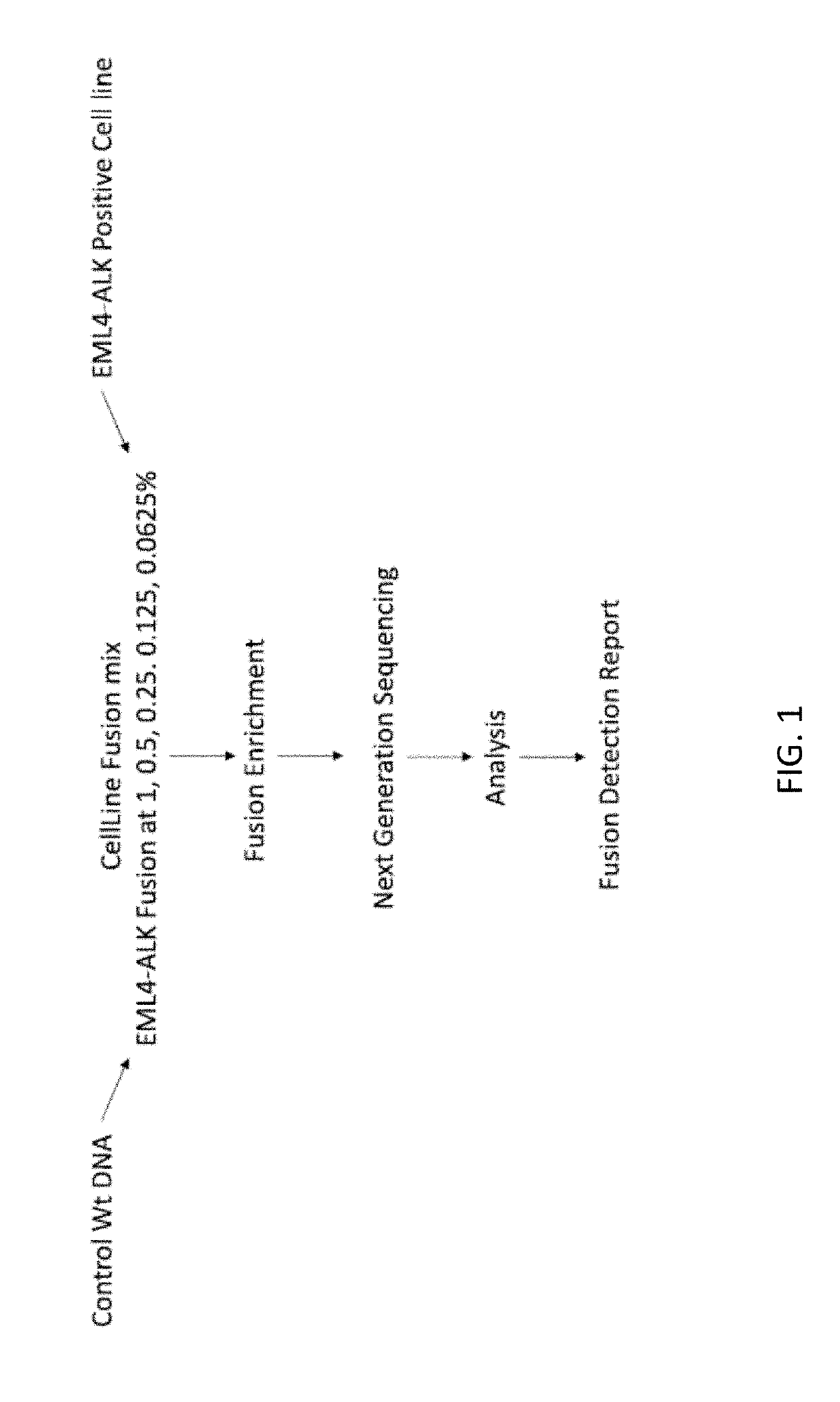
[0498]A custom cell free DNA reference standard containing an EML4-ALK fusion of sequence GAAGTTCCTATACTTTCTAGAGAATAGGAACTTC (SEQ ID NO: 1) at an allelic fraction of 2.5% was obtained from Horizon Discoveries. This reference standard was diluted in sheared (average 188 bp) human placental DNA (Bioline) to achieve allelic fractions of 1%, 0.5%, 0.25%, 0.125% and 0.0625%. Three samples were created at each allelic fraction.

[0499]Each sample was split into two replicates, each containing a total of 4000 input copies. PCR amplification was performed on two replicates using the ALK primer panel (table 1). Each PCR contained 25 uL DNA, 27.5 uL Platinum SuperFi 2× Master Mix (Invitrogen) and 2.5 uL of the ALK primer pool (for primer concentration see table 1). PCR cycling was followed using manufacturer' instructions. The PCR product was cleaned up using SPRIselect reagent (Beckman Coulter B23319) using the manufacturers protocol. DNA was ...

example 2

of ROS1-CD74 Variant

[0501]A synthetic gBlock containing a ROS1 fusion sequence (based on a sequence reported in the literature: Seki, Mizukami and Kohno, Biomolecules, 2015, 5, 2464-2476) was synthesized by IDT and was sheared using the covaris to achieve an average size of 150 bp. The gBlock was added to sheared (average 188 bp) human placental DNA (Bioline) to achieve an allelic fraction of 1%.

[0502]Each sample was split into two replicates, each containing a total of 4000 input copies. PCR amplification was performed on two of the replicates using the ROS1 primer panel (table 2). Each PCR contained 25 uL DNA, 27.5 uL Platinum SuperFi 2× Master Mix (Invitrogen) and 2.5 uL of the ROS1 primer pool (for primer concentration see table 2). PCR Cycling was followed using manufactures instructions. The PCR product was cleaned up using SPRIselect reagent (Beckman Coulter B23319) using the manufacturers protocol. DNA was eluted in 18 uL and a second PCR using Indexed illumina primers was p...

example 3

of ROS1-CD74 Variant Using Sequential Amplification

[0504]The same synthetic ROS1 fusion gBlock at 1% allelic fraction as was used in Example 2 was tested.

[0505]Each sample was split into two replicates, each containing a total of 4000 input copies. Linear amplification of the template was performed on two of the replicates using only the ROS1 forward primer panel. Each reaction contained 25 uL DNA, 27.5 uL Platinum SuperFi 2× Master Mix (Invitrogen) and 2.5 uL of the ROS1 forward primer pool. Cycling was followed using manufactures instructions. The PCR product was cleaned up once using SPRIselect reagent (Beckman Coulter B23319) using the manufacturers protocol. DNA was eluted in 18 uL and a first PCR using a i5 adapter forward primer and the ROS1 reverse primer pool was performed. Each PCR contained 10 uL DNA, 25 uL Platinum SuperFi 2× Master Mix (Invitrogen), 2.5 ul of the i5 adapter forward primer and 2.5 uL of the ROS1 reverse primer pool. Cycling was followed using manufacture...


PUM

PropertyMeasurementUnit
pHaaaaaaaaaa
fluorescence in situ hybridizationaaaaaaaaaa
gel electrophoresisaaaaaaaaaa

Abstract

The present disclosure relates to methods for detecting and targeting genomic rearrangements, in particular gene fusion events, by targeting a DNA molecule of interest with a set or pool of primers, wherein the forward primers and reverse primers produce a PCR amplification product when a genomic rearrangement is present. The present disclosure also relates to methods of bioinformatic analysis to determine whether or not the detection of an amplification product from the selective PCR is actually indicative of the presence of a gene fusion. The present disclosure also related to related methods of diagnosis and treatment of diseases and conditions associated with such genomic rearrangements, in particular cancers, such as lung cancer.

Description

CROSS-REFERENCING[0001]This application claims the benefit of GB1709675.1, filed on Jun. 16, 2018, which application is incorporated herein in its entirety for all purposes.BACKGROUND[0002]The present disclosure relates to methods for detecting genomic rearrangements, in particular gene fusion events, as well as related methods of diagnosis and treatment of diseases and conditions associated with such genomic rearrangements, in particular cancers, such as lung cancer.[0003]Genetic or chromosomal rearrangements are a type of chromosomal abnormality in which the normal order of the genetic code has been altered. A common genomic rearrangement that is associated with cancer is genetic fusion. A gene fusion event may occur in cancerous or pre-cancerous cells and can be detected in patients to help classify the cancer and determine appropriate treatments.[0004]Existing methods for detecting gene fusions include fluorescence in situ hybridization (FISH), RT-PCR, long range PCR and hybridi...

Claims


Application Information

Patent Timeline
Patent Type & Authority Applications(United States)
IPC IPC(8): C12Q1/6886C12Q1/6806C12Q1/686
CPCC12Q1/6886C12Q1/6806C12Q1/686C12Q2600/16G16B20/00G16B30/00C12Q1/6853C12Q2525/155C12Q2525/161C12Q2531/113C12Q2535/122C12Q2537/159C12Q2600/156C12Q2600/106
Inventor WOODHOUSE, SAMUELLENSING, STEFANIEFORSHEW, TIMPLAGNOL, VINCENTSMITH, MATTHEW EDWARDHOWARTH, KARENEPSTEIN, MICHAEL
Owner INIVATA LTD
Learn More