Ultrasound shear wave vibro-elastography of the abdomen

a technology of ultrasound and vibroelastography, applied in the field of medical imaging, can solve problems such as difficulties in the presence of the ribcage, and achieve the effect of large region or volume of interes

Active Publication Date: 2019-06-27
THE UNIV OF BRITISH COLUMBIA
View PDF0 Cites 10 Cited by
  • Summary
  • Abstract
  • Description
  • Claims
  • Application Information

AI Technical Summary

Benefits of technology

[0013]This invention provides apparatus and methods useful for shear-wave elastography. Specific example mechanical and electrical designs which allow for deep, consistent shear wave transmission into the tissue, thro...

Problems solved by technology

A range of issues arise when quantitative ultrasound shear wave vibro-elastography images are required in deep tissue that is under the ribcage...

Method used


Image

  • Ultrasound shear wave vibro-elastography of the abdomen
  • Ultrasound shear wave vibro-elastography of the abdomen
  • Ultrasound shear wave vibro-elastography of the abdomen

Examples


Embodiment Construction

[0065]Throughout the following description, specific details are set forth in order to provide a more thorough understanding of the invention. However, the invention may be practiced without these particulars. In other instances, well known elements have not been shown or described in detail to avoid unnecessarily obscuring the invention. Accordingly, the specification and drawings are to be regarded in an illustrative, rather than a restrictive sense. The specific details disclosed herein are not to be interpreted as limiting, but rather as a representative basis for teaching one skilled in the art how to employ the present invention in virtually any detailed system, structure, or manner.

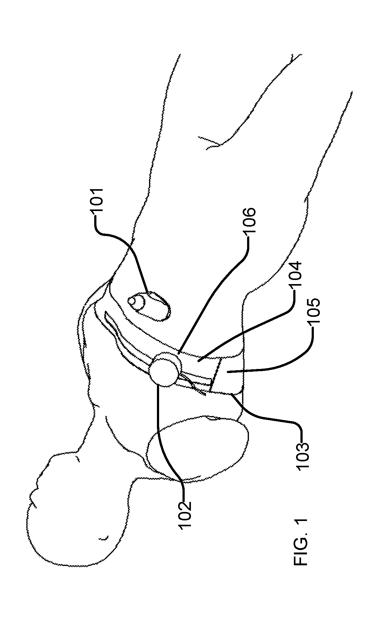
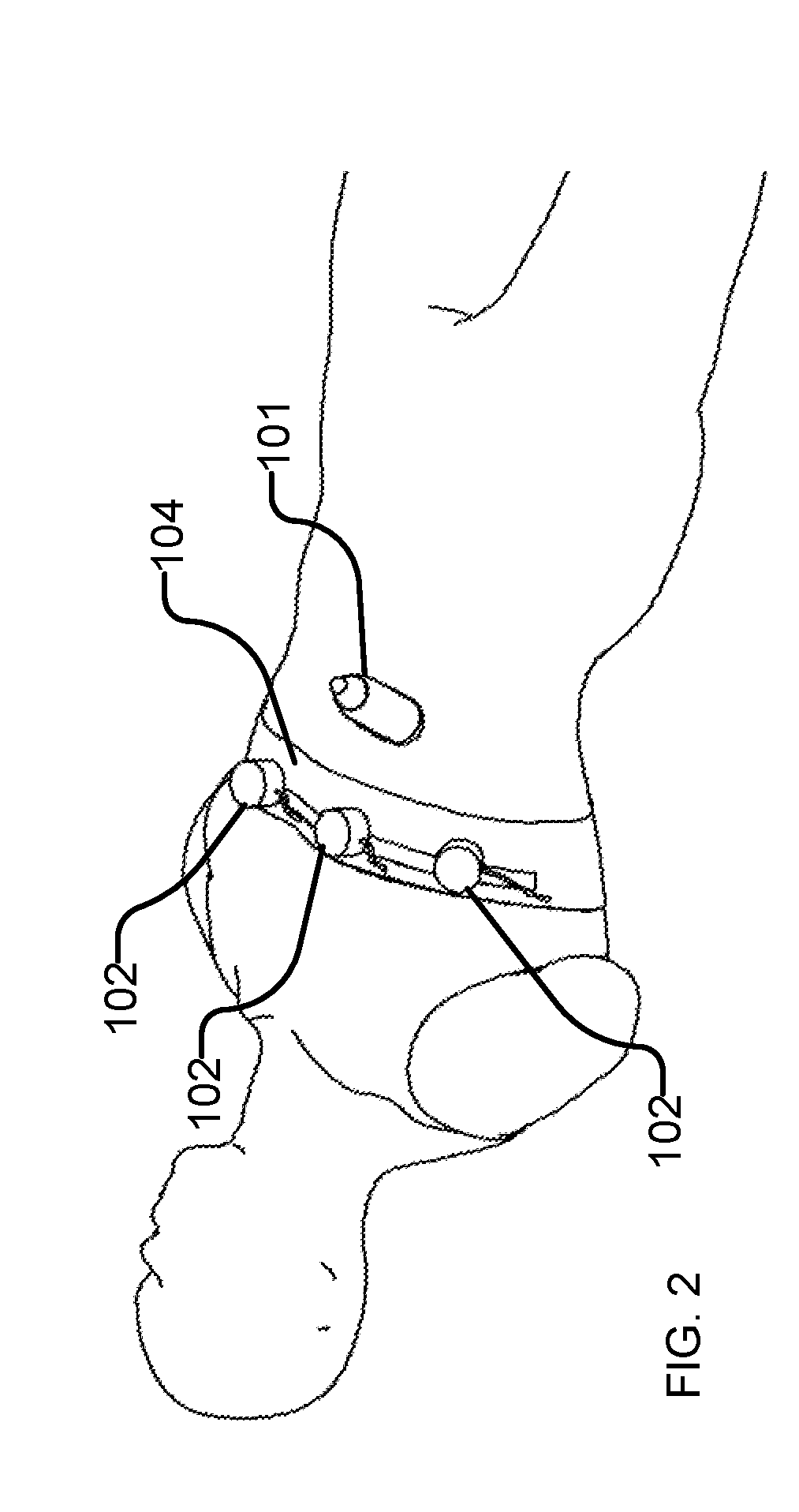
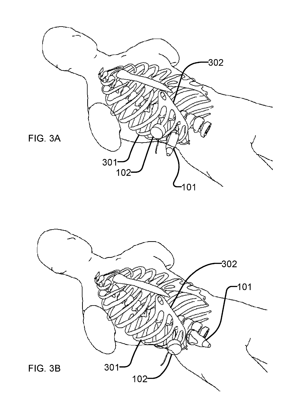
[0066]The invention may be applied to image the abdomen of a person or animal although the invention can also be used to image other parts of the body. The liver is used as an illustrative example of an organ that can be imaged with this invention.

[0067]Throughout this document: “exciter” refers to...


PUM


Abstract

A system useful for performing ultrasound elastography of organs such as the liver allows efficient and robust data acquisition. The system may be applied to perform real-time, non-invasive ultrasound imaging of the liver in humans. Steady-state, shear wave absolute elastography is used to measure the Young's modulus of the liver tissue. This method involves the use of an external exciter or vibrator to shake the tissue and generate a shear wave. Accurate placement of an ultrasound transducer facilitates measurement of the tissue motion due to the shear wave. The stiffness of tissues in the region being imaged may be computed from the measured tissue motions. The following innovations address both vibrator and transducer placement, as well as some specific methods to ensure adequate wave propagation, in order to obtain accurate and consistent measurements.

Description

CROSS-REFERENCE TO RELATED APPLICATIONS[0001]This application is a continuation of PCT International Application No. PCT / CA2017 / 050804 filed 30 Jun. 2017, which claims the benefit under 35 U.S.C. § 119 of U.S. Application No. 62 / 357254 filed 30 Jun. 2016, both of which are entitled ULTRASOUND SHEAR WAVE VIBRO-ELASTOGRAPHY OF THE ABDOMEN and both of which are hereby incorporated herein by reference for all purposes.FIELD OF THE INVENTION[0002]This invention relates to medical imaging, in particular, medical imaging that applies shear-wave elastography. The present technology may provide information characterizing the mechanical properties of abdominal organs such as the liver, kidneys, spleen, bladder wall etc. Example applications of the present technology include imaging of the abdomen, including, for example, the liver.BACKGROUND OF THE INVENTION[0003]Medical imaging is used in many applications to determine the composition of tissues. In medical images the intensity and / or color ...

Claims


Application Information

Patent Timeline
IPC IPC(8): A61B8/08A61B8/14A61B8/00
CPCA61B8/485A61B8/14A61B8/54A61B8/4218A61B8/085A61B8/461A61B8/4209A61B8/4227A61B8/483G01S7/52042A61B5/0051A61B8/4245A61B8/52A61B8/08
Inventor SALCUDEAN, SEPTIMIUROHLING, ROBERTHONARVAR, MOHAMMADLOBO, JULIO RAULSCHNEIDER, CAITLIN MARIE
Owner THE UNIV OF BRITISH COLUMBIA
Why Eureka
Try Eureka