Image simulation system to covisualize electrodes and cortical vessels on 3D brain and its method

a brain structure diagram and image simulation technology, applied in image enhancement, tomography, instruments, etc., can solve the problems of not providing electrode integration and electrode image technology, traditional ways cannot simultaneously learn relative position information of brain structures, blood vessel distribution and electrode position

Inactive Publication Date: 2019-04-04
CHUNG SHAN MEDICAL UNIV HOSPITAL
View PDF1 Cites 1 Cited by
  • Summary
  • Abstract
  • Description
  • Claims
  • Application Information

AI Technical Summary

Benefits of technology

The present invention provides an image simulation system and method for accurately showing the positions of electrodes and blood vessels in a 3D brain structure diagram. This helps to avoid bleeding and other complications during medical surgery. The invention solves the problem of conventional technology where the relative positions of electrodes and blood vessels in brains could not be known at the same time.

Problems solved by technology

In addition, although a method disclosed by Taiwan patent No. 201225922 titled “An Angiography Method for Visualization of Trans-Bone Blood Vessels” includes angiography using contrast media, no technique of electrode integration and electrode images is provided in the above mentioned patent.
Especially for patients with epilepsy, when electrodes have been intracranially implanted, traditional ways cannot simultaneously learn relative position information of brain structures, blood vessel distribution and electrode positions.

Method used


Image

  • Image simulation system to covisualize electrodes and cortical vessels on 3D brain and its method
  • Image simulation system to covisualize electrodes and cortical vessels on 3D brain and its method
  • Image simulation system to covisualize electrodes and cortical vessels on 3D brain and its method

Examples


Embodiment Construction

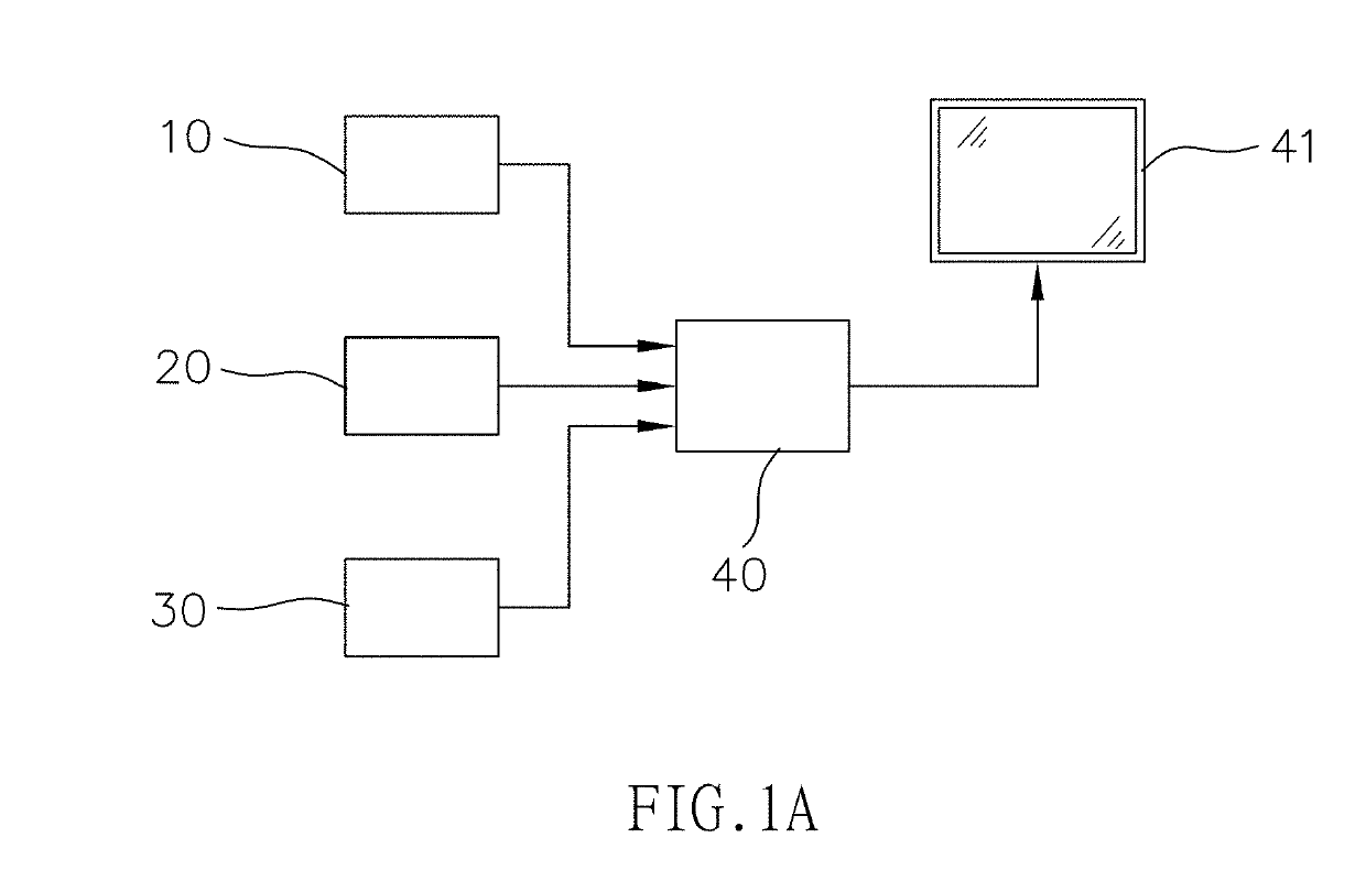
[0044]Referring to FIGS. 1A, 1B, 2, 3, 4 and 5, the present invention is related to an image simulation system and method for covisualizing electrodes and cortical vessels on a three dimensional (3D) brain structure diagram. The image simulation system comprises a first image capturing device 10, a second image capturing device 20, a third image capturing device 30, and a processing device 40.

[0045]The first image capturing device 10 is used to capture a plurality of first images (for example, the 146th to 149th layers, referring to FIGS. 7A, 7B, 7C and 7D) of a brain 90 in advance. The plurality of first images are completely used to display a three-dimensional structure of the brain 90.

[0046]The second image capturing device 20 is used to capture a plurality of second images (for example, the 146th to 149th layers, referring to FIGS. 8A, 8B, 8C and 8D) of the brain 90 in advance. The plurality of second images are completely used to display blood vessels 91 of the brain 90 (referr...


PUM


Abstract

A method comprises steps of capturing first images for displaying a three-dimensional brain structure, capturing second images for displaying blood vessels of the brain and capturing third images displaying implanted electrodes in the brain. These second images are set as standards to respectively adjust and align the first images and the third images. Contrast of the blood vessels is enhanced and the blood vessels are colored with a first color. Contrast of the electrodes is enhanced and the electrodes are colored with a second color. The aligned first images, the colored second images and the colored third images are integrated to obtain integrated viewable information of a brain, (intracranial) electrodes and blood vessels for medical reference. So, spatial positions of implanted electrodes and blood vessels of brains can be confirmed before conducting a medical surgery. And, the physicians can avoid bleeding problem due to accidental touch on blood vessels.

Description

BACKGROUND OF THE INVENTION1. Field of the Invention[0001]The present invention is related to an image simulation system and method for covisualizing electrodes and cortical vessels on a three dimensional (3D) brain structure diagram, and more particularly to an image simulation system and method for covisualizing electrodes and cortical vessels on a 3D brain structure diagram having advantages that spatial positions of implanted electrodes and blood vessels of brains can be confirmed in advance at the time of medical surgery, and that physicians can avoid bleeding problem due to accidental touch on blood vessels.2. The Related Arts[0002]Although a conventional method disclosed by Taiwan patent No. 1318874 titled “A Method to Construct a Three-Dimensional Image of Human Tissues” discloses a method of constructing a three-dimensional structure of brains, no technique of displaying electrodes and blood vessels in the brains is provided at the same time.[0003]In addition, although a me...

Claims


Application Information

Patent Timeline
IPC IPC(8): G06T7/00G06F17/50G16H30/40
CPCG06T7/0016G06F17/5009G16H30/40A61B5/055A61B5/0042A61B6/032G06T2207/30016G06T2207/10028G06T2207/10088G06T2207/10081A61B5/7425A61B5/061A61B5/489G01R33/5635A61B2576/026G01R33/285G01R33/5608A61B6/501A61B6/5247A61B2090/364G16H50/50G16H20/40G06F30/20A61B34/10
Inventor HSIN, YUE-LOONGPENG, SYU-JYUNCHANG, CHENG-SIUSHIH, FU-YUAN
Owner CHUNG SHAN MEDICAL UNIV HOSPITAL
Why Eureka
Try Eureka