System comprising indicator features in high-resolution micro-ultrasound images

a high-resolution micro-ultrasound and indicator feature technology, applied in the field of medical imaging and diagnostics, can solve the problems of obstructing the urinary tract, trus is not reliable enough to be used solely as a biopsy template, and the accuracy of trus was only 52%, so as to facilitate the diagnosis of the tissue

Active Publication Date: 2018-11-22
EXACT IMAGING INC
View PDF18 Cites 10 Cited by
  • Summary
  • Abstract
  • Description
  • Claims
  • Application Information

AI Technical Summary

Benefits of technology

[0002]This invention provides a system and method for providing Indicator Features in high resolution micro-ultrasound prostate images, wherein the ultrasound device operates at a center frequency of 15 MHz and above. These Indicator Features can be used to train a linear or non-linear classifier, which can be used to classify patient images of the prostate. These Indicator Features can be used to guide a clinician as to where to take a biopsy core from the prostate, as well as assist in diagnosis of the tissue.

Problems solved by technology

Hyperplastic enlargement of the prostate gland, or enlargement due to abnormal but benign multiplication of the cells thereof, often leads to compression of the urethra thereby resulting in obstruction of the urinary tract.
TRUS however, is not reliable enough to be used solely as a template for biopsy.
Furthermore, in PSA screened populations, the accuracy of TRUS was only 52% due to false-positive findings encountered.
Efficacy of color and power Doppler ultrasound for targeting cancer has not been demonstrated, probably due to limited resolution and small flow velocities.
These modalities are helpful, but have drawbacks and limitations.
MRI is expensive and not mobile.
Nuclear scintillation is expensive, provides low resolution planar images, and there are problems with radiotracer excretion through the kidneys.
Both these modalities are not available for general use.
However, conventional ultrasound technology has limitations that make it unsuitable for some applications.
For example, ultrasound waves do not pass well through certain types of tissues and anatomical features, and ultrasound images typically have weaker contrast and lower spatial resolution than X-Ray and MRI images.
Also, ultrasonic imaging has difficulties distinguishing between acoustically homogenous tissues (i.e. tissues having similar ultrasonic properties).
In essence, conventional ultrasound images only demonstrate the possibility of revealing dark spots (hypoechoic areas), which correlate very poorly with cancer.
When it is visible (hyper or hypoechoic), it is not possible to say with certainty if it is cancer or benign because many other non-cancer conditions such as prostate atrophy, inflammation of the prostate gland, and benign tumors may also look similar in appearance on ultrasound examination.
More importantly, taking targeted biopsies of only these areas has a high false negative rate, missing clinically significant cancer in up to 48% of patients with proven disease on radical prostatectomy (Carter H B et al: J. Urol. 1989, 142: 1008).
Biopsies are uncomfortable and bleeding may result as a complication.
Because of poor lesion detection, even the current prostate biopsy techniques miss approximately 30% of prostate cancer.
Utility of color flow and power Doppler in conjunction with gray scale ultrasound has been explored, but has not achieved wide-spread use.
While they provide some level of consistency and are very likely to find larger tumors, smaller cancers and atypically located cancers may be missed.
These patients often undergo repeat or saturation biopsies, often in combination with more costly and specialized imaging such as MRI.
These further procedures come with increased costs, complexity, and an increased risk of morbidity both due to the repeated procedures themselves and to potential delays in diagnosis which can lead to decreased opportunity for cure.

Method used


Image

  • System comprising indicator features in high-resolution micro-ultrasound images
  • System comprising indicator features in high-resolution micro-ultrasound images
  • System comprising indicator features in high-resolution micro-ultrasound images

Examples


Embodiment Construction

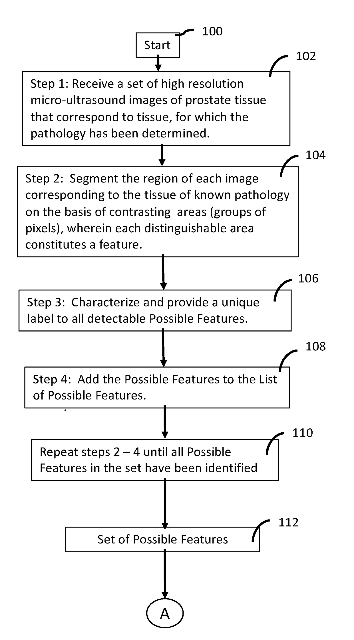
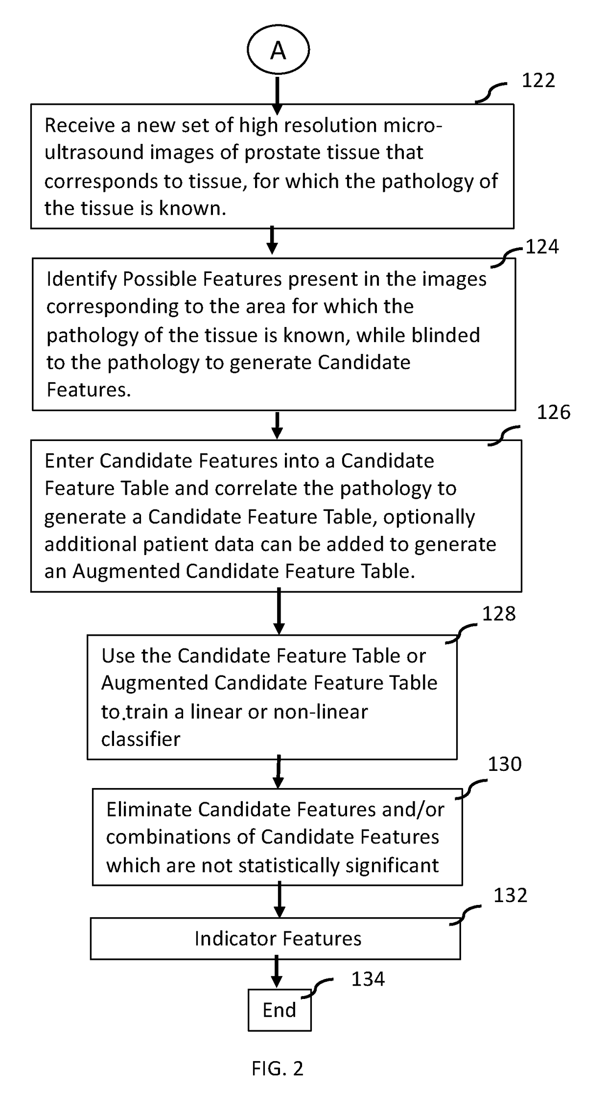
[0037]This invention provides a method of generating Indicator Features, Indicator Features and their use in a system comprising high resolution micro-ultrasound to determine a degree of risk for cancer in prostate tissue. Indicator Features are provided in high resolution micro-ultrasound images and are used to train a classifier, which can be used to determine a risk of cancer in other images, or can be grouped into a multiclass classifier, which can be used to determine a risk of cancer.

[0038]A Method of Generating Indicator Features

[0039]With conventional ultrasound, there was essentially only one feature available, a dark spot (hypoechoic region), which was used in the images as a possible indicator of cancer. As described in the Background, this feature correlated poorly with cancerous tissue. Thus, there was little confidence that ultrasound technology in general would be useful for identifying risk of cancer. Initially, once the prostate was able to be visualized with high r...


PUM


Abstract

This invention provides a system and method for providing Indicator Features in high resolution micro-ultrasound prostate images, wherein the ultrasound device operates at a center frequency of 15 MHz and above. Indicator Features are features, which have been identified alone and/or in combination with other features in high resolution micro-ultrasound images, and have been determined to be significantly statistically correlated to either benign tissue or some grade of cancerous tissue on the basis of predictive probabilities. These Indicator Features can be used to train a linear or non-linear classifier, which can be used to classify patient images of the prostate. These Indicator Features can be used to guide a clinician as to where to take a biopsy core from the prostate, as well as assist in diagnosis of the tissue.

Description

FIELD OF THE INVENTION[0001]The present disclosure relates generally to medical imaging and diagnostics, and more specifically to Indicator Features in high resolution micro-ultrasound images of the prostate.SUMMARY[0002]This invention provides a system and method for providing Indicator Features in high resolution micro-ultrasound prostate images, wherein the ultrasound device operates at a center frequency of 15 MHz and above. These Indicator Features can be used to train a linear or non-linear classifier, which can be used to classify patient images of the prostate. These Indicator Features can be used to guide a clinician as to where to take a biopsy core from the prostate, as well as assist in diagnosis of the tissue.COPYRIGHT AND TRADEMARK NOTICE[0003]A portion of the disclosure of this patent document contains material which is subject to copyright protection. The copyright owner has no objection to the facsimile reproduction of the patent document or the patent disclosure, a...

Claims


Application Information

Patent Timeline
Patent Type & Authority Applications(United States)
IPC IPC(8): A61B8/08G06T7/11G06K9/62G06K9/46G06T7/00A61B8/12
CPCA61B8/5223G06T7/11G06K9/6257G06K9/6277G06K9/46G06T7/0012G06K9/628A61B8/481A61B8/488A61B8/485A61B8/5246A61B8/483A61B8/085A61B8/12G06T2207/10132G06T2207/20081G06T2207/20084G06T2207/30081G06T2207/20076G06K2209/051G06T2207/30096G06T7/0014G06F16/00G16H30/40G16H40/63G16H50/20G16H50/70G06V2201/031G06F18/2148G06F18/2415G06F18/2431
Inventor WODLINGER, BRIANMCGRATH, THERESAWEN, JERROLD
Owner EXACT IMAGING INC
Why Eureka
Try Eureka