
Medical Device For The Selective Separation Of A Biological Sample

a biological sample and selective separation technology, applied in the field of medical devices and biological sample separation methods, can solve the problems of inability to sort the most fertile sperm from the sample, inability to perform in vitro and particularly inability to achieve in vitro and in situ treatment of infertility, etc., to achieve efficient separation or enrichment, reduce separation and/or preparation time, and optimal

Active Publication Date: 2018-10-04
MY123BABY MEDICAL LTD
View PDF0 Cites 4 Cited by
  • Summary
  • Abstract
  • Description
  • Claims
  • Application Information

AI Technical Summary

Benefits of technology

The patent describes a method for enriching high-quality sperm cells using a simple and cost-effective device. The device has layers that help separate biological samples, resulting in a higher quality product. The method involves a single-step process that eliminates potential damage to sperm cells and minimizes oxidative stress. This method can be used in fertility clinics, research laboratories, or at home to treat male infertility.

Problems solved by technology

Unfortunately, the success rate of ARTs is generally only about 20 to 30 percent and often requires 4 to 5 fertilization rates.
Furthermore, assisted reproduction had mainly focused on increasing the female reproduction potential, focusing primarily on egg retrieval and cultivation due to the common misbelief that sperm quality had no or not a substantial effect on fertility.
In vivo and particularly in situ treatment of infertility is rarely possible.
In some current techniques, due to time limitations or restrictions, no actual sperm separation, but rather only a sperm purification process is performed.
This means that such sperm preparation methods may not sort the most fertile sperm from a sample.
However, while high density gradient centrifugation and / or swim-up assays provide for an increased concentration and retrieval of sperm cells and have been found to generally select sperm populations with increased DNA integrity, the quality of the sperm cells is often (still) impaired.
Furthermore, said methods do not resemble the natural in vivo process and the selected sperm population is often contaminated with poorly motile sperm and somatic cells (e.g. leukocytes), generally requiring a visual inspection.
In addition, the selected sperm population may suffer iatrogenic injury (e.g. oxidative injury) due to centrifugation and / or prolonged and repeated processing, particularly reducing DNA quality.
Such methods hence do not provide a desirable outcome.
Furthermore, known selection techniques often depend on professional skills and technician experience in selecting sperm and are based on multi-step methods, rendering the method potentially error-prone.
In addition, centrifugation also requires both expensive equipment and materials and is time-extensive, limiting infertility services to middle and high income households.
Despite these adverse effects, clinical spermatozoa preparation techniques have not significantly improved in the past 20 years.
However, such methods are not applicable for e.g. sperm separation techniques, wherein a large number of cells is required and furthermore require professional skill for handling.
By the same token, from the patent application US 2009 / 0123961 A1 a method for testing of chemotaxis is known using a fluid gradient, which furthermore provides no barriers and may hence not be applicable to or effective for highly motile cells.
Hence, in most cases, the complexity of using microfluidic devices and the need for supporting infrastructures decrease effectiveness while increasing costs and hence currently do not allow for a clinical implementation, scaling, and / or commercialization.
Moreover, the low sample volume characteristic of microfluidics proves to be a disadvantage in the context of sperm selection due to the inherent milliliter-scale size of human samples.

Method used


Image

  • Medical Device For The Selective Separation Of A Biological Sample
  • Medical Device For The Selective Separation Of A Biological Sample
  • Medical Device For The Selective Separation Of A Biological Sample

Examples


Embodiment Construction

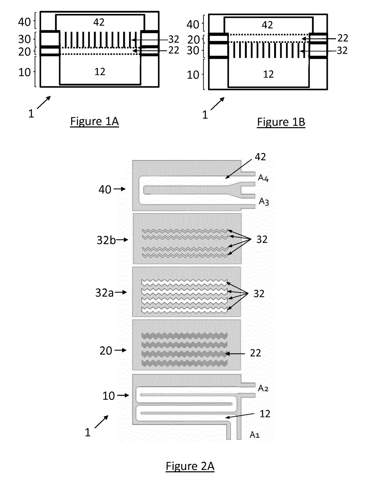
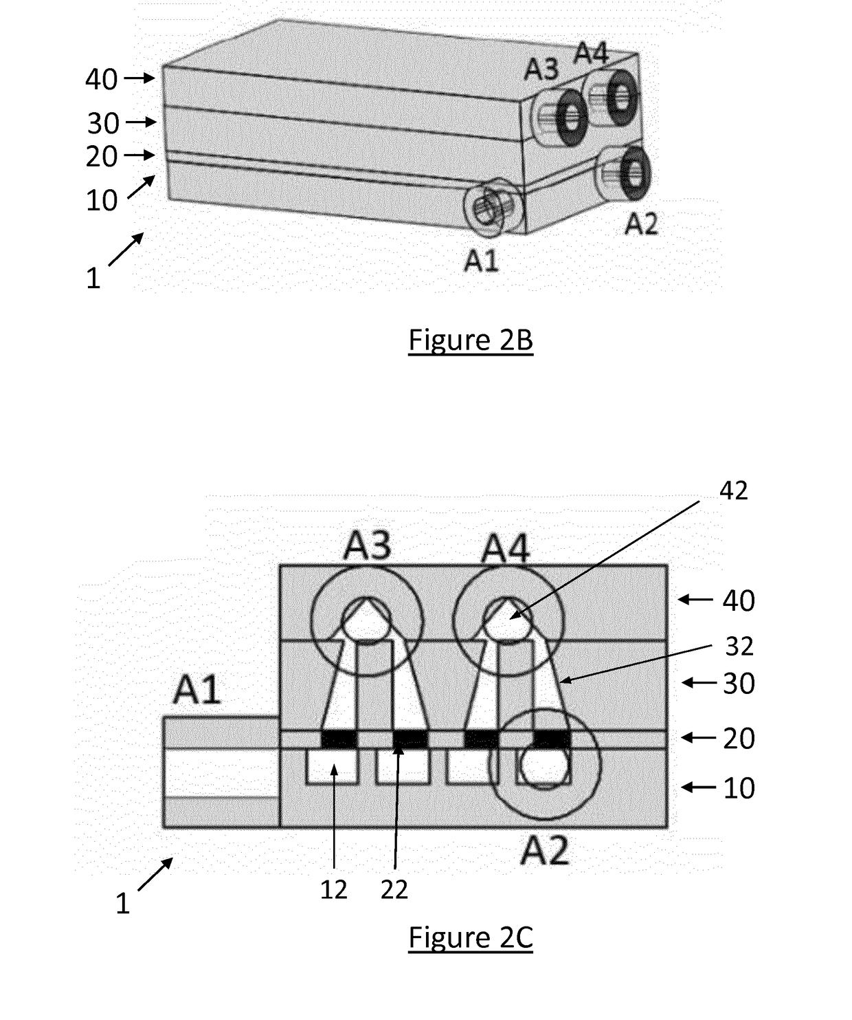
[0090]In FIG. 1A, a medical device 1 is depicted for the selective separation of a biological sample of a mammal into a first portion and a second portion. The medical device 1 generally comprises four layers. The bottom layer 10 comprises a first reservoir 12 for receiving the biological sample. As depicted in FIG. 1, the first reservoir 12 may comprise the majority of the first layer 10 and furthermore is in fluid communication with the fourth layer 20 as indicated by the dashed line. However, other configurations, e.g. a larger first layer relative to the first reservoir, may be provided for e.g. stability and handling purposes.

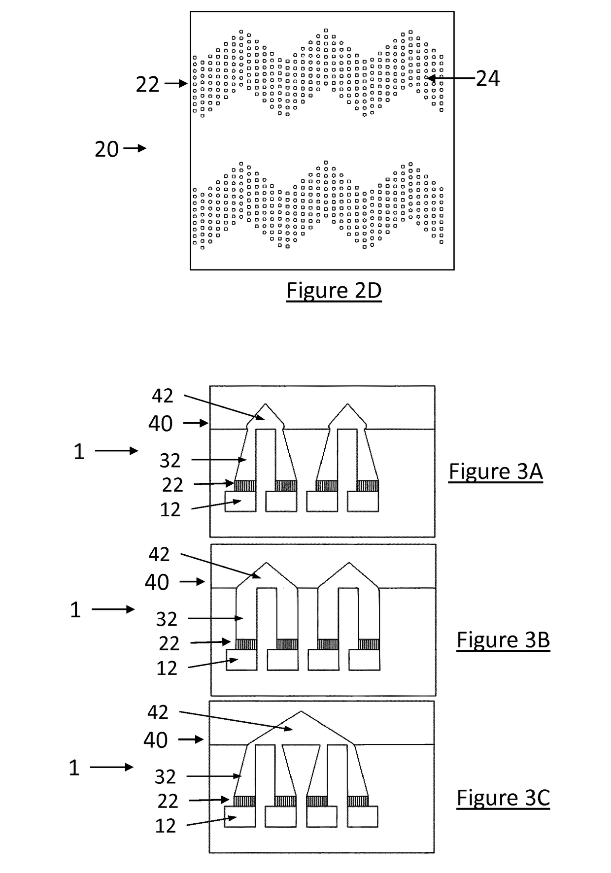
[0091]The fourth layer 20 comprises a separation layer 22, which preferably comprises a porous structure, i.e. allowing compounds with a certain size or molecular mass to pass through the porous structure to the third layer 30. For example, the fourth layer may be a cellulose-based membrane, comprising pores which are large enough for spermatozoa to pass t...


PUM


Abstract

The present invention provides a medical device and a method for the selective separation of a biological sample of a mammal into a first portion and a second portion. It comprises a first layer comprising a first reservoir for receiving the biological sample and for retaining the first portion of the sample, and a second layer comprising a second reservoir for receiving the second portion of the sample. Between the first layer and the second layer a third layer is provided, wherein the third layer comprises a plurality of channels configured to provide a fluid communication between the first reservoir and the second reservoir. Furthermore, between the first layer and the second layer a fourth layer adjacent to the third layer is provided, wherein the fourth layer comprises and / or is configured as a separation layer. At least the third and fourth layer are configured to selectively separate the biological sample between the first reservoir and the second reservoir into the first portion and the second portion of the sample. According to the invention, the layers are to be understood to be stackable in a substantially vertical plane, forming a three-dimensional layered structure.

Description

TECHNICAL FIELD[0001]The present invention relates to a medical device and a method for separating a biological sample of a mammal, preferably semen, into a first and second portion. In particular, the invention relates to a medical device and a method for a motility-based separation of biologically active sperm cells for the enrichment of qualitative sperm.TECHNOLOGICAL BACKGROUND[0002]Infertility is an increasing phenomenon leading to prevalence rates ranging between 11 and 16 percent in both industrialized and developing countries worldwide. Couples coping with these problems in many cases seek medical assistance to artificially enhance their pregnancy and live birth potential. Known technologies include e.g. Assisted Reproductive Technologies (ART) and Intrauterine Insemination (IUI). Unfortunately, the success rate of ARTs is generally only about 20 to 30 percent and often requires 4 to 5 fertilization rates. Furthermore, assisted reproduction had mainly focused on increasing t...

Claims


Application Information

Patent Timeline
IPC IPC(8): C12M3/00C12N5/071C12M1/00B01L3/00
CPCC12M21/06C12N5/0612C12M47/02B01L3/502753B01L2200/0652B01L2300/0887B01L2400/06B01L2300/0681B01L2300/1805B01L2400/0638B01L2400/0666
Inventor VOLLMER, MARION
Owner MY123BABY MEDICAL LTD
Learn More