


Quick Research
Generate reliable direction feasibility study reports for your R&D in just a few steps.

Technical Q&A
Discover and master advanced knowledge NOW. Basics, ideas, possibilities, all at once.

Find Solutions
As an expert in R&D theories, this can generate solutions to your technical problems instantly.

Evaluate Feasibility
Analyze your overall solution with one click, know your potential R&D risks in advance.

Monitor Landscape
Get weekly tech updates, stay abreast of the latest tech innovations and key insights.
In vivo detection of a xenon-binding cage molecule
a molecule and cage technology, applied in the field of in vivo detection of xenon-binding cage molecules, can solve the problem that xenon biosensors have yet to be detected in living animal models following intravenous injection
- Summary
- Abstract
- Description
- Claims
- Application Information
AI Technical Summary
Benefits of technology
Problems solved by technology
Method used
Image

Examples
Embodiment Construction
[0032]Unless defined otherwise, all technical and scientific terms used herein have the same meaning as commonly understood by one of ordinary skill in the art to which the invention belongs. Although any methods and materials similar or equivalent to those described herein can be used in the practice or testing of the present invention, the preferred methods and materials are now described. All publications mentioned hereunder are incorporated herein by reference.
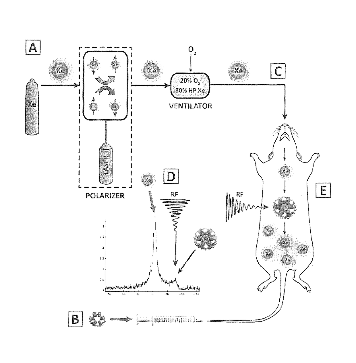
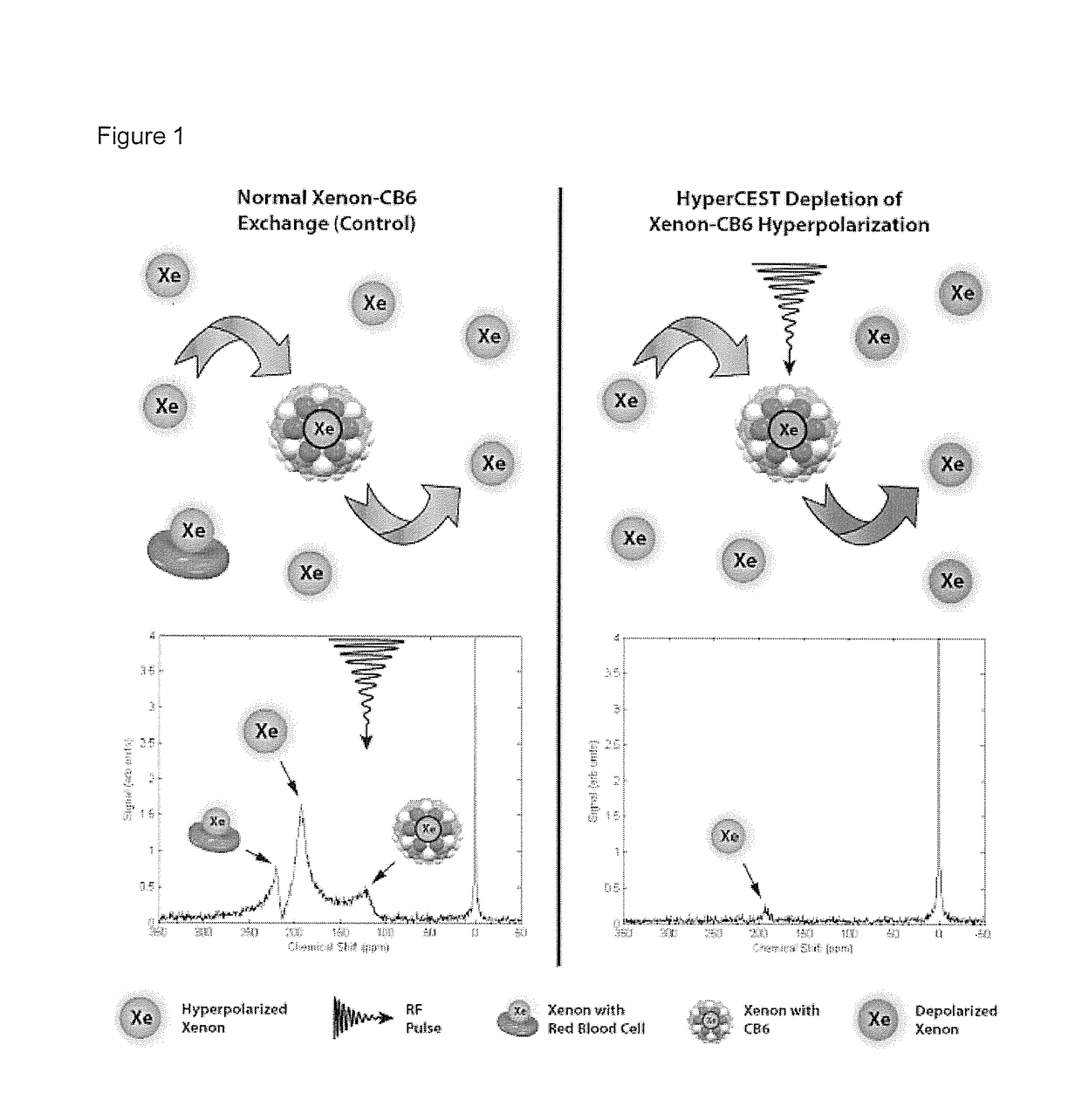
[0033]Herein, we demonstrate the first example of the in vivo detection of a HP gas MRI contrast agent using HyperCEST-enhanced 129Xe MRI, of the cucurbit[6]uril (CB6) cage molecule, within the vasculature of a live rat. By having the rat breathe xenon gas, which dissolves in the blood and interacts with the injected CB6 cages circulating in the vasculature, we were able to successfully detect the presence of CB6 in the brain, heart, aorta, carotid arteries, kidneys, and eventually followed its renal clearance into the bla...
PUM
| Property | Measurement | Unit |
|---|---|---|
| chemical shift | aaaaa | aaaaa |
| chemical shift | aaaaa | aaaaa |
| chemical shift | aaaaa | aaaaa |
Abstract
Description
Claims
Application Information
 - R&D Engineer
- R&D Manager
- IP Professional
- Industry Leading Data Capabilities
- Powerful AI technology
- Patent DNA Extraction
Browse by: Latest US Patents, China's latest patents, Technical Efficacy Thesaurus, Application Domain, Technology Topic, Popular Technical Reports.
© 2024 PatSnap. All rights reserved.Legal|Privacy policy|Modern Slavery Act Transparency Statement|Sitemap|About US| Contact US: help@patsnap.com