Irreversible electroporation to create tissue scaffolds

a tissue scaffold and electroporation technology, applied in the field of biomedical engineering, can solve the problems that the use of ire in generating tissue scaffolds for tissue engineering has not been recognized

Active Publication Date: 2017-07-27
VIRGINIA TECH INTPROP INC
View PDF4 Cites 40 Cited by
  • Summary
  • Abstract
  • Description
  • Claims
  • Application Information

AI Technical Summary

Benefits of technology

The present invention provides a method of making engineered tissue scaffolds using irreversible electroporation to decellularize natural tissue. This method uses a controlled, precise way to destroy cells of a tissue or organ while leaving the underlying ECM, including vascularization, neural tubes, and other gross morphological features of the original tissue intact. The decellularized scaffolds are then suitable for seeding with cells of the appropriate organism. This technology addresses long felt needs in the tissue engineering field and provides a novel means for achieving that goal.

Problems solved by technology

However, the usefulness of IRE in generating tissue scaffolds for tissue engineering has not been recognized.

Method used


Image

  • Irreversible electroporation to create tissue scaffolds
  • Irreversible electroporation to create tissue scaffolds
  • Irreversible electroporation to create tissue scaffolds

Examples


example 1

eshly Excised Mouse Tissue to Create a Scaffold: IRE Scaffold Test Protocol

[0080]A single mouse was sacrificed via CO2 asphyxiation and the liver was surgically removed. Two round sections (1 experimental, 1 control) were removed from the liver using a 5 mm core borer. A straight edge was then cut into each section to facilitate orientation and imaging. The first section was subjected to eighty 100 μs 2500V / cm pulses at 4 Hz. The second section was not treated and left as control. Each section was then divided into 5 samples using a scalpel. The outer samples were discarded, leaving three samples from the experimental section and three samples from the control section.

[0081]One sample from the experimental section and one sample from the control section were subjected to sonication for 2 hours at 37° C. One sample from the experimental section and one sample from the control section were subjected to agitation via stir bar for 2 hours at 37° C. with a rotational rate of 60-300 rpm. ...

example 2

rmance Indicia

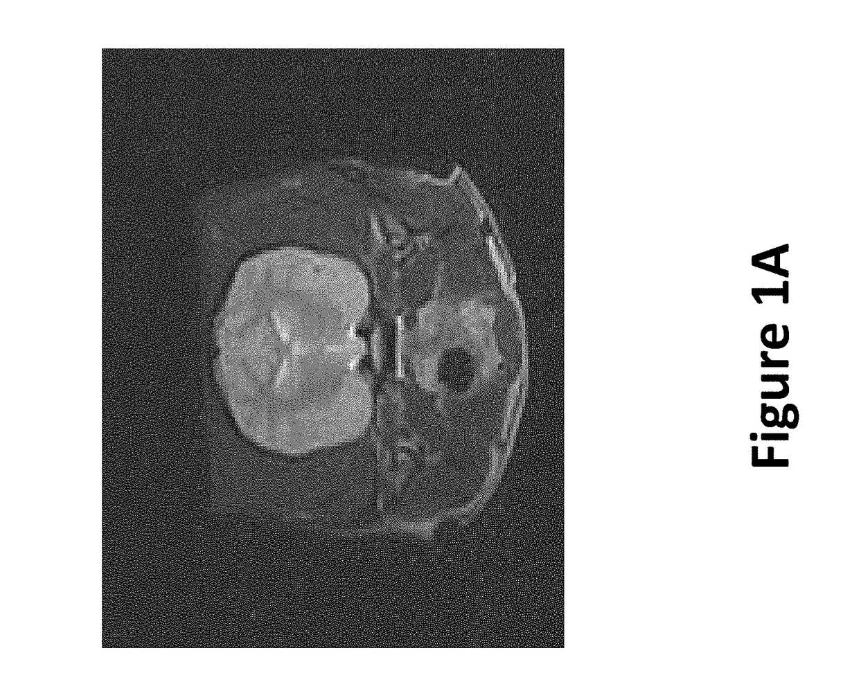
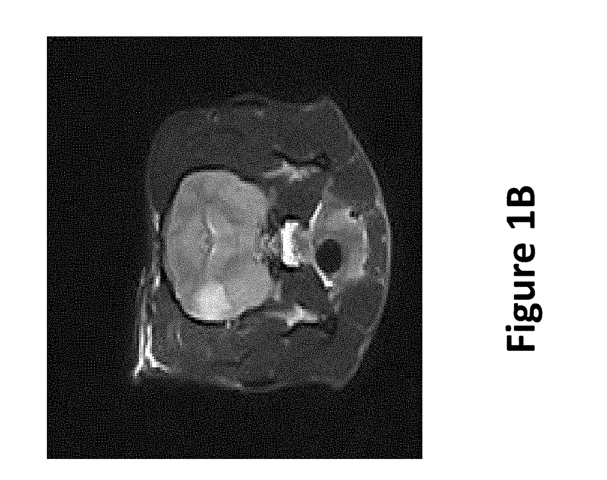
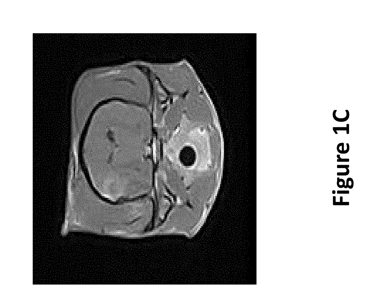
[0084]To illustrate 1) the possibility to monitor creation of the scaffold in real-time using imaging techniques, 2) the variety of tissues that can be used, and 3) how to preserve vasculature, a healthy female purpose bred beagle was used. Nine sets of ten pulses were delivered with alternating polarity between the sets to prevent charge build-up on the electrode surfaces. The maximum voltage-to-distance ratio used was 2000 V / cm because the resulting current did not exceed 2 amps. The charge that was delivered to the brain during the IRE procedure was 22.5 mC, assuming ninety pulses (50 μs pulse durations) that would result from a maximum hypothetical current of 5 amps.

TABLE 1IRE pulse parametersEXPOSUREGAPVOLTAGE TOPULSEELECTRODELENGTHDISTANCEVOLTAGEDISTANCEDURATIONTYPE[mm][mm][V]RATIO [V / cm]PULSES[μs]1 mm5550010009050BipolarStandard7160020009050

[0085]Method: After induction of general anesthesia, a routine parietotemporal craniectomy defect was created to expose the...


PUM


Abstract

The present invention provides engineered tissue scaffolds, engineered tissues, and methods of using them. The scaffolds and tissues are derived from natural tissues and are created using non-thermal irreversible electroporation (IRE). Use of IRE allows for ablation of cells of the tissue to be treated, but allows vascular and neural structures to remain essentially unharmed. Use of IRE thus permits preparation of thick tissue scaffolds and tissues due to the presence of vasculature within the scaffolds. The engineered tissues can be used in methods of treating subjects, such as those in need of tissue replacement or augmentation.

Description

CROSS-REFERENCE TO RELATED APPLICATIONS[0001]The present application is a Divisional of U.S. patent application Ser. No. 12 / 432,295 filed Apr. 29, 2009, which published as U.S. Patent Application Publication No. 2009 / 0269317 on Oct. 29, 2009, and which relies on the disclosure of and claims the benefit of the filing date of U.S. Provisional Patent Application No. 61 / 125,840, filed Apr. 29, 2008. The entire disclosures of each of these applications are hereby incorporated herein by reference.BACKGROUND OF THE INVENTION[0002]Field of the Invention[0003]The present invention relates to the field of biomedical engineering. More specifically, the invention relates to methods of producing naturally-derived scaffolds for creation of tissues for medical uses, and tissues created from those scaffolds.[0004]Description of Related Art[0005]Tissue engineering holds great promise for treating some of the most devastating diseases of our time. Because engineered tissue and organ replacements can ...

Claims


Application Information

Patent Timeline
Patent Type & Authority Applications(United States)
IPC IPC(8): A61L27/36C12N13/00A61L27/38A61K35/12
CPCA61L27/3691A61L27/3625A61L27/3604A61L2430/40A61L27/38C12N13/00A61L2430/28A61L27/3633A61K35/12C12N5/0068C12N2533/90
Inventor DAVALOS, RAFAEL V.
Owner VIRGINIA TECH INTPROP INC
Why Eureka
Try Eureka